Actinic keratosis and premalignant skin lesions
Actinic keratosis and other premalignant skin and mucosal lesions: Bowen disease, keratoacanthoma, leukoplakia and field cancerisation.
- Actinic keratosis is a rough, scaly patch of sun-damaged skin that is a precancerous (premalignant) keratinocyte lesion: a small but real proportion progress to cutaneous squamous cell carcinoma if left untreated, while a similar proportion regress on their own.
- It rarely occurs alone. Most patients have field cancerisation, a wider area of sun-damaged skin containing visible and invisible dysplastic change, which is why treatment often targets a whole field, not just the obvious spot.
- Related premalignant lesions in this topic are Bowen disease (squamous cell carcinoma in situ of the skin), keratoacanthoma, and leukoplakia (mainly oral, strongly linked to tobacco), each with its own scale and management.
- Management is a choice between lesion-directed treatment (for a few isolated lesions, most often cryotherapy) and field-directed therapy (for multiple lesions: 5-fluorouracil, imiquimod, photodynamic therapy, and others), always combined with lifelong sun protection.
- Guidelines agree on the categories (American Academy of Dermatology 2021; European guidance 2015 and 2024; British Association of Dermatologists 2017) while doses and regimens vary by product and country.
By the end of this page you should be able to:
- Recognise actinic keratosis and field cancerisation and explain how chronic ultraviolet (UV) light produces them.
- Place Bowen disease, keratoacanthoma, keratoacanthoma-like lesions and leukoplakia in the spectrum of premalignant disease and distinguish each from invasive squamous cell carcinoma.
- Choose and describe first-line lesion-directed and field-directed treatments with their typical adult regimens, expected clearance and common adverse effects.
- Use the management algorithm, including when to biopsy, escalate or refer.
- State the prognostic numbers that matter (risk of progression, regression, recurrence) and the special cautions for immunosuppressed and high-risk patients.
- Explain sun protection, chemoprevention and low-resource management of these lesions.
This topic covers the common (and clinically paired) premalignant lesions of the skin and mucosal surfaces. The centrepiece is actinic keratosis, the everyday work of dermatology and primary care clinics, together with the concept of field cancerisation that surrounds it. Bowen disease, keratoacanthoma and leukoplakia are then placed beside it so that a medical student can move comfortably between "a scaly spot on a sun-exposed forearm", "a red plaque on a bald scalp", "a dome-shaped crater on a sun-damaged hand" and "a white patch inside the mouth".
Throughout, national and regional guidance differs. British practice is shaped by the British Association of Dermatologists (BAD) care guideline (2017 edition), National Health Service primary-care pathways and the National Institute for Health and Care Excellence (NICE) Clinical Knowledge Summaries, while much published trial data and the 2021 guideline that quotes doses below comes from the American Academy of Dermatology (AAD). European guidance exists in both a 2015 evidence- and consensus-based S3 guideline (European Dermatology Forum) and a European consensus-based interdisciplinary guideline (2024, on behalf of European Association of Dermato-Oncology, European Dermatology Forum, European Academy of Dermatology and Venereology, and Union of Medical Specialists). Where guidelines differ, both values are given rather than a single "correct" one.
DefinitionActinic keratosis
A premalignant keratinocyte lesion (also called solar keratosis) that develops on skin with long-term exposure to ultraviolet radiation. Histologically, the atypical keratinocytes are confined to the lower part of the epidermis rather than full-thickness dysplasia.
The AAD defines an actinic keratosis more tightly as a keratinocyte neoplasm on chronically UV-exposed skin.[1] In practice the two phrases describe the same lesion.
Field cancerisation is not a separate lesion but a state of the skin: multiple clinical and subclinical (visible and invisible) areas of dysplasia and keratinocyte change within one continuous region of chronic sun damage.[7] The concept was first introduced studying tissue around oral squamous cell carcinoma in 1953 and is now applied to sun-damaged skin, where one or more actinic keratoses sit in a carpet of photodamaged epidermis.[7] This explains why a single cryotherapy freeze can clear the frozen spot while new lesions appear beside it a year later, and why field-directed therapy is recommended even when only a handful of lesions are visible.[7]
Bowen disease is squamous cell carcinoma in situ: malignant-appearing squamous (keratinocyte) cells occupy the full thickness of the epidermis but have not yet broken through the basement membrane.[5] It is named after the American dermatologist John T. Bowen, who described it in 1912.[5] Although premalignant, the risk of progression to invasive squamous cell carcinoma is low, reported as about 3% to 5%.[5] A genital form, erythroplasia of Queyrat, carries a higher risk.
Keratoacanthoma is a low-grade, rapidly growing (1 to 2 cm), dome-shaped skin tumour with a central keratin plug, originating from the hair follicle (pilosebaceous) unit.[4] It passes through three stages: rapid proliferation for about 6 to 8 weeks, a maturation phase, and finally involution to a scar.[4] It is benign but shares histology with squamous cell carcinoma and is now often reclassified histologically as squamous cell carcinoma, keratoacanthoma type, so it is treated as premalignant/malignant in practice.
Leukoplakia is a white patch or plaque on the oral mucosa that cannot be diagnosed clinically or pathologically as any other specific condition.[6] The World Health Organization (WHO) groups it among potentially malignant disorders. Homogeneous leukoplakia (uniformly white, usually asymptomatic) has a lower risk of transformation; non-homogeneous (irregular, nodular or speckled red-and-white) has a greater probability of malignant change; proliferative verrucous leukoplakia is the most aggressive variant.[6] Cutaneous (actinic) lip lesions and actinic cheilitis follow the same sun-driven logic as actinic keratosis and are often managed on the same pathway, although the AAD AK guideline deliberately did not cover premalignant neoplasia on non-keratinising epithelium such as actinic cheilitis.[2]
Other lesions that sit on sun-damaged skin and are managed alongside actinic keratosis include solar lentigulae, cutaneous horns (often superimposed on an underlying keratoacanthoma or squamous carcinoma), and lichenoid or pigmented variants of actinic keratosis. Bowenoid lesions and intraepidermal carcinoma are discussed above. Dysplastic naevi belong to a different pathway (melanoma) and are not covered here.
| Lesion | Typical site | Appearance | Malignant potential (reported) | Usual first approach |
|---|---|---|---|---|
| Actinic keratosis | Sun-exposed skin (face, scalp, forearms, hands) | Rough, scaly macule/papule or plaque on erythematous base (often better felt than seen) | about 0.025% to 16% per lesion per year in immunocompetent patients; under 0.1% to 20% cited in series[1][3][7] | Cryotherapy for few; field therapy for many |
| Field cancerisation | Whole photo-exposed area around AK | Diffuse erythema, telangiectasia, mottled pigmentation, scattered scaly lesions | Drives new AKs and squamous cell carcinoma risk | Photoprotection plus field-directed therapy |
| Bowen disease | Sun-exposed skin, trunk, digits | Single or multiple persistent red, scaly patches or plaques; on glans penis as erythroplasia | about 3% to 5% to invasive squamous cell carcinoma (higher in genital form) | Excision or field topical/PDT |
| Keratoacanthoma | Sun-exposed, hair-bearing skin; digits, lips | Rapid 1 to 2 cm dome with central keratin crater | Benign but histologically overlaps squamous cell carcinoma; treated as malignant | Excision with 4 mm margin [4] |
| Leukoplakia (oral) | Oral mucosa | White patch or plaque not removable; homogeneous or non-homogeneous | 1% to 9% (reported 0.1% to 17.5% globally) | Remove cause; excise moderate/severe dysplasia |
| Actinic cheilitis | Lower lip | Diffuse scaling, fissuring, atrophy of lip vermilion | Treat like actinic keratosis (not covered by the 2021 AK guideline) | Photoprotection, field therapy, biopsy of suspicious areas |
Actinic keratosis is extremely common in pale-skinned populations. Reported prevalence in Australia is close to 60%, compared with around 20% in the United States.[3] Incidence rises with age and is higher in men because of greater cumulative outdoor exposure and less use of photoprotection. Immunosuppressed patients, for example after solid organ transplantation, develop AKs at younger ages and in larger numbers.
Squamous cell carcinoma in situ (Bowen disease) affected about 14.9 per 100,000 white patients annually in the early 1990s in Minnesota, with incidence recorded as rising ten-fold within a few years.[5] It is most common in middle-aged and older adults and rare in darkly pigmented skin.[5]
Oral leukoplakia has a global prevalence variously estimated at 2.6%, with a malignancy conversion rate reported between 0.1% and 17.5%.[6] In India, where betel-nut and tobacco chew use is common, prevalence has been reported from 0.2% to 5.2% with malignant transformation of 0.13% to 10%.[6] Anywhere from 1% to 9% of people with oral leukoplakia go on to develop invasive cancer in the lesion.[6] Keratoacanthoma occurs most often between 50 and 69 years of age, affects men about twice as often as women, and is rare before age 20.[4]
The dominant cause of nearly everything on this page is chronic ultraviolet injury, principally to fair skin.
- Ultraviolet radiation. Cumulative UV (sunlight, sunbeds) is the independent risk factor that mutates epidermal keratinocytes. UV is a tumour initiator and promoter.[7] Sunburn counts, but so does lifelong low-dose exposure; outdoor occupations and sports add risk.
- Skin phenotype. Fitzpatrick phototypes I and II (pale, light eyes, red or blonde hair, poor tanning, easy burning) carry the highest risk.[3][4]
- Age. Risk accumulates over decades of exposure.
- Sex. AK and keratoacanthoma are more common in men; Bowen disease affects men and women roughly equally.[3][4][5]
- Immunosuppression. Organ-transplant recipients, people living with HIV, and those on long-term immunosuppressive or biologic therapy develop more, larger, faster-progressing AKs and squamous carcinoma at a younger age.[3]
- Prior skin cancer or AK. A history of skin malignancy or AK marks both genetic susceptibility and cumulative UV load.[3]
- Geography. Prevalence rises as one moves toward the equator and at altitude.[3]
- Tobacco and betel nut are the dominant causes of oral leukoplakia, both smoked and smokeless.[6] Reverse smoking (smoke held in the mouth) and HIV also contribute.[6]
- Other causes of Bowen disease. Chronic sun is by far the commonest cause; arsenic ingestion and some viral infections (including human papillomavirus in periungual and genital disease) are also implicated.[5]
- Other triggers for keratoacanthoma. Chemical carcinogens, immunosuppression, trauma (including surgery or laser at the site), BRAF and hedgehog pathway inhibitors, and mutations in p53 or H-Ras have been reported.[4]
UV (especially UVB) directly damages keratinocyte DNA. The mutations that matter are the classic "UV fingerprint" C to T and CC to TT changes, and the gene most often hit is TP53, whose p53 protein normally drives repair or apoptosis of damaged cells.[1] Other repeatedly mutated genes in AK include p16, RAS family members, CDKN2A and TNF-alpha.[1] Reactive oxygen species (largely UVA-driven) contribute further oxidative damage. The result is loss of normal differentiation control and intraepidermal proliferation of dysplastic keratinocytes.
The important clinical ladder is therefore:
normal epidermis → sun-damaged epidermis with scattered atypical cells → actinic keratosis (atypia largely confined to the lower third) → squamous cell carcinoma in situ/Bowen disease (full-thickness atypia) → invasive squamous cell carcinoma.
A given lesion may step forward along that ladder, sit still, or step backward. At a population level the average risk of an individual actinic keratosis progressing in an immunocompetent person is about 8%, with reported per-lesion annual risks ranging from 0.025% to 16%.[1][3][7] However, the crude estimates across studies range from under 0.1% to 20%.[1] At the same time, 15% to 63% of actinic keratoses regress spontaneously each year, and of those that regress, up to 50% recur within the first year.[1] Over 60% of invasive squamous cell carcinomas contain a contiguous residual actinic keratosis, which is why actinic keratosis is treated as an early stage of the same carcinogenic pathway.[1]
Field cancerisation extends the picture: instead of one mutated cell clone, there is a patch of skin carrying independent clones with different but overlapping mutations, all driven by the same carcinogen. New visible lesions are the tip of an iceberg of subclinical change.
Most actinic keratoses cause no symptoms. On examination they are rough, scaly macules, papules or plaques on an erythematous base sitting on visibly sun-damaged skin (mottled pigmentation, telangiectasia, wrinkles, atrophy). Most patients describe them as "rough patches the doctor can feel but I can barely see". They feel gritty or sandpaper-like on palpation, and scale can obscure or accentuate them. Any lesion that is tender, bleeding with minor trauma, rapidly growing, thickened or ulcerated must be reassessed for squamous cell carcinoma rather than reassured.
Sites are the sites of greatest cumulative exposure: face, bald or thinning scalp, ears and neck, dorsal forearms and hands, and the lower legs in women. Variants include atrophic, hypertrophic or hyperkeratotic (with thick scale or cutaneous horn) and pigmented or bowenoid (a dark, more infiltrated plaque) types. Clinically, lesions are sometimes graded as mild, moderate and severe on the 3-grade Olsen scale, but grading is not part of most clinicians' routine workflow, and no gold-standard classification exists (histologic grading systems co-exist).[1]
Dermoscopy helps, particularly on the face: non-pigmented facial actinic keratosis can show a "strawberry pattern" of an erythematous pseudo-network of vessels, prominent follicular openings and surrounding white haloes.[3]
Bowen disease usually looks like a solitary or few persistent erythematous scaly patches or plaques on sun-exposed skin of the head, neck or trunk, and on the backs of the hands or lower legs. Early lesions are red scaly patches; as they progress they become thicker nodules or plaques that may ulcerate. Pigmented variants occur, and the lesion can be mistaken for eczema, psoriasis or a fungal infection, or for melanoma when pigmented. It often persists for months or years and may coexist with actinic keratosis in the same field. A minority (about 3% to 5%) progress to invasive squamous cell carcinoma, and the figure is higher for the genital variant.[5]
Keratoacanthoma begins as a small, round, pink or skin-coloured papule and grows rapidly over about 6 to 8 weeks into a dome-shaped nodule 1 to 2 cm across with a central keratin plug that gives a crateriform ("crater-like") outline.[4] It occurs most often on sun-exposed, hair-bearing areas such as the face, neck, scalp margin, forearm and hand; trunk lesions are uncommon. It is also seen on mucosal, subungual or perianal sites in rarer subtypes. It may be itchy or tender. Regional lymph nodes should be examined because, although rare, invasion and metastasis have been reported.[4] Dermoscopy cannot reliably distinguish keratoacanthoma from squamous cell carcinoma; biopsy depth matters (see Investigations).
Oral leukoplakia is discovered as an incidental white patch or plaque during examination; it cannot be scraped off and is not attributable to another cause such as lichen planus, candidiasis, chronic bite trauma or lupus. Patients often have no symptoms. On palpation it may be flat or indurated. The homogeneous form is flat, sharply demarcated and white; the non-homogeneous form is thick, nodular or verrucous, sometimes speckled red and white (erythroleukoplakia), with a distinctly higher malignant potential. Proliferative verrucous leukoplakia is multifocal, persistent, common in older women, and carries the highest rate of malignant transformation. Tobacco and betel nut are the dominant risk factors; idiopathic (non-tobacco) leukoplakia on the tongue or floor of the mouth in women behaves aggressively.[6]
Scaly or keratotic lesions on sun-damaged skin are a common source of diagnostic error. The table gives the feature that most usefully separates each possibility.
| Possibility | How it is distinguished from actinic keratosis / premalignant lesions |
|---|---|
| Solar lentigo | Flat, evenly pigmented brown macule without scale or induration; no roughness on palpation |
| Seborrhoeic keratosis | Warty, "stuck-on", greasy lesion; tan to black; occurs on trunk as well as face; does not sit in a uniformly sun-damaged field |
| Verruca vulgaris / seborrhoeic wart | Discrete hyperkeratotic papule with thrombosed capillary dots; often on hands |
| Squamous cell carcinoma | Indurated, thicker, faster-growing, tender or bleeding nodule or plaque; may ulcerate; often arises in a previously stable lesion |
| Basal cell carcinoma | Pearly papule with telangiectasia and rolled edge; pigmented variants mimic melanoma |
| Cutaneous horn | Hyperkeratotic projection on a base that must be excised or biopsied to exclude carcinoma |
| Hypertrophic lichen planus | Pruritic, polygonal, violaceous, Wickham-striae plaques, not confined to sun-exposed areas |
| Discoid lupus erythematosus | Erythematous scaly plaques with follicular plugging, scarring and pigment change; often in younger patients |
| Bowen disease | Larger, well-demarcated, persistent plaque; may be on trunk or digits; histology shows full-thickness atypia |
| Keratoacanthoma | Rapidly enlarging dome with central keratin; may involute without treatment |
| Melanoma / amelanotic melanoma | Asymmetry, border irregularity, colour change, evolution; on dermoscopy vascular structures may differ |
| Oral lichen planus / candidiasis / bite trauma | Not adherent scrapable white patches or non-scrapable but clearly attributable lesions in the mouth |
| Leukoplakia mimics | Candidiasis, chemical burn, leukoedema, lupus, psoriasis, white sponge naevus |
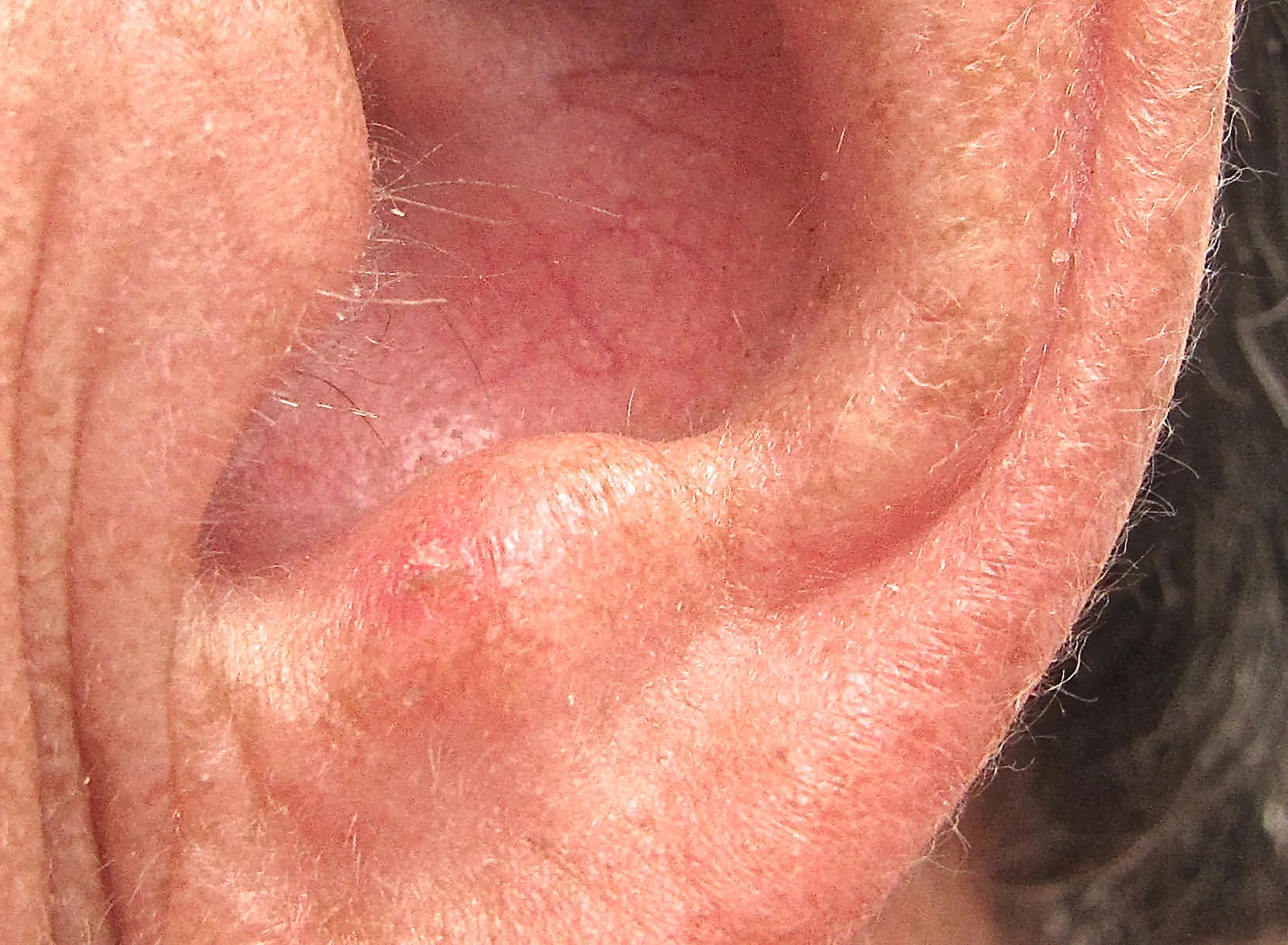
Figure 1Actinic keratosis on ear
Long description
Actinic keratoses are precancerous lesions common on sun-exposed areas of the skin, and this image shows a very common presentation of AKs on an ear.

Most actinic keratoses and Bowen disease lesions are diagnosed clinically on a sun-damaged background. Investigations are used selectively.
- Full-skin examination should be done at least once in anyone with one or more AK, because the same patient often has other pre-cancers and skin cancers.
- Dermoscopy supports the clinical diagnosis and helps select which lesion to biopsy. On non-pigmented facial lesions, the strawberry pattern described above is typical. Dermoscopy cannot reliably distinguish keratoacanthoma from squamous cell carcinoma.[3][4]
- Biopsy is reserved for: lesions that fail to respond to appropriate treatment; any suspected squamous cell carcinoma (firm, enlarging, bleeding, ulcerated, tender); when the diagnosis is uncertain; and when a biopsy result would change management. For oral lesions, excisional biopsy is preferred for small lesions and incisional biopsy (including adjacent normal tissue) for large ones.[3][6]
- Keratoacanthoma specifically needs an excisional biopsy: a shave biopsy is often too superficial to separate keratoacanthoma from invasive squamous cell carcinoma. If the lesion is subungual, radiograph the digit to assess osteolysis.[4]
- Bowen disease is confirmed by biopsy with full thickness of the lesion.
- Oral leukoplakia needs biopsy to exclude dysplasia or carcinoma, followed by surveillance even when excision is performed.
There are no useful blood tests for actinic keratosis itself. In patients who are immunosuppressed or in whom malignancy is suspected, the work-up follows the cancer pathway rather than the keratosis pathway.
There is no single validated scoring system used everywhere.
- AK clinical severity (Olsen and colleagues) grades each lesion from mild (flat, slightly palpable, better felt than seen) through moderate (readily palpable and visible) to severe (thick, obvious hyperkeratosis, possibly with an early cutaneous horn). The AAD guideline noted this scale is used in research but grading is not part of most clinicians' workflow, and no validated gold-standard classification system exists.[1]
- Field cancerisation severity can be quantified (for example with the Actinic Keratosis Area and Severity Index (AKASI) score) and followed with photography, dermoscopy and reflection confocal microscopy in specialist centres.[7]
- Oral epithelial dysplasia (oral leukoplakia) is graded by the fraction of epithelium involved: mild (less than a third), moderate (one third to two thirds) and severe (more than two thirds); carcinoma in situ involves the full thickness without breaching the basement membrane.[6]
- Bowen disease is itself the diagnostic term for carcinoma in situ on clinical grounds supported by histology.
Treatment of actinic keratosis is either lesion-directed (for a few discrete lesions) or field-directed (for multiple lesions and the surrounding field), and the best plan usually combines both. Shared decision-making is essential: patients must tolerate the inflammation, pain and pigment change that accompany any effective treatment, and non-treatment can be appropriate when life expectancy is short or treatment morbidity exceeds benefit.[1][2] [4]
There are no emergencies specific to actinic keratosis. Prompt attention is needed when a lesion is numerous, bleeding, painful or growing rapidly, because these are the features that suggest squamous cell carcinoma rather than keratosis.[3] In those cases, biopsy first.
Rigorous sun protection is the first step, and the American Academy of Dermatology (AAD) 2021 guideline made a strong (good practice) recommendation for it.[1][2] Measures with evidence behind them include avoiding both natural and artificial UV, avoiding exposure at peak hours, wearing sun-protective clothing, wide-brimmed hats and sunglasses, and using broad-spectrum sunscreen that blocks both UVA and UVB.[2][3] European guidance specifically recommends sunscreens containing liposome-encapsulated DNA photolyase alongside high UVA and UVB filters, which repair cyclobutane pyrimidine dimers and have been shown to reduce new actinic keratoses and to be useful after treatments such as photodynamic therapy.[7] The AAD 2021 review reported a vehicle-controlled 6-month study of sunscreen (sun protection factor 17) in which users developed fewer new actinic keratoses (rate ratio 0.62; 95% CI 0.54 to 0.71) and had greater odds of remission of existing ones (odds ratio 1.53; 95% CI 1.29 to 1.80), and that daily sunscreen users over two years had a 24% lower count of actinic keratoses than discretionary users.[1] [4]
Chemoprevention is an adjunct, not a substitute, for treatment:
- Nicotinamide (vitamin B3) 500 mg twice daily has been shown to be safe and effective in reducing actinic keratoses and new non-melanoma skin cancers in high-risk patients.[3][7]
- Acitretin 25 mg daily (or oral retinoids in selected patients) has the strongest evidence for chemoprophylaxis and treatment of actinic keratosis in solid organ transplant recipients.[7]
- Topical photolyase-containing products can be used as monotherapy or after procedural therapy in patients with field cancerisation.[7]
- Polypodium leucotomos extract as an oral photoprotective supplement is sometimes used as an adjunct in highly sun-exposed patients.[7]
These target individual lesions. They are appropriate when there are only a few actinic keratoses, or for a keratoacanthoma, an atypical lesion or a thick plaque.
- Cryotherapy (liquid nitrogen) is the most widely used lesion-directed method and is strongly recommended by the AAD 2021 guideline as good practice.[1][2] It is quick and office-based. Clearance varies with freeze time: in a prospective multicentre spray-cryosurgery study, complete clearance was 39% with freezes of under 5 seconds, 69% with 5 to 20 seconds, and 83% with over 20 seconds. Reported overall cure rates across series range from 57% to 98.8%.[1] Shorter freezes lessen pain and dyspigmentation but lower cure rates; longer freezes on thick facial lesions risk scarring. Cryotherapy treats the lesion, not the subclinical field around it.
- Curettage and electrodesiccation, or a shave biopsy, remove the lesion and provide tissue for histology. They are useful for hyperkeratotic lesions that fail other therapy, and for keratoacanthoma on the extremities when it is under 2 cm.[3][4]
- Surgical excision is required when the diagnosis is uncertain, when squamous cell carcinoma is suspected, or for keratoacanthoma. Keratoacanthoma is treated by excision with a 4 mm margin.[4] Larger or cosmetically sensitive tumours can need tissue-sparing surgery (Mohs micrographic) and aggressive (over 2 cm) or perineural tumours are an indication for Mohs surgery.[4] Subungual keratoacanthoma requires radiographic follow-up of the digit.[4]
- Laser ablation (CO2, erbium:YAG) and chemical peels (30% to 45% trichloroacetic acid) are alternative field or lesion-destructive techniques; a single session of Jessner solution plus 35% TCA can approach the results of 5-fluorouracil twice daily for 3 weeks, while medium to deep peels are generally needed for field disease. In the AAD 2021 review, 3-month clearance was 78.2% with cryosurgery versus 72.4% with laser ablation.[1][3][7]
These treat multiple and subclinical lesions across a whole field and are the treatment of choice whenever more than a few actinic keratoses are present in one region.
| Agent | Typical concentration and regimen | Key published figures | AAD 2021 strength |
|---|---|---|---|
| 5-fluorouracil (5-FU) | 5% cream twice daily for 4 weeks; or 0.5% cream once daily for 1, 2 or 4 weeks | 4-week 5% 5-FU: complete clearance 38% vs 17% with vehicle at 6 months; 0.5% also superior to vehicle; over 90% have local irritation with 0.5% 5-FU/salicylic acid | Strong, moderate-quality evidence [1] |
| Imiquimod | 5% cream two to three times weekly for 1 to 2 courses of 4 weeks (often 4 to 8 weeks total); 3.75% daily for two 3-week blocks; 2.5% with similar scheduling | 5%: average complete clearance 29.3% (9–24 doses) and 40.8% (32–56 doses); discontinuation due to adverse events 1.6% to 33.3%; 3.75%: 34% to 35.6% complete clearance; influenza-like symptoms up to 10.3% | Strong, moderate-quality evidence [1] |
| Diclofenac 3% in 2.5% hyaluronic acid gel | Twice daily for 60 to 90 days (some protocols run 60 to 168 days) | 60-day: 33% vs 10% clearance; 90-day: 42% vs 14% complete clearance; local adverse events in 29% at 24 weeks vs 4.7% with vehicle; discontinuation 15% vs 4% | Conditional, low-quality evidence [1] |
| Tirbanibulin 1% ointment | Once daily for 5 days (face/scalp) | Complete clearance 44% vs 5% (trial 1) and 54% vs 13% (trial 2); 47% recurrence at 1 year among complete responders | Not in 2021 guideline (approved since) [1] |
| 5-FU plus calcipotriene (calcipotriol) | Applied twice daily for 4 days (emerging) | 87.8% vs 26.3% reduction on face and 27% vs 0% complete clearance (face) in one trial | Outside the scope of the 2021 guideline [1] |
| Diclofenac + cryosurgery, imiquimod + cryosurgery | Sequential field-directed then cryotherapy | Modestly improved clearance; extra burden and cost | Conditional recommendations against/for addition vary |
| Photodynamic therapy (PDT) | ALA 10% gel, 1 to 4 hours incubation (AAD 2021), then red light; or daylight PDT with 30 minutes incubation and 2 hours daylight on the face/scalp | ALA-red light PDT: 89.1% vs 32.7% lesion clearance at 12 weeks; complete clearance 77.1% vs 16.6%; long-term skin cancer at treated site 3.6% vs 5.0% at 1 year | Conditional, low-quality (one session) to moderate (two sessions) [1][7] |
| Chemical peels (TCA 30–45%) | Office procedure, often repeated | Effectiveness around 75%; medium/deep peels match field data | Not a formal AAD recommendation; British guidance includes peels [3] |
| Laser resurfacing (CO2/erbium:YAG) | Office procedure | Clearance similar to cryosurgery (72.4% at 3 months) but less widely available | Conditionally favour cryosurgery over laser |
| Ingenol mebutate 0.05–0.15% | Previously 2 to 3 days | Withdrawn from many markets (European and Canadian) over a possible excess skin cancer signal compared with imiquimod at 3 years | Not recommended (unavailable) |
Imiquimod activates toll-like receptor 7, driving a type 1 (interferon-rich) response against dysplastic keratinocytes. 5-fluorouracil is an antimetabolite that preferentially kills rapidly dividing cells. Diclofenac, a non-steroidal anti-inflammatory drug, is gentler but much less effective and carries the boxed warnings of the class for cardiovascular thrombotic events and gastrointestinal bleeding, ulceration or perforation.[1] PDT works photochemically: a topically applied prodrug (aminolevulinic acid or methyl aminolevulinate) is converted in dysplastic cells to protoporphyrin IX, which is activated by light to generate reactive oxygen species. Because PDT also targets subclinical change, it is particularly useful for field cancerisation, although in the United States the methyl aminolevulinate formulation is not available and only aminolevulinic acid protocols are used.[1][7]
A useful pearl from European clinical experience is the 12-day uninterrupted course of imiquimod 5% (rather than intermittent three nights weekly) as an adherence-friendly alternative, and the use of a 7-day pretreatment with topical 5-fluorouracil before daylight photodynamic therapy on the limbs to improve clearance.[7]
- Bowen disease is treated predominantly by destructive or topical means: local excision or mapping biopsy with complete excision is standard, with topical 5-fluorouracil and imiquimod, photodynamic therapy and (for those with multiple or inaccessible lesions) radiotherapy as accepted options.[5]
- Keratoacanthoma is managed surgically first-line. Excision with a 4 mm margin is the treatment of choice; lesions under 2 cm on the extremities can be treated with curettage and electrodesiccation, with Mohs surgery reserved for aggressive, large or perineurally invasive tumours. Nonsurgical intralesional methotrexate (regression in 83% to 100% of small series) and intralesional 5-fluorouracil (40 to 75 mg weekly for 3 to 8 weeks; response reported around 98%) exist for cosmetically sensitive or poor-surgical-candidate cases, but rest on small series. Topical 5-fluorouracil, topical imiquimod and oral isotretinoin have case-level data only.[4]
- Oral (and cutaneous) leukoplakia management begins with removing the cause (smoking cessation, stopping betel-nut chewing, treating candidiasis), plus biopsy. Lesions on high-risk sites (ventral tongue, floor of mouth, soft palate, oropharynx) or with moderate or severe dysplasia are excised or laser-ablated; a homogeneous buccal lesion elsewhere may be watched closely with periodic review. Proliferative verrucous leukoplakia and erythroleukoplakia should always be excised.[6] Recurrence after excision is not uncommon and outcome surveillance continues lifelong if dysplasia was present.
The AAD 2021 guideline issued 18 evidence-based recommendations.[1] In summary:
- 5-fluorouracil plus cryosurgery is preferred over cryosurgery alone for actinic keratosis (conditional).
- Imiquimod plus cryosurgery is preferred over cryosurgery alone, though on lower quality evidence.
- Diclofenac or topical adapalene should not be added to cryosurgery.
- Imiquimod should not be routinely added after photodynamic therapy (modest extra lesion reduction, no difference in complete clearance, and added cost and burden).
Clinicians in practice combine and sequence approaches frequently (for example 5-fluorouracil before daylight photodynamic therapy on the limbs), and the approach should be tailored to the size of the field and to what the patient can tolerate.[7]
Anyone with actinic keratosis should be taught self-surveillance of the skin and reviewed periodically; the frequency depends on the number of lesions and other risk factors. Refer promptly, or biopsy rather than treat, when:
- the diagnosis is uncertain, or the lesion is firm, indurated, rapidly growing, bleeding or ulcerated;
- there are many lesions or a large field in an immunosuppressed patient;
- field treatment fails or lesions recur;
- the patient is at high risk (transplant recipient, xeroderma pigmentosum, significant immunosuppression);
- a pigmented lesion changes, or any lesion has features suggesting melanoma.
Suspected invasive squamous cell carcinoma or melanoma should enter a suspected-cancer (two-week-wait) referral pathway according to national rules.
In resource-limited settings, the principles do not change, only the toolkit shrinks.
- Clinical diagnosis is sufficient for typical actinic keratoses; biopsy only the changing, atypical or bleeding lesion.
- Liquid nitrogen cryotherapy and surgical excision are the backbone of lesion-directed care and should be preserved.
- Where topical 5-fluorouracil or imiquimod is unavailable, the AAD 2021 guideline noted that 5-fluorouracil preparations can sometimes be formulated locally in a petrolatum or base cream, and trichloroacetic acid peels and dermabrasion require simple equipment.[7]
- Sun protection education is the cheapest and most powerful intervention; local, high-UV settings (equatorial, high-altitude) make it essential.
- In East Africa, people with albinism carry a very high burden of actinic keratosis and squamous carcinoma and should be screened regularly and offered photoprotection from childhood.
- Photodynamic therapy, fractional lasers and proprietary dermatologic devices are often unavailable; secondary or tertiary referral and excision for suspected carcinoma remain the priority.
- Immunosuppressed patients have more and more aggressive lesions; evidence at the time of the AAD 2021 guideline was insufficient to issue specific treatment recommendations for transplant recipients or other immunosuppressed patients, and decisions are individualised, often with dermatology-led programmes (acitretin, reduced immunosuppression where safe).
- Older adults may reasonably be offered observation for limited-life-expectancy patients when the morbidity of treatment exceeds benefit.[2]
- Children with actinic keratosis are rare and warrant a search for a genodermatosis (xeroderma pigmentosum, albinism) or an immunodeficiency.
- Pregnancy and breastfeeding: ablative treatments are preferred and topical cytotoxics avoided where possible; management should follow local specialist advice.
- Bowen disease of the nipple/anogenital area needs specialist management and exclusion of an underlying carcinoma.
- Malignant transformation to squamous cell carcinoma, usually signalled by induration, bleeding, ulceration or rapid growth.
- Recurrence and new lesions in the same and adjacent fields, including after regression.
- Keratoacanthoma complications: metastasis and perineural spread are rare but reported; subungual lesions can cause osteolysis.
- Treatment adverse effects: blistering, erosion, crusting, pain, pruritus, erythema and oedema (universal across all active treatments); dyspigmentation (especially with cryotherapy and imiquimod); influenza-like symptoms with imiquimod; scarring with prolonged or aggressive freezes; rarely severe cardiovascular or gastrointestinal events with topical diclofenac (class warning). No active treatment is risk-free.[1][3]
- Cosmetic and psychologic effects: pigment change, scarring and the emotional burden of a visible chronic disease.
Prognosis for an individual actinic keratosis is good but unpredictable: the AAD 2021 pooled estimates gave an average progression risk of about 8% (range 0.025% to 16% in immunocompetent patients) and a crude range of under 0.1% to 20% across studies; 15% to 63% regress each year; 50% of those that regress recur within a year.[1][3] European authors quote 0.025% to 16% progression per lesion per year.[7] These widely differing figures reflect small, short, heterogeneous studies: what matters clinically is that progression is possible, not its exact rate.
Keratoacanthoma has an excellent prognosis after surgical excision but signals a skin cancer field, so ongoing surveillance is needed. Bowen disease has a low progression rate (3% to 5%, higher for genital disease) and is controlled by simple office measures. Oral leukoplakia carries an overall malignancy risk of 1% to 9% (up to 17.5% in older series, with much higher rates for proliferative verrucous and non-homogeneous forms).[5][6] Successfully treated patients without immunosuppression and with good photoprotection do well.
Primary prevention is reducing cumulative UV from childhood onward: shade, clothing, hats, sunglasses, avoidance of tanning beds, and broad-spectrum sunscreen. Secondary prevention is detecting and treating lesions early. Patients with actinic keratosis should have periodic full-skin checks and be taught to self-monitor for the "ABCDE of the single rough scaly spot": any lesion that becomes firm, tender, bleeds, grows or fails to heal. High-risk groups (transplant recipients, people with albinism, xeroderma pigmentosum, severe immunosuppression) need structured surveillance. In the AAD 2021 executive summary, a vehicle-controlled trial showed sun protection also reduced the incidence of new actinic keratoses, and the guideline strongly recommended shared decision-making and observation as appropriate options.[2]
- Actinic keratosis is a sun-driven premalignant keratinocyte lesion: average progression risk about 8% (reported range 0.025% to 16%, crude 0.1% to 20%), but 15% to 63% regress annually, and half of regressions recur within a year.
- It is almost always part of field cancerisation, so clearing the obvious spot is not the whole plan; photoprotection and, when needed, field-directed therapy are essential.
- Distinguish actinic keratosis from Bowen disease (carcinoma in situ, 3% to 5% progression), keratoacanthoma (benign but histologically overlaps carcinoma, treated surgically) and oral leukoplakia (1% to 9% malignancy, excise dysplastic lesions).
- First-line for a few lesions: cryotherapy (freeze longer than 5 seconds for acceptable clearance; 5 to 20 seconds clears about 69%, over 20 seconds about 83%).
- First-line field therapy: 5-fluorouracil 5% twice daily for 4 weeks or 0.5% daily for 1, 2 or 4 weeks; imiquimod 5% (two to three times weekly, 4 to 8 weeks) or 3.75% daily in two 3-week blocks; both strongly recommended in the AAD 2021 guideline.
- Diclofenac 3% gel twice daily for 60 to 90 days is gentler but less effective; photodynamic (aminolevulinic acid, 1 to 4 hours incubation then red light) and daylight photodynamic therapy are useful for fields; tirbanibulin 1% ointment for 5 days is effective where available.
- Always combine treatment with sun protection: avoidance, clothing and broad-spectrum sunscreen; a sun protection factor 17 sunscreen reduced new lesions by a rate ratio of 0.62 in one trial and daily use gave a 24% lower lesion count. [1]
- Chemoprevention helps high-risk patients: nicotinamide 500 mg twice daily, acitretin in transplant recipients, photolyase-containing photoprotectors.
- Urgent referral or biopsy, not topical therapy, is required for an indurated, bleeding, rapidly growing, non-healing or treatment-resistant lesion.
- Keratoacanthoma is treated by excision with a 4 mm margin; Bowen disease by excision or field therapy; oral leukoplakia by cause removal and excision when moderate or severely dysplastic.
- Guidelines (AAD 2021; European S3 2015 and European consensus 2024; BAD 2017) agree on the categories but differ in doses and emphasis; always check the local formulary.
Answer each one in your head first, then open the answer.
A 62-year-old outdoor worker has ten rough scaly patches on his forearms and hands, with general sun damage between them. What is the most appropriate overall management?
Show answerHide answer
Lesion-directed plus field-directed treatment: cryotherapy for any thick or symptomatic individual lesions, plus a field-directed agent such as 5-fluorouracil cream or imiquimod across the whole damaged area, or photodynamic therapy. Combine this with rigorous photoprotection (avoidance, clothing, broad-spectrum sunscreen) and review; biopsy anything that is tender, bleeding, indurated or fails to respond.
Which topical agent is strongly recommended for field treatment of actinic keratosis in the 2021 American Academy of Dermatology guideline, and which is only conditionally recommended?
Show answerHide answer
Strongly recommended: 5-fluorouracil and imiquimod, plus cryosurgery and ultraviolet protection as good-practice statements. Conditionally recommended: photodynamic therapy and diclofenac. (Ingenol mebutate is withdrawn/unavailable.)
A 70-year-old man has a rapidly enlarging, 1.5 cm dome-shaped nodule with a central keratin plug on the dorsum of his hand; it appeared 6 weeks ago. Diagnosis and treatment?
Show answerHide answer
Keratoacanthoma. Treatment of choice is surgical excision with a 4 mm margin. A shave biopsy is insufficient; if excision is not immediately possible, intralesional 5-fluorouracil or methotrexate (case series only) may be considered. Because keratoacanthoma is histologically close to squamous cell carcinoma, referral and histology are essential.
What is field cancerisation, and why does it change management even when only three actinic keratoses are visible?
Show answerHide answer
Field cancerisation is a contiguous area of sun-damaged skin containing visible and subclinical dysplastic change. Multiple independent clones sit in the same field, so treating only the visible lesions leaves subclinical disease that will produce new keratoses. Field-directed therapy and photoprotection address the whole field.
How long should 5-fluorouracil be applied, and what is the expected clearance?
Show answerHide answer
5% 5-fluorouracil cream is applied twice daily for 4 weeks. In a large placebo-controlled trial, complete clearance at 6 months was 38% versus 17% with vehicle. The 0.5% formulation is used once daily for 1, 2 or 4 weeks and is also superior to vehicle. Local irritation is almost universal (over 90% with the salicylic acid combination) and expected.
A woman with immunosuppression after renal transplantation has numerous actinic keratoses on her face and forearms. What is special about her management?
Show answerHide answer
Immunosuppressed patients develop more numerous and more aggressive lesions and the 2021 guideline had insufficient evidence for specific recommendations in this group. Management should be specialist-led and intensified, with a low threshold for biopsy; chemoprevention such as acitretin, careful UV protection, and sometimes reduction of immunosuppression are considered. They are also entering their skin cancer surveillance programme.
How would you distinguish Bowen disease from a plaque of actinic keratosis?
Show answerHide answer
Bowen disease (squamous cell carcinoma in situ) is a larger, well-demarcated, persistent erythematous plaque, sometimes on the trunk or digits, with full-thickness atypia on histology and a 3% to 5% progression rate to invasive squamous cell carcinoma (higher for the genital form). Actinic keratosis is a smaller, rough, scaly lesion with atypia confined to the lower epidermis and a lower average progression risk per lesion, although squamous carcinoma often arises in or beside one.
What simple, cheap interventions matter most for actinic keratosis in a resource-limited clinic?
Show answerHide answer
Accurate clinical diagnosis with biopsy only for atypical or changing lesions; liquid nitrogen cryotherapy or simple surgical excision; strong counselling on sun avoidance, clothing and sunscreen; and early referral of suspicious or treatment-resistant lesions. Nicotinamide 500 mg twice daily is an inexpensive adjunct for high-risk patients.
- Squamous cell carcinoma of the skin and its management
- Sun protection and skin cancer prevention
- Basal cell carcinoma
- Melanoma and dysplastic naevus syndrome
- Human papillomavirus and anogenital lesions
- Dermoscopy and skin biopsy techniques
- Immunosuppression and cancer surveillance
- Oral cancer and oral mucosal disease
- Eisen DB, Asgari MM, Bennett DD, et al. Guidelines of care for the management of actinic keratosis. J Am Acad Dermatol 2021;85(4):e209–e233. doi:10.1016/j.jaad.2021.02.082. Accessed .
- Eisen DB, Asgari MM, Bennett DD, et al. Guidelines of care for the management of actinic keratosis: Executive summary. J Am Acad Dermatol 2021;85(4):945–955. doi:10.1016/j.jaad.2021.05.056. Accessed .
- Marques E, Chen TM. Actinic Keratosis. StatPearls [Internet]. Treasure Island (FL): StatPearls Publishing; 2023. Updated 2023 Aug 17. Accessed .
- Zito PM, Scharf R. Keratoacanthoma. StatPearls [Internet]. Treasure Island (FL): StatPearls Publishing; 2023. Updated 2023 Aug 8. Accessed .
- Charifa A, Volner K, Oakley AM. Intraepidermal Carcinoma. StatPearls [Internet]. Treasure Island (FL): StatPearls Publishing; 2023. Updated 2023 Sep 4. Accessed .
- Mohammed F, Fairozekhan AT. Oral Leukoplakia. StatPearls [Internet]. Treasure Island (FL): StatPearls Publishing; 2023. Updated 2023 Jul 17. Accessed .
- Piquero-Casals J, Morgado-Carrasco D, Gilaberte Y, et al. Management pearls on the treatment of actinic keratoses and field cancerization. Dermatol Ther (Heidelb) 2020;10(5):903-915. doi:10.1007/s13555-020-00425-4. Accessed .
Figure credits
Educational content for medical students and doctors. It is not a substitute for clinical judgement or local guidelines.