Fungal skin infections: tinea, candida, sporotrichosis
Fungal skin infections summarised and treated: tinea versicolor, dermatophytosis by site, cutaneous candidiasis and sporotrichosis.
- Fungal skin disease is not one condition: it is three separate problems that share a microscope. Dermatophytes (ringworm, "tinea") live on keratin in skin, hair and nails. Malassezia yeasts cause pityriasis (tinea) versicolor. Candida causes moist-fold (intertriginous) rash. Sporothrix is a deeper infection from a thorn or splinter.
- A scaly, itchy, annular plaque with a raised active border on ordinary skin is a dermatophyte until proved otherwise. A moist red rash with satellite pustules in a fold is Candida. Fine, non-itchy, hypo- or hyper-pigmented patches on the trunk are pityriasis versicolor.
- Most infections are cured with the right topical cream or shampoo used long enough. Nails and scalp need oral treatment, and a boggy, painful scalp (kerion) is urgent.
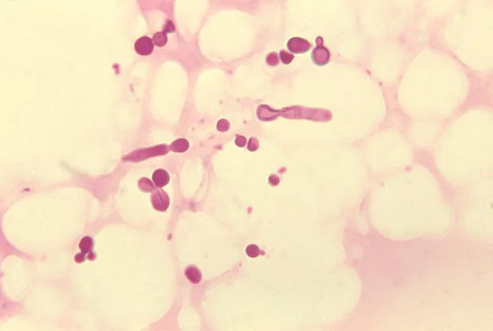
- Confirm with potassium hydroxide (KOH) when you can: hyphae mean dermatophyte; pseudohyphae plus budding yeast mean Candida; short stubby hyphae mixed with round spores ("spaghetti and meatballs") mean pityriasis versicolor.
- Avoid combination steroid-antifungal creams: they mask the fungus, produce "tinea incognito", and do not speed cure [5].
By the end of this page you should be able to:
- Classify the major superficial fungal skin infections by organism, site and depth.
- Recognise dermatophyte, Candida, pityriasis versicolor and sporotrichosis lesions and name their mimics.
- Perform and interpret a KOH preparation and Wood's lamp examination.
- Choose topical, oral and surgical treatment by site, with drug, dose and duration, and know when to refer.
- Advise patients on hygiene, animal contacts and prevention of recurrence.
- Adjust management for children, pregnancy and older adults.
DefinitionFungal skin infection
Dermatophytes are moulds that need keratin. They are the only common fungi that can digest keratin, so they confine themselves to stratum corneum, hair and nails; they do not invade living tissue unless the host is seriously immunocompromised. [5] Three genera cause almost all human disease: Trichophyton, Microsporum and Epidermophyton. Their species are described by where they normally live: anthropophilic (human to human, usually least inflamed), zoophilic (from animals, more inflamed) and geophilic (from soil). [2]
Yeasts are different. Candida species, especially Candida albicans, are commensals of skin folds and mucosae; disease is overgrowth pushed by moisture, occlusion, antibiotics, diabetes or immunosuppression. [4] Malassezia species are lipid-loving commensals of human skin that cause pityriasis versicolor when they switch from the round yeast form to short hyphae; they are not dermatophytes, despite the old name "tinea versicolor". [1] [8]
Sporothrix schenckii is a dimorphic fungus: mould in the environment at room temperature, yeast at body temperature. [3] Infection is acquired by inoculation - a rose thorn, splinter, sphagnum moss, or the bite or scratch of an infected cat - and the fungus travels along lymphatics.
Dermatophyte infections are the single most common group of superficial mycoses and occur worldwide. [5] Patterns depend on site and place. Trichophyton rubrum dominates in Europe and North America. In UK cities most scalp infection is caused by Trichophyton tonsurans, while in mainland Europe and rural parts of the UK Microsporum canis (from cats and dogs) is the usual culprit. [14]
- Tinea pedis (athlete's foot) is the commonest dermatophyte in adults. It favours warm, occlusive footwear, public showers, communal pools and sharing towels. Interdigital infection can serve as a reservoir for nail and groin infection. [5]
- Tinea capitis is mainly a disease of pre-pubertal children; in many settings it is the commonest dermatophytosis of childhood. Infection is transmitted by direct contact and by fomites such as combs, hats and pillowcases. [10] [14]
- Tinea cruris ("jock itch") is commoner in adult men and in hot, humid climates; it usually starts from a toe-web infection carried by hand or clothing.
- Tinea unguium (onychomycosis) rises with age and is the commonest fungal infection seen in older adults in many series. Diabetes, peripheral vascular disease and nail trauma increase risk. [5]
- Tinea manuum is often unilateral ("two feet, one hand") and should prompt a search for tinea pedis.
Pityriasis versicolor is common wherever the climate is warm and humid. It affects adolescents and young adults most, and can remain year-round in the tropics. Family clustering is reported, and warm, sweaty, occluded skin encourages overgrowth. [1] [6]
Cutaneous candidiasis is a disease of skin folds and of the host. Risk factors are moisture and maceration: obesity, incontinence, infancy (napkin candidiasis), skin folds under the breast or in the groin, occlusive dressings and warm climates. Systemic or local immunosuppression, broad-spectrum antibiotics, corticosteroids and diabetes predispose. [4] [11]
Sporotrichosis is occupational and environmental. It clusters in gardeners, florists, nursery workers and forestry workers, and in people who handle sphagnum moss, rose thorns or splinters. In Brazil and increasingly elsewhere it is a zoonosis transmitted by infected domestic cats. [3] Diabetes, alcohol misuse and immunosuppression raise the risk of disseminated disease. [3]
Dermatophytes bind to keratin and release keratinases that digest it. Growth occurs at the advancing edge of the lesion, which explains the raised, scaly active border and the healing centre that gives the "ring". Inflammation varies from almost none (anthropophilic chronic infection) to a brisk, sometimes pustular response (zoophilic infection). [9] A distant, "id" or dermatophytid reaction - an allergic, sterile vesicular rash on the hands or trunk - can appear when the fungus is elsewhere. [5] Topical corticosteroids blunt this inflammation without killing the fungus, producing atypical tinea incognito: larger, less scaly, often pustular lesions that are hard to recognise. [5] [13]
Candida is a normal inhabitant of folds. Disease occurs when its micro-environment changes - warmth, moisture, loss of bacterial competition after antibiotics, hyperglycaemia - and the yeast converts to a pseudohyphal form that invades the stratum corneum and provokes the characteristic sharply demarcated erythema with satellite lesions. [4]
Pityriasis versicolor is not infection from an outside source. The Malassezia yeast already on the skin undergoes a temperature- and humidity-driven phase change to the mycelial form; the resulting metabolites (including dicarboxylic acids that inhibit tyrosinase) cause the pigment change. Because the organism is a commensal, the infection is not contagious. [1] [8]
Sporothrix is dimorphic. Conidia in the environment are inoculated into subcutaneous tissue, convert to yeast at 37 °C and spread along lymphatic channels to form a chain of nodules that may ulcerate. [3]
Tinea corporis. Well-demarcated, mildly erythematous, scaly patch or plaque that is annular or oval with a raised, active, sometimes pustular leading edge and central clearing; pruritus is common. Lesions occur on the trunk, neck, arms and legs. [9] Atypical shapes follow prior steroid use (tinea incognito). [5] [9]
Tinea cruris. Pruritic, half-moon-shaped erythematous patches in the groin and inner thighs, often with a raised scaly border; the scrotum is usually spared, which helps distinguish it from candida intertrigo. [5]
Tinea pedis. Three main patterns: interdigital maceration and fissuring between the fourth and fifth toes; a "moccasin" pattern of fine scale on the soles, heels and sides of the foot; and a vesicular/bullous type on the instep. Chronic plantar infection can look like eczema. [5]
Tinea capitis. Scaling, patchy hair loss with broken hairs ("black dots") for anthropophilic species; there may be pustules and crusting. A kerion is a painful, boggy, purulent swelling with matted hair and regional lymphadenopathy - a hypersensitivity reaction that can scar and cause permanent alopecia if untreated. [10] [14]
Tinea manuum and tinea faciei. Tinea manuum is often a single dry, scaly hand. Tinea faciei produces annular facial plaques that flare with sun exposure. Tinea barbae is a deep, often pustular infection of bearded skin in men.
Tinea unguium (onychomycosis). Thickening, discoloration (yellow, white or brown), onycholysis and subungual debris; often starting at the distal lateral corner of the nail. Nails grow slowly, so cure is slow. [5]
Dermatophytid ("id") reaction. A sterile, symmetrical, vesicular eruption on the hands, feet or trunk in a patient with active tinea elsewhere. It worsens when the underlying infection is treated abruptly and resolves with cure of the primary lesion. [5]
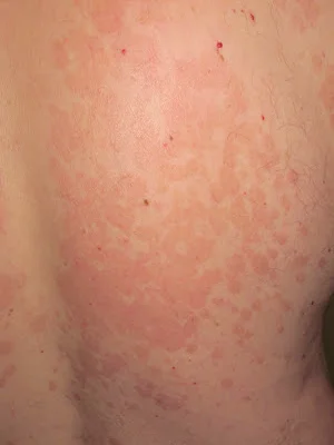
Fine, slightly scaly macules or patches that are hypo- or hyper-pigmented relative to surrounding skin, typically on the upper trunk, neck, shoulders and upper arms; lesions can join into larger patches. Pruritus is absent or mild. [1] [6] In a tanned person the affected skin does not tan, so the patches become obvious in summer. After successful treatment the colour takes weeks to months to settle. [12]
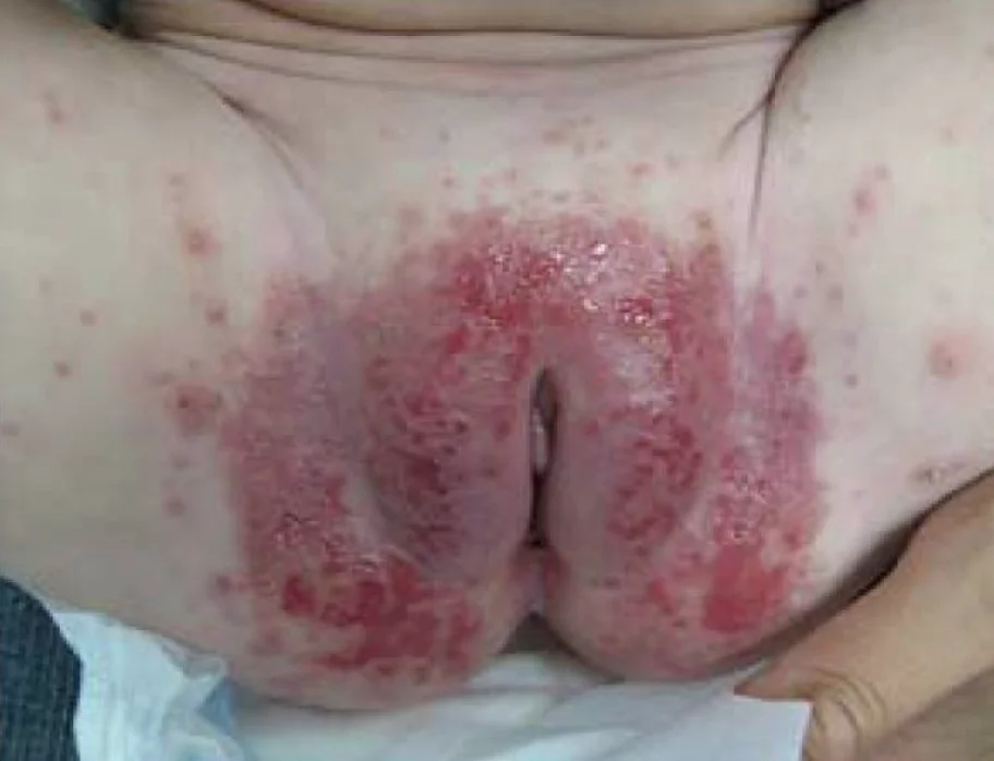
A red, moist, glazed or macerated patch in a body fold, with a scalloped border and satellite papules or pustules at the edge. Sites are the groin, gluteal cleft, axillae, inframammary folds, abdominal overhang and nappy area in infants. Itching, soreness and burning are common. Candidal paronychia produces a tender, swollen, retracted nail fold. [4] [11]
The cutaneous (fixed) form is a firm, pink or violaceous nodule or ulcer at the site of inoculation, often on the hand or finger, that may resemble an insect bite, pyoderma or mycobacterial infection. The more common lymphocutaneous form adds a linear chain of subcutaneous nodules and ulcers along draining lymphatics - the "sporotrichoid" pattern. Disseminated (cutaneous) disease occurs in immunocompromised hosts and can destroy cartilage. [3]
- Map every lesion and its site, then check the feet, groin, nails and scalp even if the presenting complaint is elsewhere; treating only the visible lesion invites recurrence.
- Look for the active border and central clearing of tinea; for satellite pustules of Candida; for follicular pustules of tinea barbae; for a boggy kerion and broken hairs of tinea capitis.
- Wood's lamp. In tinea versicolor the affected skin shows a yellowish-green fluorescence. [6] Dermatophytes as a rule do not fluoresce, with the important exception of Microsporum canis and M. audouinii in scalp infection, which give a blue-green fluorescence. Erythrasma fluoresces coral red and, if seen, changes the diagnosis.
- Evoked scale sign: gently scraping the lesion makes scale more apparent in pityriasis versicolor. [8]
| Presentation | Likely diagnosis | Key distinguishing feature |
|---|---|---|
| Annular, scaly, itchy plaque with raised active border | Tinea corporis | Hyphae on KOH; central clearing; zoophilic cases more inflamed.[9] |
| Nummular eczema | Patch is uniformly scaly and inflamed, no active border; KOH negative. | |
| Psoriasis (plaque or guttate) | Sharper, silvery scale; elbows/knees/sacrum; Koebner; KOH negative. | |
| Erythema annulare centrifugum | Trailing scale inside the ring, not at the leading edge. | |
| Granuloma annulare | Non-scaly, smooth papules in a ring; no active border. | |
| Erythrasma (groin/feet) | Brown scaly patches with coral-red Wood's lamp fluorescence; KOH negative. | |
| Intertrigo (candida) | Moist fold with satellite pustules; pseudohyphae on KOH. [4] | |
| Inverse psoriasis / seborrhoeic dermatitis | Sharply defined red plaques in folds; KOH negative; often other sites. | |
| Vitiligo | Chalk-white, totally depigmented, non-scaly; Wood's lamp shows bright white. | |
| Pityriasis alba | Ill-defined hypopigmented patches on face of children; little scale. | |
| Pityriasis rosea | Herald patch, collarette scale, Christmas-tree distribution. | |
| Alopecia areata | Smooth, non-scaly patches; exclamation-mark hairs; no broken stubs. | |
| Trichotillomania | Irregular patches with broken hairs of varying length. | |
| Seborrhoeic dermatitis (scalp) | Greasy scale, no broken hairs, no lymphadenopathy. | |
| Lupus vulgaris / cutaneous TB | Verrucous or ulcerated plaques; biopsy and culture diagnostic. | |
| Sporotrichoid nodule chain | Mycobacterial or Nocardia infection, leishmaniasis, cat scratch disease, tularemia. |
Figure 1Disease known as ringworm, or tinea capitis
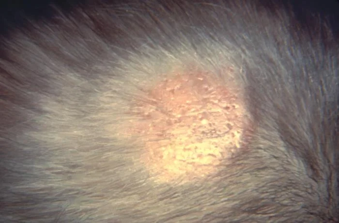
Long description
PHIL image ID 24235 is a 1960 close clinical photograph of a patient's scalp displaying symptoms of ringworm (tinea capitis) caused by the dermatophyte Microsporum nanum, with the PHIL page identifying tinea capitis as an infection of the scalp caused by dermatophyte fungi.

Figure 2Ringworm or Tinea cruris of fungal infection
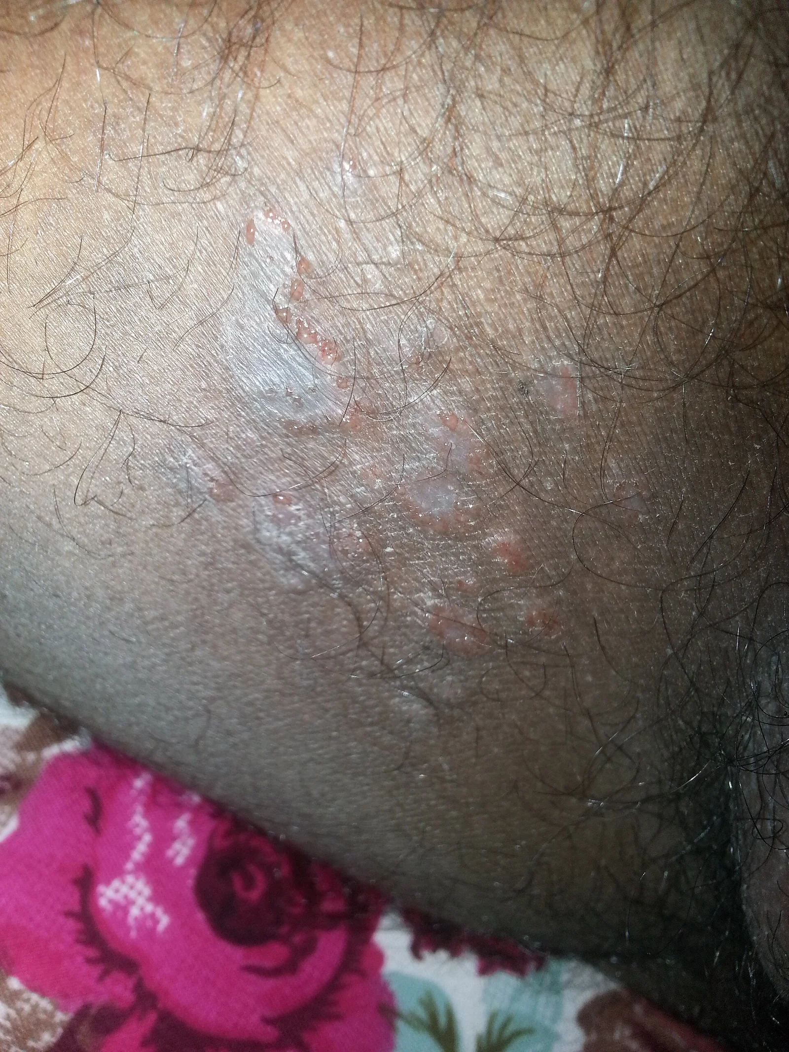
Long description
A clinical photograph of tinea cruris (jock itch) showing a red, itchy, often scaly rash in the groin and upper thigh region.

Figure 3Intertrigo-1
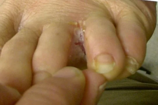
Long description
A clinical photograph credited as 'Athlete's foot or Tinea pedis lesion, a mycosis caused by a dermatophyte fungus', described as intertrigo between the toes.

Figure 4Yeartinfection
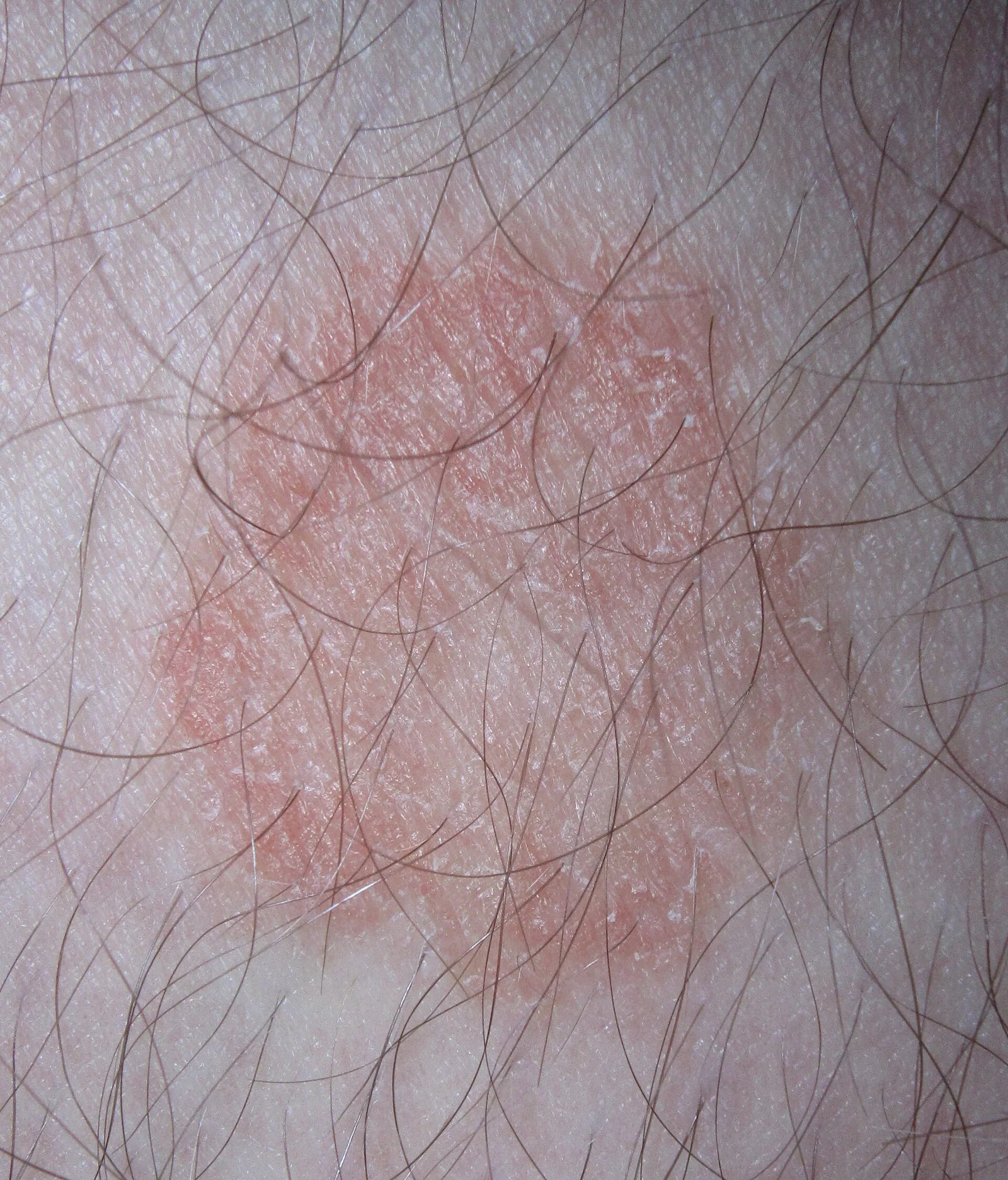
Long description
A close-up clinical photograph of dermatophytosis/ringworm described as ringworm with a mildly raised border and central clearing.

Figure 5DiaperRashCandida
Long description
A clinical photograph of diaper dermatitis complicated by Candida, reproduced as Figure 19 (Candida culture-positive diaper dermatitis) from the open-access article "Diagnosis of Atopic Dermatitis: Mimics, Overlaps, and Complications".

Figure 6Malassezia furfur in skin scale from a patient with tinea versicolor PHIL 3938 lores
Long description
CDC PHIL image 3938 shows yeast-like fungal cells and short hyphae of Malassezia furfur in skin scale from a patient with tinea versicolor.

Figure 7Pitiriasis Versicolor en la Espalda
Long description
A clinical photograph titled in Spanish 'Fungus: Pityriasis Versicolor on the Back' showing pityriasis versicolor on the back.

| Test | When to use | What a positive result means |
|---|---|---|
| KOH preparation | Any suspected tinea, candida or versicolor | Dermatophyte: branching septate hyphae. Candida: pseudohyphae plus budding yeasts. Versicolor: short stubby hyphae mixed with round spores ("spaghetti and meatballs"). [1] [9] |
| Wood's lamp | Scalp ringworm, versicolor, erythrasma | Blue-green fluorescence with M. canis/M. audouinii; yellow-green with versicolor; coral red with erythrasma. |
| Fungal culture (Sabouraud or DTM) | Uncertain diagnosis, severe/extensive disease, immunocompromise, treatment failure, nail or scalp infection | Speciates the organism; DTM turns red for dermatophytes. [10] |
| Periodic acid-Schiff (PAS) stain | Skin or nail biopsy when other tests fail | Shows fungal elements in tissue. |
| PCR for dermatophytes | When confirmation is needed quickly or culture has failed | Faster than culture and useful when confirmation is needed quickly. [5] |
| Blood tests (glucose/HbA1c, HIV) | Recurrent or widespread candida; severe or recurrent dermatophytosis | Look for predisposing disease that must be treated. [11] |
| Mycologic culture and microscopy for sporotrichosis | Any nodular/ulcerative lesion after plant exposure | Culture is the gold standard; [3]. |
Choose the drug from the organism and site, not the brand. The sensible default is a topical allylamine for a limited dermatophyte infection and a topical imidazole for candida and versicolor. Oral treatment is driven by the tissue the fungus has invaded: hair shafts and nails cannot be reached by creams.
| Class | Examples | Use | Typical regimen |
|---|---|---|---|
| Allylamines (fungicidal) | Terbinafine 1% cream, butenafine, naftifine | Tinea corporis, cruris, pedis | Once or twice daily for 1-2 weeks. [13] [9] |
| Imidazoles (fungistatic) | Clotrimazole 1% cream, miconazole cream, ketoconazole, econazole | Candida, versicolor and any tinea | Twice daily, continued for 10 days after lesions heal. [13] [11] |
| Other topical agents | Ciclopirox, tolnaftate, selenium sulfide, zinc pyrithione | Versicolor shampoos/soaps, mild tinea | Selenium sulfide or ketoconazole shampoo for versicolor as needed; [6] [8] |
| Anti-inflammatory adjunct | Hydrocortisone 1% (mild) | Marked inflammation with an antifungal, never alone | Once or twice daily for up to 7 days. [11] |
| Drug | Adult dosing | Main indications | Notes |
|---|---|---|---|
| Terbinafine | 250 mg once daily | Tinea capitis, onychomycosis, extensive or refractory tinea | First-line for tinea capitis and onychomycosis. [5] Children under 25 kg: 125 mg; 25-35 kg: 187.5 mg; over 35 kg: 250 mg daily. [5] |
| Griseofulvin | Microsize 20-25 mg/kg/day (max 1 g); ultramicrosize 10-15 mg/kg/day (max 750 mg) | Tinea capitis (especially Microsporum) | Give with a fatty meal; colour may change little in adults; contraindicated in pregnancy. [5] |
| Itraconazole | 100-200 mg daily | Extensive tinea, onychomycosis, versicolor, sporotrichosis | Many drug interactions (CYP3A4); check QT and interacting medicines. [1] [3] [7] |
| Fluconazole | 150 mg weekly or 50 mg daily | Versicolor, candidiasis, tinea | Useful in candidiasis and in patients who cannot take terbinafine. [1] [11] |
- Tinea corporis, cruris and pedis with limited disease: give a topical allylamine or azole for 1-2 weeks after clearing; [9] [13]. If infection is extensive, multiple, deep, recurrent, chronic, or the patient is immunocompromised, use oral terbinafine or another oral azole. [9] [5]
- Tinea cruris: treat the groin and the feet together, otherwise the toe web re-seeds the groin. Keep the area dry and cool; avoid tight occlusive clothing. [5]
- Tinea pedis: drying between toes, open footwear if possible, and treating concurrent onychomycosis are the real cure. Recurrence after topical therapy alone is common. [5]
- Tinea capitis: this always needs oral treatment (topical alone will not clear the hair shaft). Terbinafine is preferred for most modern (Trichophyton) infection, griseofulvin for Microsporum. [5] [10] Add an antifungal shampoo during treatment to reduce carriage and spread, and examine and sample close contacts. [14] Refer the same week if a kerion is present, and do not squeeze it. [14]
- Tinea manuum, barbae and faciei with facial/hair involvement deserve oral therapy if they are inflammatory or deep.
- Onychomycosis: confirm the diagnosis first. Oral terbinafine (250 mg daily for 6 weeks for fingernails, or 12 weeks for toenails) is preferred where there are no contraindications; topical lacquer or device therapy is reserved for limited disease or when oral drugs are unsuitable. [5]
Treatment clears the organisms but the colour fades slowly. Topical therapy is the treatment of choice because it is effective, cheap and has fewer interactions. [8]
- Small areas: clotrimazole or another imidazole cream twice daily for a few weeks after clearing. [12]
- More widespread: ketoconazole 2% shampoo applied to affected skin once daily for 5 days, lathered and rinsed. [12]
- Very extensive, thick or recurrent disease, or failure of topical therapy: oral itraconazole or fluconazole may be used in non-pregnant adults; refer children and pregnant or breastfeeding women. [12]
- Prophylaxis: in patients with frequent recurrence, intermittent maintenance therapy before exposure reduces relapse; relapse rates can reach 80% within 2 years without prophylaxis. [12]
Treat the organism and the moisture at the same time.
- Limited, non-widespread infection in an adult or child who is not significantly immunocompromised: a topical imidazole (clotrimazole, econazole, miconazole or ketoconazole) or terbinafine; clotrimazole, econazole and miconazole are licensed in children and adults. [11]
- Marked inflammation or itch: add a short course of hydrocortisone 1%, once or twice daily for 7 days, stopping if symptoms resolve, continuing for a further 7 days if there is significant improvement, and reassessing if there is no response. [11]
- Widespread, treatment-resistant or immunocompromised patients 16 years or older: fluconazole 50 mg daily for 2 weeks, then review; extend for a further 2 weeks if not resolved, after which refer, obtain swabs, seek specialist advice or refer to dermatology. [11]
- Children under 16 or where fluconazole is contraindicated: seek specialist advice. [11]
- Measures that actually prevent recurrence: keep folds dry, use emollient soap substitute, change incontinence pads and dressings before they are saturated, reduce occlusion from tight or non-breathable clothing, and address obesity and diabetes. [11]
- Treat paronychia with topical antifungal and protect the nail fold from moisture.
Most cutaneous and lymphocutaneous disease responds to oral itraconazole, which is first-line therapy. [3] Treatment usually continues for weeks to months and is extended for a while after all lesions have healed. [3] Refractory, severe, immunocompromised or pulmonary/disseminated disease may require higher-dose itraconazole, amphotericin B or combination regimens under specialist care, and the nodules are sometimes excised or cryotherapy used for persistent fixed lesions. [3] Prolonged high-dose therapy has been needed in some Sporothrix brasiliensis infections. [7]
Refer to dermatology or infectious disease promptly for: extensive, refractory or frequently recurring infection; tinea capitis with kerion; onychomycosis before starting systemic therapy if diagnosis is unproven; suspected candida in a significantly immunocompromised patient; sporotrichosis (for treatment planning and notification where it is endemic); and any infection where malignancy or a mimic cannot be excluded. [9] [11] [13]
- Bacterial superinfection (cellulitis, impetigo) can arise from scratched dermatophyte lesions; deeper infection and osteomyelitis are risks in diabetes and peripheral vascular disease. [5]
- Permanent scarring alopecia follows a neglected kerion. [14]
- Onychomycosis causes pain, difficulty walking and secondary bacterial infection and does not usually clear without treatment. [5]
- Tinea incognito from inappropriate topical steroids makes diagnosis harder and treatment longer. [5]
- Candida can disseminate in seriously immunocompromised hosts. [4]
- Sporotrichosis can destroy cartilage and disseminate in immunocompromised patients; hyperkeratotic, verrucous or ulcerated lesions can become colonised or secondarily infected. [3]
- Adjunctive hydrocortisone used without an antifungal increases the risk of fungal proliferation and tinea incognito. [11]
Most superficial fungal infections clear with the correct drug and adequate duration, but recurrence is common unless predisposing factors are fixed and, for dermatophytes, the reservoir (feet, nails, contacts, pets) is treated. Pityriasis versicolor has a good prognosis with treatment but is chronic by nature; skin colour takes weeks to months, occasionally years, to normalise, and relapse can reach 80% within 2 years. [12] Tinea capitis is curable if treated; a kerion that is missed can scar. Candidiasis usually resolves once moisture and glucose control are addressed. [11]
There is no population screening. Prevention is practical:
- Keep skin folds dry; use an emollient soap substitute and dry thoroughly, especially between toes and under breasts. [11]
- Do not share towels, combs, hats, bedding or footwear; wash towels and bedding at 60 °C; treat or replace contaminated personal items after scalp infection clears. [10]
- Treat tinea pedis and onychomycosis when present, to stop auto-inoculation to the groin. [5]
- Examine and sample household contacts and pets during a scalp outbreak; carriers can be treated with shampoo to reduce shedding. [14]
- Wear gloves and long sleeves for gardening, handling moss, roses and soil, and thongs in communal showers and changing rooms. [3]
- Avoid unnecessary antibiotics and topical steroids on undiagnosed rashes.
- With frequent versicolor, prophylactic antifungal shampoo before summer can reduce relapse. [8] [12]
Pregnancy. Prefer topical therapy. Topical clotrimazole, miconazole and econazole are licensed for adults and children. [11] Oral terbinafine and azoles are generally avoided in pregnancy because of limited safety data; ketoconazole shampoo has minimal systemic absorption but expert advice is prudent. NICE CKS recommends that children and pregnant or breastfeeding women with pityriasis versicolor be referred for specialist management. [12] Topical corticosteroids are used with caution and for short courses.
Children. Tinea capitis is predominantly a childhood disease and is treated with oral antifungals, with griseofulvin or terbinafine chosen by the organism and local guidance; terbinafine dosing is by weight. [5] For tinea capitis with a kerion, refer urgently. [14] Candidal nappy rash is managed topically, with oral treatment only on specialist advice for children under 16. [11] A child with a dermatophyte infection of the body is usually treated topically unless the disease is extensive.
Older adults. Onychomycosis is very common; oral therapy needs thought about liver and renal function, drug interactions (particularly itraconazole and fluconazole), and adherence over weeks. [5] Confirm before treating nails, because dystrophic nails from trauma or psoriasis are common and will not respond.
Immunocompromised patients. Expect widespread, atypical or refractory disease; lower the threshold for culture, systemic therapy and referral; disseminated Candida and Sporothrix are possible. [4] [3]
Use this as a quick reminder at the bedside.
| Condition | Typical look | Usual first step |
|---|---|---|
| Tinea corporis | Annular scaly plaque, active border, central clearing | Topical allylamine 1-2 weeks [9] |
| Tinea cruris | Groin half-moon, scrotum spared | Topical allylamine or azole, treat feet too [13] |
| Tinea pedis | Toe-web maceration; moccasin scale; vesicles | Topical allylamine, drying, socks [5] |
| Tinea capitis | Scalp scale, broken hairs, black dots, kerion | Oral therapy plus shampoo; contacts [10] |
| Onychomycosis | Thick, yellow, lifted nail | Confirm, then oral terbinafine [5] |
| Pityriasis versicolor | Fine scaly hypo/hyperpigmented patches, trunk | Ketoconazole shampoo or topical azole [12] |
| Cutaneous candidiasis | Moist red fold rash with satellite pustules | Topical imidazole plus dryness [11] |
| Sporotrichosis | Thorn-track nodule chain along lymphatics | Oral itraconazole, weeks to months [3] |
A suspected fungal skin infection
- Lesion with scale, itch or colour changeNote site, shape, border, satellite lesions, hair or nail involvement. Check feet, groin, nails, scalp.
- Question: What does it look like?
Annular scaly plaque, active border
- Tinea corporis/cruris/pedis likelyKOH hyphae; if mild and limited, use a topical allylamine or azole.
- Extensive, deep, immunocompromise or failure?If yes, take culture or PCR and use oral therapy.
Moist fold rash with satellite pustules
- Cutaneous candidiasis likelyKOH pseudohyphae; topical imidazole and dryness; oral fluconazole if widespread or immunocompromised.
Fine, hypo- or hyper-pigmented patches, trunk
- Pityriasis versicolor likelyWood's lamp yellow-green fluorescence; imidazole or ketoconazole shampoo; oral therapy only for extensive disease.
Scalp scale, broken hairs or boggy swelling
- Tinea capitisSample hair; oral therapy; add antifungal shampoo; urgent referral if kerion.
Nail thickening/discolouration
- Confirm onychomycosis firstKOH or PAS; oral terbinafine if confirmed and appropriate.
Nodule or ulcer after plant/soil inoculation
- SporotrichosisCulture; oral itraconazole under specialist guidance.
- Negative or uncertain KOHTreat a limited suspected dermatophyte empirically with topical therapy for 2 weeks and reassess; culture or PCR if severe, atypical or in an immunocompromised patient.
- Refer if severe, failing treatment, immunocompromised, or diagnosis uncertain
- Most cases can be diagnosed and treated without culture: careful inspection, KOH where available, and an antifungal cream go a long way.
- Terbinafine or azole creams are inexpensive and remain first-line for limited dermatophyte disease.
- Where oral agents are unavailable, keep treatment topical and prolonged, and address moisture and predisposing disease.
- Sample and refer cases that are extensive, atypical, immunocompromised, or suspected sporotrichosis.
- The three common fungi look different and live in different places; treat the site, the organism and the moisture, not just the rash.
- A raised, active, scaly border that advances means dermatophyte; culture or PCR confirms; KOH is the quick bedside test.
- Hair shafts and nails cannot be cleared by creams: use oral therapy and confirm the diagnosis first.
- Never use antifungal-corticosteroid combinations or unsupervised topical steroids on an undiagnosed rash.
- Pityriasis versicolor is a commensal phase change, not contagion; topical therapy is first line.
- Candida skin infection is a marker of moisture, glucose and immunity as much as a treatable rash.
- Sporotrichosis is an inoculation mycosis that tracks along lymphatics; oral itraconazole is first-line.
- Treat reservoirs (feet, nails, contacts, pets) and keep the skin dry or recurrence is likely.
- Children, pregnant women and older adults need site-specific choices: griseofulvin for Microsporum capitis, avoid oral azoles in pregnancy, confirm nail disease first.
- In the era of terbinafine-resistant Trichophyton, KOH or PCR and antifungal stewardship matter more than ever. [5] [7]
Answer each one in your head first, then open the answer.
A 7-year-old with patchy hair loss has 'black dots' at scalp level, and a sibling has similar patches. What is the likely diagnosis, and what test confirms it?
Show answerHide answer
Tinea capitis, most likely due to Trichophyton tonsurans in an urban UK setting. Diagnosis is confirmed by mycological examination and culture of plucked hair (and skin), which is preferred to Wood's lamp alone because T. tonsurans does not fluoresce. [14]
A patient has a 2 cm annular, itchy, scaly plaque on the arm after brushing against rose bushes. KOH shows hyphae. Is this tinea corporis or sporotrichosis, and how would management differ?
Show answerHide answer
Why does the scrotum help you tell tinea cruris from candidal intertrigo?
A man has a dry, scaly right hand and chronic athlete's foot. What is the unifying diagnosis, and why does treatment fail if only the hand is treated?
Show answerHide answer
Tinea manuum with tinea pedis: the hand is re-seeded from the feet ("two feet, one hand"). Treating the hand alone leaves the reservoir, so both need treatment and the feet need drying. [5]
A pregnant woman in the second trimester has pityriasis versicolor. What management is appropriate, and what should be avoided?
A child in the tropics has widespread pityriasis versicolor that has not cleared after topical therapy. What is reasonable, and what is the caveat?
Related MedGrasp topics to read first or next: dermatological examination; pharmacology of antifungals; skin infections (bacterial, viral, parasitic); immunosuppression; diabetes mellitus.
- Karray M, McKinney WP. Tinea Versicolor (StatPearls). StatPearls Publishing, Treasure Island (FL). Bookshelf NBK482500. Updated 12 February 2024. Accessed .
- Yee G, Syed HA, Al Aboud AM. Tinea Corporis (StatPearls). StatPearls Publishing, Treasure Island (FL). Bookshelf NBK544360. Updated 14 February 2025. Accessed .
- Syed HA, Talati R. Sporotrichosis (StatPearls). StatPearls Publishing, Treasure Island (FL). Bookshelf NBK532255. Updated 7 July 2025. Accessed .
- Jenks JD, Tobin EH. Candidiasis (StatPearls). StatPearls Publishing, Treasure Island (FL). Bookshelf NBK560624. Updated 31 January 2026. Accessed .
- Caplan AS, Gold JAW, Smith DJ, Ely JW. Diagnosis and Management of Tinea Infections. Am Fam Physician 2025;112(4):382-392. Accessed .
- American Academy of Dermatology (AAD) patient education. Tinea versicolor: diagnosis and treatment. AAD public site, accessed 2026-11-01. Accessed .
- American Academy of Dermatology (AAD) clinical guidance. Preventing and treating Trichophyton indotineae and other emerging dermatophytoses. AAD member site, accessed 2026-11-01. Accessed .
- Leung AKC, Barankin B, Lam JM, Leong KF, Hon KL. Tinea versicolor: an updated review. Drugs Context 2022;11. doi:10.7573/dic.2022-9-2. Accessed .
- Leung AKC, Lam JM, Leong KF, Hon KL. Tinea corporis: an updated review. Drugs Context 2020;9. doi:10.7573/dic.2020-5-6. Accessed .
- Fuller LC, Barton RC, Mohd Mustapa MF, Proudfoot LE, Punjabi SP, Higgins EM. British Association of Dermatologists' guidelines for the management of tinea capitis 2014. Br J Dermatol 2014;171:454-463. doi:10.1111/bjd.13196. Accessed .
- National Institute for Health and Care Excellence (NICE). Candida - skin (NICE CKS, last revised April 2025). NICE Clinical Knowledge Summaries topic, accessed 2026-11-01. Accessed .
- National Institute for Health and Care Excellence (NICE). Pityriasis versicolor (NICE CKS, last revised March 2025). NICE Clinical Knowledge Summaries topic, accessed 2026-11-01. Accessed .
- National Institute for Health and Care Excellence (NICE). Fungal skin infection - body and groin (NICE CKS, last revised July 2023). NICE Clinical Knowledge Summaries topic, accessed 2026-11-01. Accessed .
- National Institute for Health and Care Excellence (NICE). Fungal skin infection - scalp (NICE CKS, last revised February 2025). NICE Clinical Knowledge Summaries topic, accessed 2026-11-01. Accessed .
Figure credits
- Figure 1: cdc, via Wikimedia Commons, Public domain, Public domain.
- Figure 2: Ajay Kumar Chaurasiya, via Wikimedia Commons, CC BY-SA 4.0, CC BY-SA 4.0. resized.
- Figure 3: Drgnu23 (original uploader), via Wikimedia Commons, CC BY-SA 3.0, CC BY-SA 3.0.
- Figure 4: James Heilman, MD, via Wikimedia Commons, CC BY-SA 3.0, CC BY-SA 3.0. resized.
- Figure 5: Elaine C. Siegfried1,†* and Adelaide A. Hebert2,†, via Wikimedia Commons, CC BY 4.0, CC BY 4.0.
- Figure 6: http://phil.cdc.gov/phil_images/20030610/22/PHIL_3938_lores.jpg, via Wikimedia Commons, Public domain, Public domain.
- Figure 7: Dermapixel, via Wikimedia Commons, CC BY-SA 4.0, CC BY-SA 4.0.
Educational content for medical students and doctors. It is not a substitute for clinical judgement or local guidelines.