Head and spinal cord injury in the trauma patient
Traumatic brain and spinal cord injury in the adult trauma patient: recognition, classification, imaging and evidence-based acute management.
- Traumatic brain injury (TBI) is common after head trauma; most are mild, but a few need urgent neurosurgery.
- Severity is graded with the Glasgow Coma Scale (GCS): 13 to 15 mild, 9 to 12 moderate, and 3 to 8 severe (some systems use 14 to 15 as mild).
- The main dangerous primary lesions are epidural (lens-shaped), subdural (crescent-shaped), subarachnoid, intracerebral haematoma and diffuse axonal injury.
- The goal of acute care is to prevent secondary brain injury by keeping the airway, oxygenation, blood pressure and intracranial pressure (ICP) in safe ranges.
- Traumatic spinal cord injury is assessed with the ASIA Impairment Scale; complete and incomplete syndromes are recognised, and spinal shock must be distinguished from neurogenic shock.
- Suspected cervical spine injury is cleared clinically using validated rules; when imaging is needed, CT is preferred at most centres.
By the end of this page you should be able to:
- Grade TBI severity with the GCS and state what each grade implies for care.
- Recognise the five principal brain lesions on CT and the signs of a raised ICP.
- Outline the acute management of raised ICP, step by step, and when to call neurosurgery.
- Identify skull base fracture, concussion and basilar signs, and manage a concussion.
- Explain the incomplete spinal cord syndromes and the ASIA scale.
- Distinguish spinal shock from neurogenic shock.
- Apply the NEXUS and Canadian C-spine rules and describe first-line imaging.
- Explain why corticosteroids are no longer routine and list low-resource priorities.
A head injury brought on by external force - a fall, a crash, a blow, a penetrating wound - is TBI. Most are minor: a confusing few minutes and a headache. A few are catastrophic, and many of the deaths and permanent disabilities they cause come not from the blow itself but from what happens in the minutes and hours afterwards: hypoxia, hypotension, brain swelling and bleeding inside the skull raising the pressure. This topic puts the trauma patient at the centre: the injured brain and the injured spinal cord are treated together in the first minutes, and every decision is aimed at preventing secondary injury. [1]
A patient with head and spinal trauma can have both problems at once. The brain is injured by impact, acceleration and deceleration; the spinal cord by the same forces transmitted to the neck or back, or by penetrating trauma. The priorities are the same for both: secure the airway without moving the neck, maintain oxygen and blood pressure, look for the injuries that kill or paralyse, and move to definitive care. [1] [7]
TBI is physical injury to brain tissue that temporarily or permanently impairs brain function. Injury may be open (penetrating the skull and meninges) or closed. Closed injuries, produced by acceleration-deceleration and rotation, are far more common and are the focus here. Severity on arrival is classified by the GCS score: 13 to 15 is mild TBI, 9 to 12 is moderate, and 3 to 8 is severe. [1] Some references define mild TBI as a GCS of 14 to 15, with confusion or brief loss of consciousness; the difference is one of convention, and in practice the first hour of illness matters more than the exact cut-off. [2]
The scale scores three components: eye opening, verbal response and motor response. [2]
| Component | Best response | Score |
|---|---|---|
| Eyes | Opens spontaneously | 4 |
| Eyes | Opens to voice | 3 |
| Eyes | Opens to pain | 2 |
| Eyes | None | 1 |
| Verbal | Oriented | 5 |
| Verbal | Confused | 4 |
| Verbal | Inappropriate words | 3 |
| Verbal | Incomprehensible | 2 |
| Verbal | None | 1 |
| Motor | Obeys commands | 6 |
| Motor | Localises pain | 5 |
| Motor | Withdraws from pain | 4 |
| Motor | Flexion to pain (decorticate) | 3 |
| Motor | Extension to pain (decerebrate) | 2 |
| Motor | None | 1 |
Total scores range from 3 to 15. A combined score of 8 or less is usually regarded as coma. [1] Scores after resuscitation, once hypoxia and hypotension have been corrected, are more specific for brain dysfunction than the first reading. [1]
Severity drives management. Mild TBI is observed or discharged with return precautions. Moderate TBI is admitted and watched closely because deterioration is common. Severe TBI (GCS 3 to 8) calls for airway control, intensive supportive care and treatment of raised ICP. [1]
Figure 1Trauma subdural
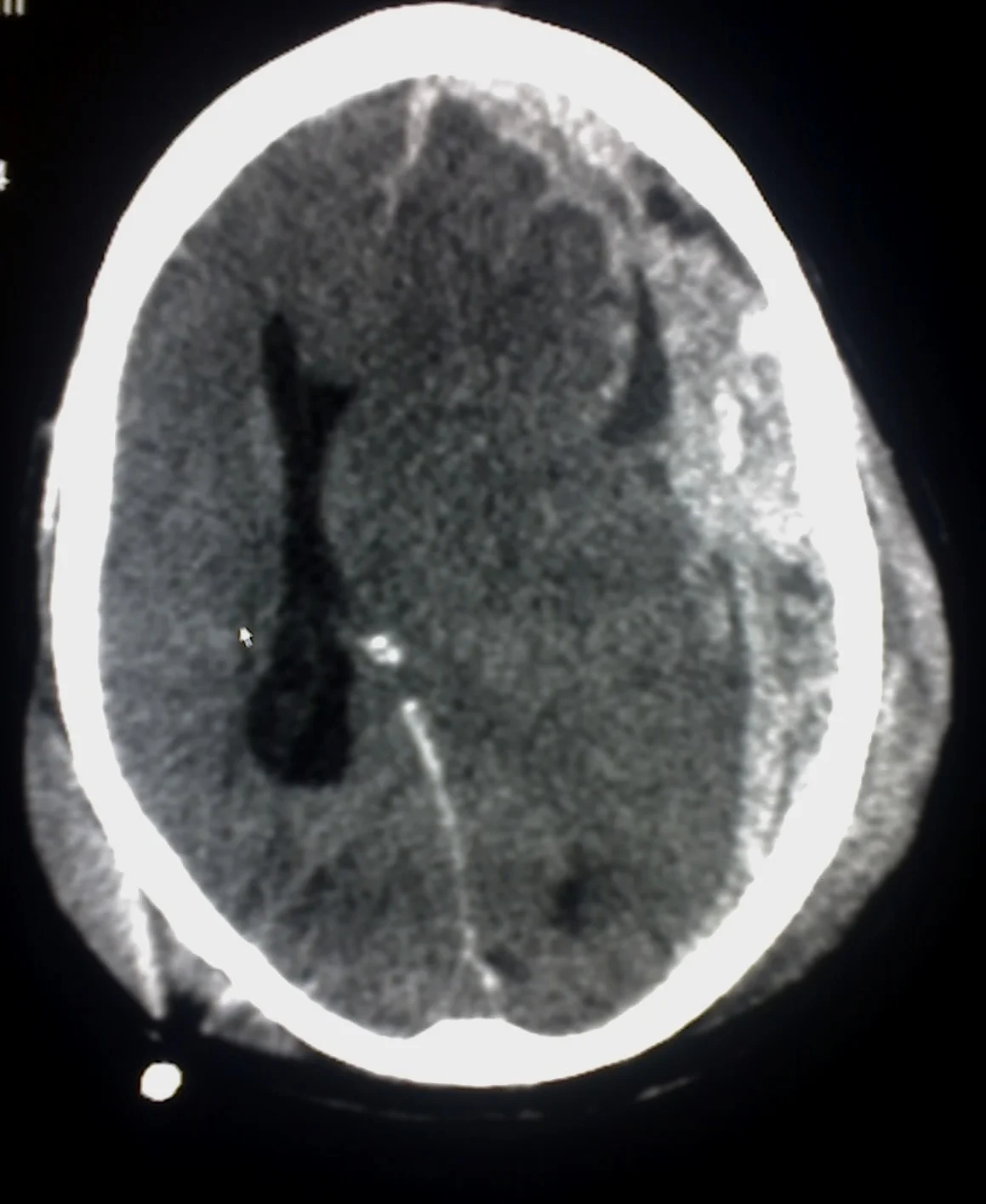
Long description
An axial CT scan of the brain described as an example of subdural haemorrhage caused by trauma, with the collection spreading as a crescent over the cerebral hemisphere.

Figure 2Grosses epidurales Haematom nach Sturz 87W - CT - 001
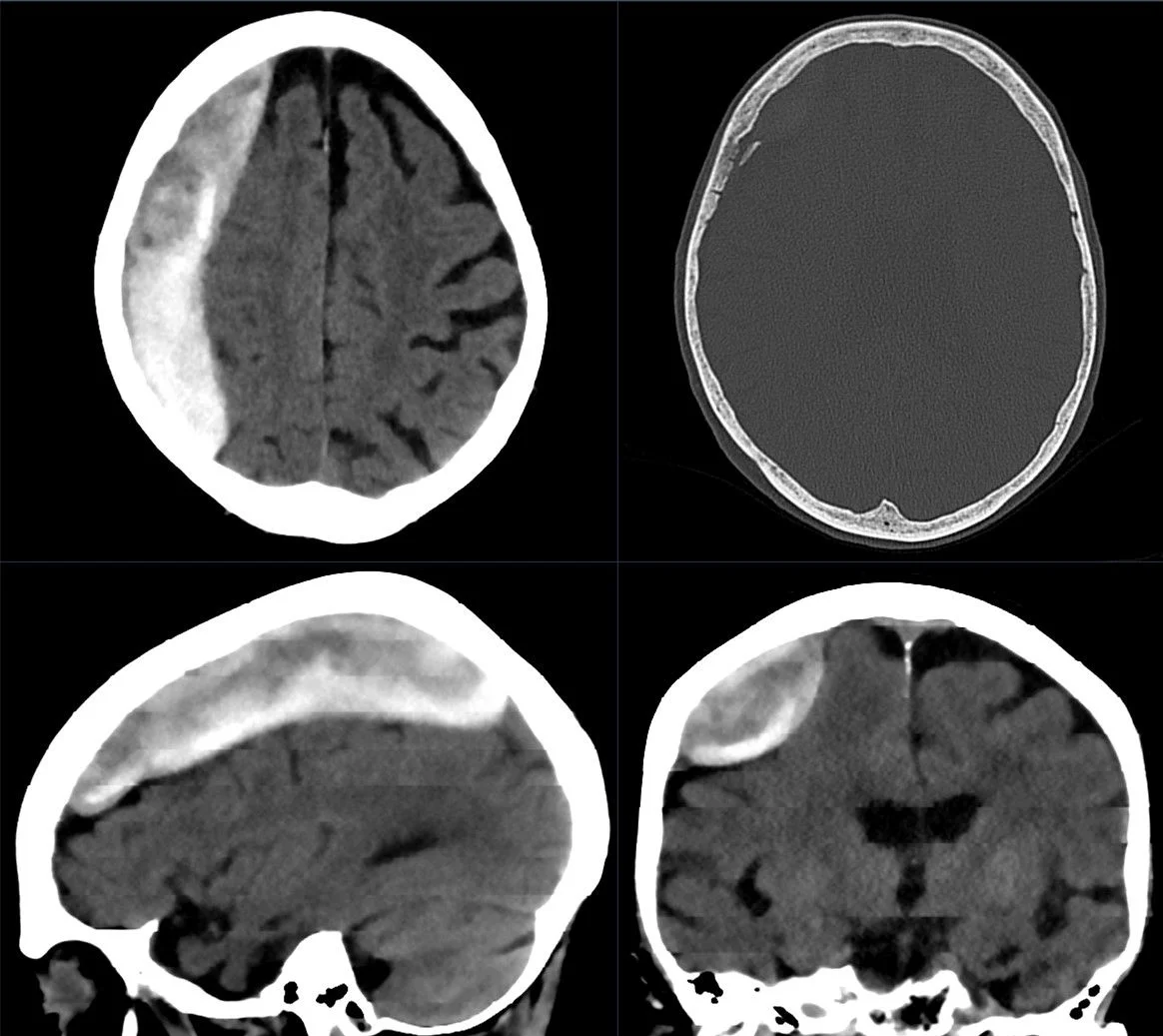
Long description
CT of a large post-traumatic epidural haematoma described as a somewhat inhomogeneous, largely clearly hyperdense mass with a convex margin against the brain parenchyma, with mass effect and initial subfalcine herniation.

Computed tomography (CT) of the head is the first imaging study in significant head trauma. It is fast, available and detects bleeding, contusions, fractures and mass effect. [1] [2]
| Lesion | Mechanism | CT appearance |
|---|---|---|
| Epidural haematoma | Arterial bleeding, often after a temporal bone fracture tearing the middle meningeal artery | Hyperdense, lens-shaped (lenticular), extra-axial collection that does not cross suture lines, often in the temporal region |
| Acute subdural haematoma | Tearing of bridging cortical veins during deceleration | Hyperdense, crescent-shaped extra-axial collection that can cross suture lines; degree of midline shift matters |
| Subarachnoid haemorrhage (traumatic) | Disruption of small pial vessels or extension of contusion | Hyperdensity lining the cerebral sulci and cisterns; traumatic SAH favours the convexities, unlike aneurysmal SAH which sits in the basal cisterns |
| Intracerebral (intraparenchymal) haematoma | Coalescence of contusions from shearing forces | Hyperdense collection within brain tissue, often with surrounding oedema; commonly frontotemporal |
| Diffuse axonal injury | Rotational acceleration-deceleration shearing axons | May be normal initially; often small petechial hyperdensities at the grey-white junction, corpus callosum, basal ganglia or brain stem; MRI is far more sensitive |
Reference 1 is the principal source for this table. [1]
- An acute subdural haematoma may appear hypodense or isodense when chronic or subacute, and in severe anaemia an acute one can even be isodense, so look for displaced sulci and ventricular compression. [1]
- A subarachnoid haemorrhage from trauma is a marker of severe injury and raises the risk oflater deterioration. [1]
- Diffuse axonal injury (DAI) is suspected when loss of consciousness lasts longer than 6 hours with altered consciousness in the absence of a mass lesion, often with GCS below 9. [1] [2]
- A midline shift of more than 5 mm is generally regarded as an indication for surgical evacuation of a haematoma. [1]
The skull is a rigid box (Monro-Kellie doctrine). Any swelling, blood or cerebrospinal fluid increase raises the pressure. [2]
- Early: headache, vomiting, altered consciousness, focal deficits.
- Markedly raised ICP of 25 mm Hg or more classically produces the Cushing triad: hypertension with a widened pulse pressure, bradycardia and irregular or slow respiration. [1]
- One StatPearls source puts the threshold for ICP concern at more than 20 mm Hg, with normal ICP below 15 mm Hg. [2] Both conventions are in use; this page treats 20 to 25 mm Hg as the treatment threshold, as the clinical guidelines do. [1]
Intracranial pressure monitoring is recommended for patients with severe TBI who cannot follow simple commands, especially with an abnormal head CT, and for those with a GCS of 3 to 8 after resuscitation. [1] Ventricular catheters are the gold standard for measuring ICP; parenchymal monitors measure a local pressure and cannot be recalibrated in place. [2]
Secondary brain injury is the damage that follows the first blow, driven by hypoxia, hypotension, hypercapnia and raised ICP. [1] [2]
General measures:
- Secure the airway and ventilate adequately: keep oxygen saturation above 90% (StatPearls says greater than 90%) and PaO2 above 60 mm Hg, and the PaCO2 at 35 to 45 mm Hg. [1] [2]
- Maintain blood pressure: in the Merck guidance a systolic pressure greater than 90 mm Hg and mean arterial pressure greater than 80 mm Hg; StatPearls suggests SBP greater than 110 mm Hg and MAP greater than 90 mm Hg for transfer. [1] [2] Both figures exist in current guidance and local protocols should be followed.
- Keep the patient warm (avoid both hypothermia and hyperthermia). [2]
- Control bleeding, replace volume, and avoid hypotonic fluids such as 5% dextrose in water, which worsen brain oedema. [1]
- Check for coagulopathy and correct it, including in anticoagulated patients. [2]
Raised ICP control:
- Elevate the head of the bed to 30 degrees and keep the head midline. [1]
- Sedation to avoid agitation and coughing; short-acting agents such as propofol have rapid onset. [1]
- Drain CSF via an external ventricular drain when a catheter is in place. [1]
- Osmotherapy: hypertonic saline (3% or 23.4% as bolus or infusion) is the more effective osmotic agent, with a target serum osmolality of 295 to 320 mOsm/kg and serum sodium kept at 155 mEq/L or below. [1]
- 20% mannitol as an alternative (used cautiously in renal or cardiac failure, since it expands the blood volume and is excreted by the kidney). [1]
- Furosemide may help reduce total body water, particularly when the volume loading of mannitol is undesirable. [1]
- Hyperventilation: prophylactic hyperventilation to a PaCO2 of 25 to 35 mm Hg is not recommended, because vasoconstriction reduces cerebral perfusion and potentiates ischaemia. It is reserved for the first several hours and for ICP unresponsive to other measures, targeting a PaCO2 of 30 to 35 mm Hg. [1]
- Neurosurgery for mass lesions, a midline shift above 5 mm, or ICP refractory to medical treatment (decompressive craniectomy, evacuation of haematoma). [1]
Traumatic brain injury: raised ICP pathway
- Primary survey and resuscitationAirway with cervical spine control, breathing, circulation; SaO2 >90%, PaO2 >60, PaCO2 35-45
- Head CT scanDetects haematoma, contusion, fracture, shift; midline shift >5 mm suggests surgical evacuation
- Question: Severe TBI or raised ICP suspected?
Yes
- Admit critical careIntubate and ventilate; sedate; keep SBP and MAP at guideline targets; warm
- ICP-directed treatmentHead of bed 30 degrees, midline, ventricular drainage, osmotherapy, brief hyperventilation only as a bridge
- NeurosurgeryMass lesion evacuation or decompressive craniectomy for refractory ICP
No (mild to moderate)
- Observe on ward or discharge with return precautionsRepeat CT if symptoms persist; escalate if any deterioration
A fracture of the skull base is diagnosed more by its signs than by plain films; CT with thin cuts can reveal fractures that are otherwise invisible. [1] [2]
Signs:
- Leakage of cerebrospinal fluid from the nose or the ear (rhinorrhoea, otorrhoea).
- Blood behind the eardrum (haemotympanum) or in the external ear canal.
- Battle sign: bruising behind the ear, over the mastoid.
- Raccoon eyes: periorbital ecchymosis (bruising in the periorbital area).
- Loss of smell and loss of hearing, which may be unnoticed until consciousness returns.
- Facial nerve impairment, immediate or delayed.
- [1] [2]
These signs are highly associated with intracranial haemorrhage and should prompt head CT and close observation. [2] Cerebrospinal fluid leaking through the nose means the patient is at risk of meningitis from the tear in the dura; surgical repair is considered when leakage persists. [1]
Concussion (mild TBI) is a traumatically induced transient disturbance of brain function: loss of consciousness, confusion or amnesia lasting seconds to minutes, sometimes a few hours. By definition there is no acute intracranial haemorrhage, lesion or contusion visible on imaging; there may be a non-displaced or minimally displaced linear skull fracture. [1] [2]
Practical points:
- A concussion is diagnosed when a transient functional disturbance follows mechanical force and the symptoms are not explained by structural injury seen on imaging. [1]
- A score of loss of consciousness under 30 minutes and post-traumatic amnesia under 24 hours are part of the standard mild TBI definition. [2]
- Multiple concussions are thought to carry a risk of chronic traumatic encephalopathy, which produces severe brain dysfunction. [1]
- Advise against same-day return to play; graded return with rest and symptom monitoring is preferred. [1]
- Second-impact syndrome, rare, is malignant cerebral oedema after a second insult before recovery. [1]
If ICP rises further, brain tissue shifts across the tentorium or through the foramen magnum: uncal, subfalcine and transtentorial herniation, with coma, abnormal pupils, posturing and the Cushing triad. [1] [2]
A spinal cord injury is damage to the cord or its blood supply producing motor, sensory or autonomic deficit below the level of injury. Traumatic injuries are classified as complete (no motor or sensory function preserved below the lesion, including the S4-S5 sacral segments) or incomplete (some preserved function). The International Standards for Neurological Classification of Spinal Cord Injury (the ASIA Impairment Scale, or AIS) grades severity from A (complete) to E (normal). [3] [4]
The motor examination covers key muscles from C5 to S1, scored 0 to 5, with a maximum of 100; the sensory examination covers C2 to S5 for light touch and pinprick. Sacral sparing (any sensation or voluntary anal contraction at S4-S5) is what separates a complete from an incomplete injury. [4] [3]
| Grade | Meaning | Motor below injury | Sensory below injury |
|---|---|---|---|
| A | Complete | None, including S4-S5 | None, including S4-S5 |
| B | Sensory incomplete | None | Preserved below the level including S4-S5 |
| C | Motor incomplete | Preserved, but less than half of key muscles grade 3 or more | Preserved sacral function |
| D | Motor incomplete | At least half of key muscles grade 3 or more | Preserved |
| E | Normal | Normal | Normal |
The four classic incomplete patterns are recognised from the tracts damaged. [3]
- Complete transection: bilateral loss of motor, pain, temperature, proprioception, vibration and touch below the lesion. High cervical lesions can compromise respiration because of diaphragm denervation above C5. [3]
- Central cord syndrome: the most common incomplete injury. Hyperextension of the neck in a stenotic canal tends to produce more weakness in the upper limbs than the lower, often with bladder dysfunction and preserved sacral sensation. [3]
- Anterior cord (anterior spinal artery) syndrome: both sides lose movement and the sensation of pain and temperature below the level of injury, while proprioception is preserved, vibration and some touch (the dorsal columns are spared). [3]
- Brown-Sequard syndrome: hemisection of the cord. Ipsilateral loss of motor function, proprioception and vibration, with contralateral loss of pain and temperature. It is seen most often with penetrating trauma. [3]
- Posterior cord syndrome is rare in trauma (more often infectious, toxic or metabolic) and produces loss of proprioception and vibration with motor and pain and temperature spared. Conus medullaris syndrome affects the terminal cord at L1-L2 and produces sacral dysfunction. [3]
These two arise together after spinal cord injury but are different. [4]
| Feature | Spinal shock | Neurogenic shock |
|---|---|---|
| Definition | Transient flaccid paralysis with areflexia and loss of motor and sensory function below the lesion | Loss of sympathetic tone with unopposed parasympathetic activity, producing vasodilatation, hypotension and bradycardia |
| Blood pressure | No direct haemodynamic change; hypotension and bradycardia, when present, come from simultaneous neurogenic shock | Hypotension from low systemic vascular resistance, reduced venous return and reduced cardiac output |
| Level | Any level | Injury at T6 or above (neurogenic shock is expected above the mid-thoracic level) [4] |
| Motor and reflexes | Flaccid paralysis; areflexia or hyporeflexia early, hyperreflexia later | Flaccid paralysis, when present, is due to spinal shock rather than to neurogenic shock itself |
| Skin | Sweating and bladder and bowel dysfunction | Warm, dry skin, temperature dysregulation, autonomic dysreflexia later |
| Duration | Days to 4 to 6 weeks, rarely up to a year | Usually 1 to 3 weeks [4] |
| Treatment | Stabilise the injury and support; reflexes return as spinal shock resolves | Oxygen, fluids, vasopressors and temperature control |
Source: Valdez-Resendiz et al, 2025. [4]
Key clinical distinction: spinal shock is an areflexic, flaccid state that can mask an incomplete injury until it resolves; the return of the bulbocavernosus reflex marks the end of spinal shock, which typically happens in the first days. [4] Neurogenic shock is a circulation problem and a reason to give vasopressors. [3] [4]
Cervical spine imaging can be deferred in a patient who is awake and alert, not intoxicated, has no focal neurologic deficit, no midline cervical tenderness and no painful distracting injury (for example a femoral fracture). Everyone else should be imaged, preferably with CT. [7] Two validated decision instruments guide imaging of alert, stable patients:
NEXUS - a patient is at low probability of significant cervical spine injury only if all five criteria are met: no midline cervical tenderness, no focal neurologic deficit, normal alertness, no intoxication, and no painful distracting injury (a clinically apparent painful injury that might distract from neck pain). In the validation study of 34,069 patients, the rule had 99.0% sensitivity for any cervical spine injury and 99.6% for a clinically significant injury; applying it could have avoided imaging in 12.6% of cases. [5]
The Canadian C-spine rule is a three-step algorithm for alert and stable adults with a GCS of 15: first, are any high-risk factors present (age 65 or more, a dangerous mechanism, or paraesthesia in the extremities)? If yes, image. If not, are any low-risk factors present (a simple rear-end collision, sitting in the emergency department, ambulatory at any time since the injury, delayed onset of neck pain, or no midline cervical tenderness)? If no, image. If yes, the patient must actively rotate the neck 45 degrees to the left and right; if unable, image. The derived rule had 100% sensitivity (95% confidence interval 98 to 100%) for 151 clinically important injuries. [6]
Where high-risk trauma exists, CT rather than plain radiography should be obtained. [7]
- Plain radiographs can screen, but CT identifies fractures better and is faster in a standard trauma protocol. [3] [7]
- MRI is the best study for the cord and soft tissues (ligaments, discs, epidural haematoma, cord contusion or oedema, and cord compression) and for predicting prognosis. [3] [7]
- CT angiography is added when a cervical fracture or subluxation is found, to exclude carotid or vertebral artery injury. [7]
- In children, the anatomy is different (injuries often above C4, ligamentous elasticity) and MRI is used more readily; spinal cord injury without radiographic abnormality (SCIWORA) is described mostly in children. [7]
Raised ICP in head trauma is managed as above (head of the bed, midline head position, sedation, drainage, osmotherapy, brief hyperventilation as a bridge, neurosurgery). Imaging strategy: non-contrast head CT first; MRI later for subtle DAI and brain stem injury. [1] [2]
In spinal cord injury, the NASCIS trials proposed high-dose methylprednisolone for 48 hours, but the methodological quality of those studies has been widely questioned, and current guidelines do not recommend high-dose intravenous methylprednisolone in acute spinal cord injury, because there is no convincing level I or II evidence of benefit and level I to III evidence of possible harm. [4]
In brain trauma, the CRASH trial (corticosteroids after significant head injury) body of evidence shows that routine steroid use is not beneficial and probably harmful; present practice is that steroids are not given routinely for TBI. [1] [2]
- Brain: cerebral oedema, increased ICP and herniation, hydrocephalus after intraventricular blood, seizures, infection after skull base fracture, chronic subdural collection.
- Spine: pneumonia and respiratory failure (especially cervical), deep vein thrombosis (roughly 85% risk in the first 3 months without prophylaxis), pressure injury, autonomic dysreflexia after the acute phase in injuries at or above T6, spasticity, urinary sepsis. [3] [4]
- Systemic secondary insults after TBI - the 3 Hs: hypoxia, hypotension and hypercapnia or hypocapnia - are what we treat first. [1] [2]
- Older adults: falls dominate, presentation may be delayed, and chronic subdural haematoma can appear weeks later with subtle confusion. Anticoagulation and antiplatelet use raise risk. [1]
- Children: physiology and imaging differ (PECARN rules for CT, precautions about suture lines); cervical injuries are more often at C1 to C4. [1] [7]
- Pregnancy: manage the mother first according to trauma principles; imaging should be used when indicated.
- Primary survey, cervical spine control, CT when available; if no CT is available, observe for 4 to 6 hours after mild injury. [2]
- Keep oxygen saturation above 90%, avoid hypotension, keep the head of the bed elevated and give simple analgesia and anti-emetics; osmotherapy can be given if monitored.
- Mobilise safely, prevent pressure injuries, and arrange transfer for spinal injury or neurosurgical care; prophylaxis against venous thrombosis should start within 72 hours where possible. [3]
- Return precautions for discharged mild TBI are more important than any test.
Trauma primary survey and approach to the trauma patient; increased intracranial pressure; brain herniation; subdural and epidural haematoma; spinal trauma and imaging; coma and brain death; concussion and sports-related concussion; autonomic dysreflexia.
- Severity of TBI is graded by GCS: 13 to 15 mild, 9 to 12 moderate, 3 to 8 severe, with a score of 8 or below regarded as coma; a score after resuscitation is more reliable. [1]
- The principal primary lesions are epidural (lens-shaped), subdural (crescent-shaped), subarachnoid, intracerebral haematoma and diffuse axonal injury.
- Raised ICP of 25 mm Hg or more gives the Cushing triad (hypertension, bradycardia, irregular respiration); in many guidelines ICP above 20 mm Hg triggers treatment. [1]
- Airway, oxygenation, blood pressure, temperature and ICP are the secondary-insult targets; PaCO2 goal is 35 to 45 mm Hg, with hyperventilation (target 30 to 35 mm Hg) reserved for the first hours or refractory ICP. [1][2]
- Osmotherapy: hypertonic saline (3% or 23.4%) is the most effective osmotic agent; target serum osmolality 295 to 320 mOsm/kg and serum sodium 155 mEq/L or below; 20% mannitol is an alternative.
- Head of the bed at 30 degrees with the head midline lowers ICP; a midline shift above 5 mm suggests surgical evacuation. [1]
- Basilar skull fractures present with CSF rhinorrhoea or otorrhoea, haemotympanum, Battle sign, raccoon eyes, smell or hearing loss; nasogastric and nasotracheal tubes are avoided in these patients. [2]
- Concussion is a transient functional disturbance without structural lesion; referral and a graded return to activity matter.
- Spinal cord injury is graded by the ASIA Impairment Scale (A to E); central cord, anterior cord and Brown-Sequard are the classic incomplete syndromes.
- Spinal shock is a transient areflexic and flaccid state; neurogenic shock is a circulatory problem needing fluids and vasopressors, and typically occurs with injuries at T6 or above.
- Cervical spine imaging can be deferred only in alert, sober patients with no deficit, no midline tenderness and no distracting injury; CT is the usual first study and MRI the best study for the cord. [7]
- NEXUS and the Canadian C-spine rule reduce unnecessary imaging in low-risk patients.
- High-dose corticosteroids are no longer routine for spinal cord injury or for TBI.
- Secondary brain injury is prevented by treating the 3 Hs; return precautions are essential for discharged mild TBI.
Answer each one in your head first, then open the answer.
A 22-year-old man loses consciousness after a car crash, then asks repetitive questions. CT of the head is normal. What is the most likely diagnosis and how should he be managed?
Show answerHide answer
Concussion (mild traumatic brain injury). Observation, return precautions, gradual return to activity and no same-day sport. A normal CT does not exclude a functional brain disturbance.
How is a traumatic brain injury graded as mild, moderate or severe on arrival?
Show answerHide answer
By the Glasgow Coma Scale: 13 to 15 is mild, 9 to 12 is moderate and 3 to 8 is severe; a combined score of 8 or below is usually coma. Some references use 14 to 15 for mild.
What CT findings distinguish an epidural from a subdural haematoma?
Show answerHide answer
Epidural: hyperdense, lens-shaped (lenticular), does not cross suture lines, often temporal. Subdural: hyperdense, crescent-shaped, can cross suture lines, often with midline shift.
A patient with severe TBI has an ICP of 28 mm Hg despite sedation. What are the next steps?
Show answerHide answer
Continue neuroprotective care: head of the bed at 30 degrees with the head midline, drain CSF if a ventricular catheter is in place, osmotherapy (hypertonic saline targeting 295 to 320 mOsm/kg and sodium 155 mEq/L or below; or 20% mannitol), temporary hyperventilation (PaCO2 30 to 35 mm Hg) as a bridge, and urgent neurosurgical review for decompressive craniectomy or haematoma evacuation.
Why is routine prophylactic hyperventilation discouraged in TBI?
Show answerHide answer
A low PaCO2 constricts cerebral vessels and reduces intracranial pressure, but it also reduces cerebral blood flow, worsening ischaemia. It is therefore reserved for the first several hours or for ICP unresponsive to other measures.
What are the signs of a basilar skull fracture?
Show answerHide answer
Cerebrospinal fluid rhinorrhoea or otorrhoea, haemotympanum, Battle sign, raccoon eyes, loss of smell or hearing and facial nerve weakness.
Describe central cord syndrome.
Show answerHide answer
The most common incomplete spinal cord injury, after hyperextension in a narrowed canal, producing more weakness in the arms than the legs, often with bladder dysfunction and preserved sacral sensation.
A high cervical spinal cord injury produces hypotension and bradycardia. Is this spinal shock or neurogenic shock?
Show answerHide answer
Neurogenic shock: loss of sympathetic tone with unopposed parasympathetic drive causing vasodilatation, hypotension and bradycardia. Spinal shock is the flaccid, areflexic state below the lesion. They often occur together; neurogenic shock needs fluids and vasopressors.
List the NEXUS criteria for not imaging the cervical spine.
Show answerHide answer
No midline cervical tenderness, no focal neurologic deficit, normal alertness, no intoxication, and no painful distracting injury.
A 75-year-old man on warfarin has a transient loss of consciousness followed by a headache that worsens over 6 hours. What injury must you suspect and why?
Show answerHide answer
An acute subdural haematoma: elderly patients with cerebral atrophy are prone to tearing of bridging veins, and anticoagulation worsens bleeding. Urgent CT of the head is required.
- Mao G, Spain DA. Traumatic brain injury (TBI). Merck Manual Professional Edition. Full review Oct 2026; peer reviewed.. Accessed .
- Ginsburg J, Smith T. Traumatic Brain Injury. StatPearls [Internet]. Treasure Island (FL): StatPearls Publishing; updated 2025-02-17.. Accessed .
- Margetis K, Das JM, Emmady PD. Spinal Cord Injuries. StatPearls [Internet]. Treasure Island (FL): StatPearls Publishing; updated 2025-06-02.. Accessed .
- Valdez-Resendiz I, Salgado-Camarillo EN, Hernandez-Morales F, Martinez-de los Santos CA, Robba C. Perioperative management in acute and chronic spinal cord injury, narrative review. Journal of Anesthesia, Analgesia and Critical Care 2025;5:33.. Accessed .
- Hoffman JR, Mower WR, Wolfson AB, Todd KH, Zucker MI, for the National Emergency X-Radiography Utilization Study Group. Validity of a set of clinical criteria to rule out injury to the cervical spine in patients with blunt trauma. New England Journal of Medicine 2000;343(2):94-99. doi:10.1056/NEJM200007133430203. Accessed .
- Stiell IG, Wells GA, Vandemheen KL, et al. The Canadian C-spine rule for radiography in alert and stable trauma patients. Journal of the American Medical Association 2001;286(15):1841-1848. doi:10.1001/jama.286.15.1841. Accessed .
- Merck Manual Professional Edition contributors. Approach to the Trauma Patient; Spinal Trauma. Merck Manual Professional Edition; updated 2025-2026.. Accessed .
Figure credits
Educational content for medical students and doctors. It is not a substitute for clinical judgement or local guidelines.