Thoracic trauma
Managing penetrating and blunt chest injury: assessment, life-saving interventions, diagnosis, and chest wall and organ-specific care.
- Thoracic trauma is survivable when threats to airway, breathing and circulation are found first and treated immediately.
- Blunt trauma is more common than penetrating trauma and causes 20 to 25% of trauma deaths, but fewer than 10% of blunt chest injuries need surgery; up to 30% of penetrating chest injuries do.[1]
- The immediately lethal patterns are tension pneumothorax, open pneumothorax, massive haemothorax and cardiac tamponade: each is treated before imaging.
- Rib fractures hurt enough to make patients splint their breathing; poor pain control leads to atelectasis and pneumonia, so analgesia is an essential part of care.[1][6]
- A normal chest X-ray does not exclude blunt aortic injury, and a widened mediastinum is present in only 27.7% of confirmed cases.[1][3]
By the end of this page you should be able to:
- Run a primary survey for chest trauma and treat the immediately lethal injuries.
- Explain the mechanism and presentation of each major chest injury and choose the right investigation.
- Insert and manage a chest drain or needle decompression safely and interpret drainage volumes.
- Recognise blunt aortic injury, blunt cardiac injury and diaphragmatic rupture and know what to do first.
- Plan rib fracture analgesia and know when surgical stabilisation is indicated.
- State the principles of damage-control thoracotomy and resuscitation.
This page covers the diagnosis and management of thoracic trauma in adults. It treats the chest as a mechanical system: the ribs contain two spaces (pleural and pericardial), two tubes (airway and oesophagus) and two great vessels. Any break in that system produces one of a small number of problems—air where it should not be, blood where it should not be, or a chest wall that no longer moves properly. The working principle of trauma care is to find those problems in order of lethality and to treat each one as it is found, rather than waiting for a complete diagnosis.
A thoracic (chest) trauma is any injury to the chest wall, pleura, lungs, airways, mediastinum, great vessels, heart, diaphragm or oesophagus caused by blunt force, penetrating force or both. Blunt injury is caused by kinetic energy transfer without a break in the skin (road traffic collisions, falls, assaults, blast); penetrating injury is caused by an object entering the thorax (stab, gunshot, impalement).
The WSES-AAST classification separates chest injury into four grades by respiratory function and haemodynamic status: [1]
| Grade | Severity | Respiratory function | Haemodynamic status |
|---|---|---|---|
| I | Minor | Normal | Stable |
| II | Moderate | Respiratory impairment (reduced lung function without distress) | Stable |
| III | Severe | Respiratory distress (acute diffuse lung injury, severe hypoxaemia) | Stable |
| IV | Severe | Any | Unstable |
Respiratory impairment means reduced lung function without distress; respiratory distress means acute diffuse lung injury that produces severe hypoxaemia within hours. A Thoracic Trauma Severity Score between 13 and 25 predicts a high risk of developing acute respiratory distress syndrome (ARDS).[1]
Injuries are usually grouped by the structure damaged: thoracic wall, pleural, airway and lung, vascular and cardiac, and diaphragmatic. Penetrating injuries to the central chest may injure the heart or pericardium, and a "right-sided" cardiac injury with pericardial laceration can decompress into the right haemothorax, so central penetrating chest bleeding must be assumed cardiac until proved otherwise.[1]
DefinitionOccult pneumothorax
A pneumothorax that is not seen on a supine chest X-ray but is found on CT or ultrasound. Management depends on the patient's clinical condition, not on the size of the pneumothorax.[1]
Blunt chest trauma is more common (70 to 80%) than penetrating trauma and accounts for 20 to 25% of all trauma deaths.[1] In many low- and middle-income countries penetrating trauma, both stab and gunshot wounds, is at least as common as blunt trauma and is immediately lethal: over 75% of stab wounds to the heart kill the patient before hospital arrival.[1] Motor vehicle collisions are associated with higher morbidity and mortality, especially at high speed and when seat belts are not used.[1] Older patients may have a worse outcome because of frailty and comorbidity.[1]
Fewer than 10% of patients with blunt chest trauma require surgical intervention, whereas up to 30% of patients with penetrating chest trauma require surgery and have higher all-cause mortality.[1] The simple or tension pneumothorax risk in polytrauma is 20% and rises to 50% in severe chest trauma; haemothorax is more frequent in penetrating and high-energy trauma and in injuries to the ribs and sternum or flail chest, and haemothorax occurred in 30% of thoracic trauma in modern military series.[1]
East Africa
In low-resource and conflict settings penetrating chest trauma is at least as common as blunt trauma, and many patients with massive haemothorax or cardiac injury die before reaching care. The same principles apply: diagnose by examination and focused ultrasound, decompress immediately, and drain with the simplest reliable equipment. [1]
Causes are kinetic and environmental: road traffic collisions, falls from height, assaults, industrial and occupational injury, sports injuries, blasts, and impalements. The force type determines the injury pattern. Blunt trauma can be grouped into three mechanisms: rapid deceleration, compression or crush, and blast. Rapid deceleration is the predominant mechanism of blunt traumatic aortic injury and produces stretch, shear, pinch and thump forces on the thoracic aorta.[3] Penetrating injuries are divided into stab wounds, which are usually low energy and focal, and gunshot or blast injuries, which cause cavitation and multiple tract injuries.
Modifiable risks include alcohol or drug intoxication, absence of seat belts or crash helmets, unsafe working at height and manual handling. Non-modifiable risks include age and frailty, anticoagulation and bleeding tendency, poor bone quality from osteoporosis, chronic lung disease that limits reserve, and heritable connective tissue disease. Patients with Marfan syndrome (a connective tissue disorder from FBN1 variants, with an estimated prevalence of 1 in 5,000 to 10,000) or vascular Ehlers-Danlos syndrome have a fragile aortic wall and are at higher risk of arterial dissection; in these patients trauma should be minimised and, after even modest chest trauma, the aorta should be assessed carefully.[4]
The core problem in most chest trauma is loss of the negative pressure that keeps the lung expanded. Air or blood in the pleural space collapses the ipsilateral lung, shifts the mediastinum and, if pressure continues to rise, compresses the great veins and the opposite lung.
Tension pneumothorax behaves like a one-way valve: air enters the pleural space on inspiration and cannot leave on expiration. Intrapleural pressure rises, the lung collapses, the mediastinum shifts away and the vena cava and opposite lung are compressed. The result is hypoxaemia and obstructive shock: raised jugular venous pressure, hypotension, tachycardia, absent breath sounds and, eventually, cardiac arrest.[1]
Massive haemothorax is rapid bleeding into the chest, usually from intercostal vessels, the internal mammary artery, lung lacerations or, less often, the great vessels and heart. The problem is simultaneous hypovolaemia and loss of ventilatory surface; the blood also splints the lung and, if retained, becomes a nidus for empyema.
Flail chest occurs when a segment of rib cage is detached from the rest of the chest wall, commonly with multiple contiguous rib fractures or a sternal fracture. The segment moves inward on inspiration and outward on expiration, so ventilation is mechanically inefficient. Far more important are the pain, which splints breathing, and the pulmonary contusion beneath the flail segment. The WSES-AAST guideline recommends considering surgical stabilisation in all stable patients with a flail chest, because surgical fixation improves ventilator and intensive care outcomes.[1]
Pulmonary contusion is bleeding and oedema in the alveoli and interstitium after blunt force. It impairs gas exchange, increases shunt and stiffens the lung. Effective pain control and careful fluid management are the keys to preventing pneumonia and ARDS.[1]
Cardiac tamponade occurs when blood collects in the pericardium. Because the pericardium is stiff, a small volume raises pressure rapidly, compressing the heart and preventing filling. Cardiac output falls even though the muscle itself may be normal. The classic presentation is Beck's triad: hypotension, muffled heart sounds and raised jugular venous pressure, with tachycardia and pulsus paradoxus.[2]
Blunt cardiac injury ranges from myocardial contusion, which may be completely asymptomatic, to rupture and valvular injury. The right ventricle is at highest risk because it lies behind the sternum. Contusion presents with chest pain, dyspnoea and arrhythmias; right bundle branch block is a common consequence, and left bundle branch block is rare. Cardiac enzymes such as troponin and creatine kinase rise after myocardial injury.[2] In severe, uncontained rupture most patients do not reach hospital alive.[2]
Blunt traumatic aortic injury is produced by deceleration and shear at fixed points, especially the aortic isthmus near the ligamentum arteriosum. The tear may be partial and contained initially, but can progress to rupture, pseudoaneurysm or intramural haematoma. The connective tissue disorders above increase risk and can allow dissection at small aortic diameters.[3][4]
Tracheobronchial disruption is an airway tear, often near the carina. It produces massive air leak, pneumomediastinum, subcutaneous emphysema and a persistent pneumothorax despite a chest drain. Because ventilation can be lost suddenly, the initial priority is to secure the airway.[1]
Diaphragmatic rupture opens the barrier between abdomen and chest. Pressure in the abdomen is higher than in the chest, so bowel or stomach can herniate and compress the lung. Penetrating trauma usually makes a small tear; blunt trauma (high energy, such as road traffic collisions and falls) makes larger tears.[1][5]
The chest is assessed during the primary survey: airway, breathing, circulation, disability and exposure. Life-threatening chest injuries can be missed if the team waits for a full examination. On inspection look for tracheal deviation, asymmetric chest movement and open wounds; on palpation feel for surgical emphysema, crepitus and tenderness; on percussion and auscultation compare the two sides.
Tension pneumothorax presents with severe breathlessness, chest pain, tachypnoea, tachycardia, hypotension, hypoxia, absent breath sounds, hyperresonance and, in late cases, tracheal deviation away from the injured side and jugular venous distension; cyanosis and cardiac arrest follow.[1]
Massive haemothorax presents with shock, tachypnoea, decreased or absent breath sounds, dullness to percussion, asymmetric chest movement and haemoptysis in some patients.[1]
Open (sucking) pneumothorax presents with a chest wall defect that moves air audibly and prevents effective ventilation.[1]
Flail chest presents with paradoxical segment movement, severe pain, shallow breathing and often haemoptysis.[1]
Cardiac tamponade presents with Beck's triad—hypotension, muffled heart sounds and raised jugular venous pressure—plus tachycardia and pulsus paradoxus. The injured patient may not have distended neck veins if haemorrhagic shock masks the diagnosis, so bedside ultrasound is essential.[2]
Blunt cardiac injury presents with chest pain, dyspnoea, arrhythmia or hypotension; many patients are asymptomatic.[2]
Aortic injury may present with tearing interscapular or chest pain, hypertension in the arms with reduced femoral pulses, a harsh systolic murmur, neurological signs or haemodynamic collapse, but it is frequently silent and found only on imaging.[3]
Diaphragmatic rupture may present with abdominal pain and chest dyspnoea, shoulder tip pain, reduced breath sounds and bowel sounds in the chest. Left-sided injuries predominate, probably because the liver buffers the right side; right-sided ruptures are harder to see on CT because herniated liver is subtle.[5]
Tracheobronchial injury presents with dyspnoea, haemoptysis, massive subcutaneous emphysema, pneumomediastinum and a pneumothorax that does not resolve with drainage.[1]
Many chest trauma patients also have medical causes of breathlessness or chest pain. The key differentials are separated by mechanism and examination.
| Condition | Key separating feature |
|---|---|
| Primary spontaneous pneumothorax | No trauma mechanism; tall, thin young smoker; sudden unilateral pleuritic pain |
| Massive pulmonary embolism | Sudden dyspnoea, tachycardia, hypoxia, right heart strain; risk factors for thrombosis |
| Acute coronary syndrome | Crushing central pain with ECG ischaemia; may be precipitated by blunt cardiac injury |
| Oesophageal rupture (Boerhaave) | Severe pain after vomiting; mediastinal air; surgical abdomen |
| Asthma or COPD exacerbation | Wheeze, known disease; no focal signs or trauma mechanism |
| Simple rib fracture or costochondritis | Focal tenderness, normal imaging; diagnosis of exclusion in significant trauma |
For an intrathoracic mass seen on imaging after trauma, the differential includes traumatic pseudoaneurysm, mediastinal haematoma, ductus diverticulum, motion artefact, diaphragmatic hernia and pre-existing aortic disease.[3]
Figure 1Spannungspneu+Drainage
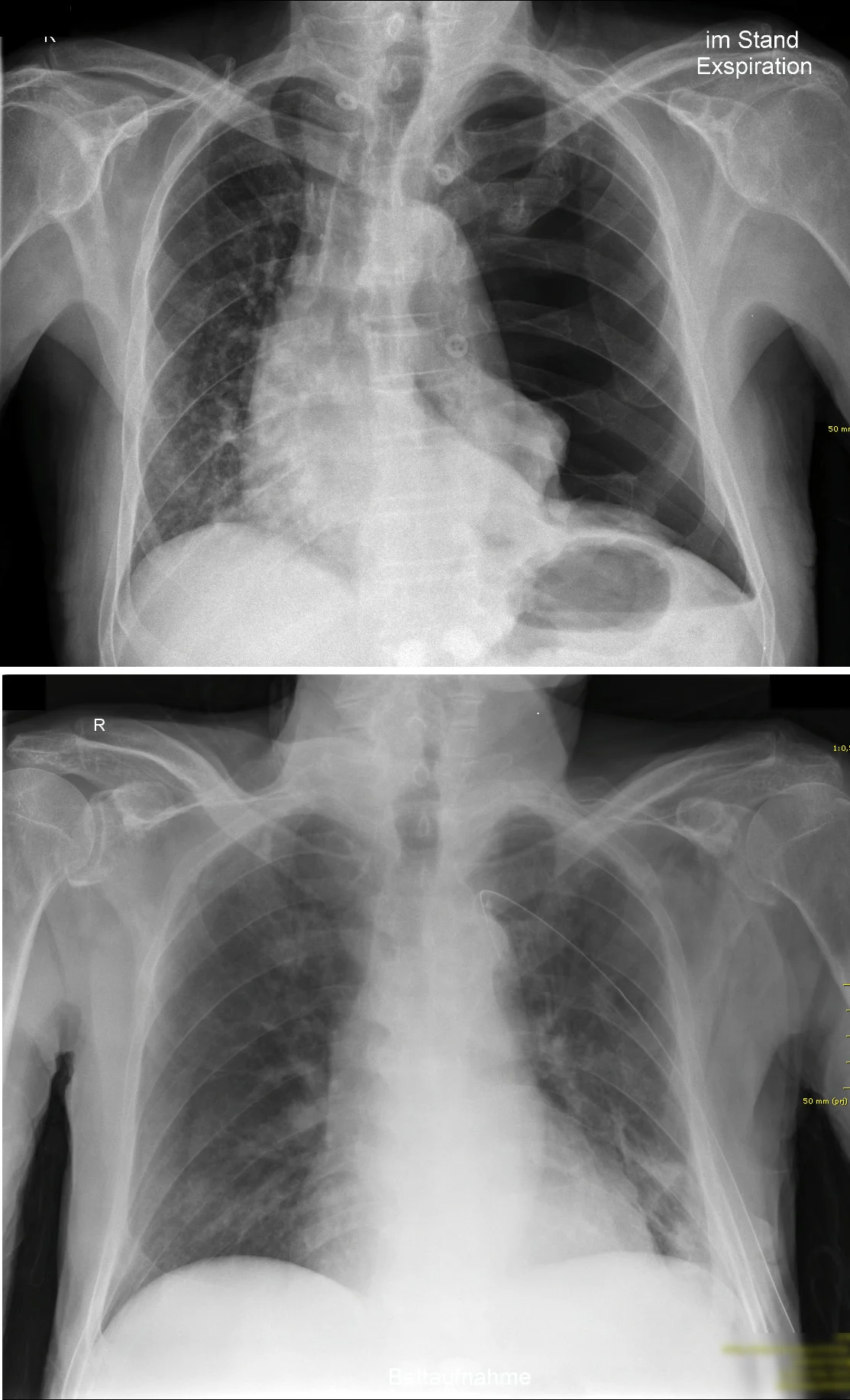
Long description
A left-sided tension pneumothorax before (top) and after (bottom) chest-drain placement, with the description stating that the displacement of the mediastinum to the healthy side is clearly visible and lung markings are absent in the pneumothorax.

Unstable patients. In unstable penetrating and blunt trauma, diagnosis should be pursued with extended FAST and/or chest X-ray. [1] Ultrasound, especially E-FAST, can achieve 98% sensitivity and 100% specificity for free fluid in skilled hands with a linear ultrasound transducer probe (5 to 10 MHz) and is more useful than chest X-ray in unstable patients for detecting pneumothorax at the bedside.[1] Arterial blood gas, haemoglobin, lactate and clotting studies are taken in parallel. E-FAST is not accurate in obesity and after subcutaneous emphysema.
Stable patients. In stable patients a contrast-enhanced CT of the chest with possible 3D reconstruction is strongly indicated to define the type and extent of injury.[1] CT angiography is the preferred imaging for vascular injury, aortic injury and pulmonary thrombosis, and for planning intervention.[1][3]
Airway injury. Bronchoscopy remains the gold standard for diagnosing tracheal and bronchial injuries because it gives direct airway visualisation.[1]
Oesophageal injury. In suspected oesophageal perforation after chest trauma, contrast CT or oesophagoscopy is recommended.[1]
Cardiac injury. Echocardiography gives functional and structural assessment after blunt cardiac injury, including valve, septal or free-wall damage and pericardial effusion. Cardiac MRI can assess myocardial contusion and regional infarction. Troponin and other cardiac enzymes may be raised after myocardial injury.[2]
Aortic injury. Aortic damage is assessed as part of the vascular survey. A widened mediastinum on chest X-ray (more than 8 cm) is present in only 27.7% of confirmed blunt aortic injuries and in 47.4% of the most severe grade 4 injuries; grade 1 to 4 injuries show it in 7.8%, 23.3%, 35.3% and 47.4% respectively.[1][3] CT angiography is the preferred imaging technique for diagnosing vascular lesions and planning management.[1][3] CTA findings such as intimal flap, intramural haematoma, pseudoaneurysm and contrast extravasation underlie the Society for Vascular Surgery grading system. Rapid deceleration is the predominant mechanism; shear at the aortic isthmus near the ligamentum arteriosum and other forces (stretch, pinch and abrupt intraluminal pressurisation) produce the tear.[3] Patients with Marfan syndrome or vascular Ehlers-Danlos syndrome have fragile arteries and can dissect at small diameters; management therefore stresses blood-pressure control, avoidance of trauma and unnecessary invasive procedures, and expert multidisciplinary follow-up of the whole arterial tree.[4]
Diaphragmatic injury. Diagnosis is by CT. Left-sided injuries predominate because the liver buffers the right side; right-sided injuries are harder to see, and pneumothorax, lower rib fractures or liver injury may be the only clues.[5] Diaphragmatic injury occurs in about 1 to 15% of blunt thoracic trauma.[5] [1]
The grading table above is the severity framework. The Thoracic Trauma Severity Score predicts ARDS risk when it lies between 13 and 25; it combines oxygenation, rib fractures, lung contusion, pleural involvement and age.[1]
For suspected oesophageal perforation, use contrast CT or oesophagoscopy.[1] For tracheobronchial injuries, bronchoscopy is the gold standard. In blunt aortic injury, grades II to IV are increasingly treated first by thoracic endovascular aortic repair (TEVAR) rather than open surgery, with open surgery reserved for instability or severe injury not amenable to endovascular repair.[1][3]
Occult pneumothorax should be treated according to the patient's condition rather than its size; a small occult pneumothorax may be observed, but requires at least 24 hours of observation.[1]
Chest trauma care is simultaneous resuscitation and investigation. Give oxygen, secure the airway with appropriate protection if needed, and control haemorrhage. Use massive transfusion protocols, tranexamic acid and damage-control surgery for major haemorrhage.[1] In pulmonary contusion avoid excessive oxygen (which increases lung complications and death) and give oxygen only to maintain adequate saturation; avoid injudicious fluid loading.[1] Give careful fluid administration and lung-protective ventilation.[1] Antibiotic prophylaxis is indicated in penetrating injuries and all surgical exploration (thoracotomy or thoracoscopy), but not for chest tube insertion in blunt or spontaneous pneumothorax.[1]
Tension pneumothorax and massive haemothorax are immediate emergencies and must be treated before imaging. Chest tube (tube thoracostomy) is the treatment of choice for simple and tension pneumothorax and haemothorax.[1] The lateral insertion site is the 4th to 6th intercostal space on the anterior or mid-axillary line. The patient is positioned with the arm raised, and the tube is passed just above the rib to avoid the intercostal neurovascular bundle. Typical sizes are small-bore catheters (8.5 to 16 Fr, or 18 to 24 Fr) for pneumothorax, and large-bore catheters (28 to 40 Fr) for haemothorax, with no benefit from sizes larger than 32 Fr. Pigtail catheters may be used for small isolated pneumothorax. [1]
Where no chest tube is available, in the prehospital or immediate setting, needle decompression in the 2nd intercostal space midclavicular line, or alternatively the 5th intercostal space midaxillary line, may be used. Because a needle may displace or be ineffective during transport, simple finger thoracostomy may work effectively to decompress the chest in an emergency.[1]
Open pneumothorax should be initially managed with a three-sided occlusive dressing, then tube thoracostomy and chest wall defect repair. [1]
Practical points:
- Do not remove a chest tube that has been inadvertently placed in an unintended organ (lung, spleen, liver, diaphragm). Obtain urgent cross-sectional imaging and arrange multidisciplinary management.[1]
- No difference exists between negative pleural suction and non-suction drainage in uncomplicated traumatic pneumothorax, haemothorax or haemo-pneumothorax. Continuous low-pressure suction after penetrating chest trauma helps evacuate blood, increases lung expansion and reduces empyema.[1]
- Antibiotic prophylaxis is not suggested for needle decompression or chest tube insertion in blunt or spontaneous pneumothorax; it is indicated for penetrating injuries, open pneumothorax and drainage of retained haemothorax.[1]
A small (no more than 2 cm) pneumothorax in a stable patient can be managed conservatively with at least 24 hours of observation.[1] Stable patients with previous thoracic surgery or chronic lung disease may be better managed with image-guided catheter drainage to avoid adhesions. [1]
A haemothorax of at least 500 ml in a stable patient should be drained, ideally within 72 hours; an unstable patient should be drained with a large chest tube regardless of size. Massive haemothorax is defined as rapid blood loss greater than 1,500 ml into the chest cavity and requires immediate recognition, decompression and blood restoration.[1] Small haemothorax (HTX) can be managed without initial drainage in a stable patient, with a repeat chest X-ray to monitor progression for up to 24 hours.[1]
Surgery is indicated for:
- haemodynamic instability with active intrathoracic bleeding; [1]
- chest tube drainage of more than 1,500 ml in 24 hours, or more than 200 ml per hour over 3 consecutive hours without another cause; [1]
- large retained haemothorax, persistent air leak, or suspected relevant injuries such as diaphragmatic rupture;
- central penetrating injuries with residual haemothorax, where a cardiac injury with pericardial laceration must be excluded;
- unstable vascular injuries, which require immediate open or hybrid exploration.[1]
In stable haemothorax with persistent bleeding, endovascular procedures, video-assisted thoracoscopy or occasionally open surgery may be required. Access is usually anterolateral thoracotomy or median sternotomy, depending on the injury and the surgeon's experience.[1] Chest tube output alone can be misleading, so close clinical monitoring is mandatory. [1]
A tension haemothorax is a life-threatening cause of obstructive shock; emergency thoracotomy can be required for aortic clamping or control of major intrathoracic haemorrhage, although this is part of damage-control resuscitation alongside massive transfusion and tranexamic acid.[1]
Figure 2Paradoxical-breathing
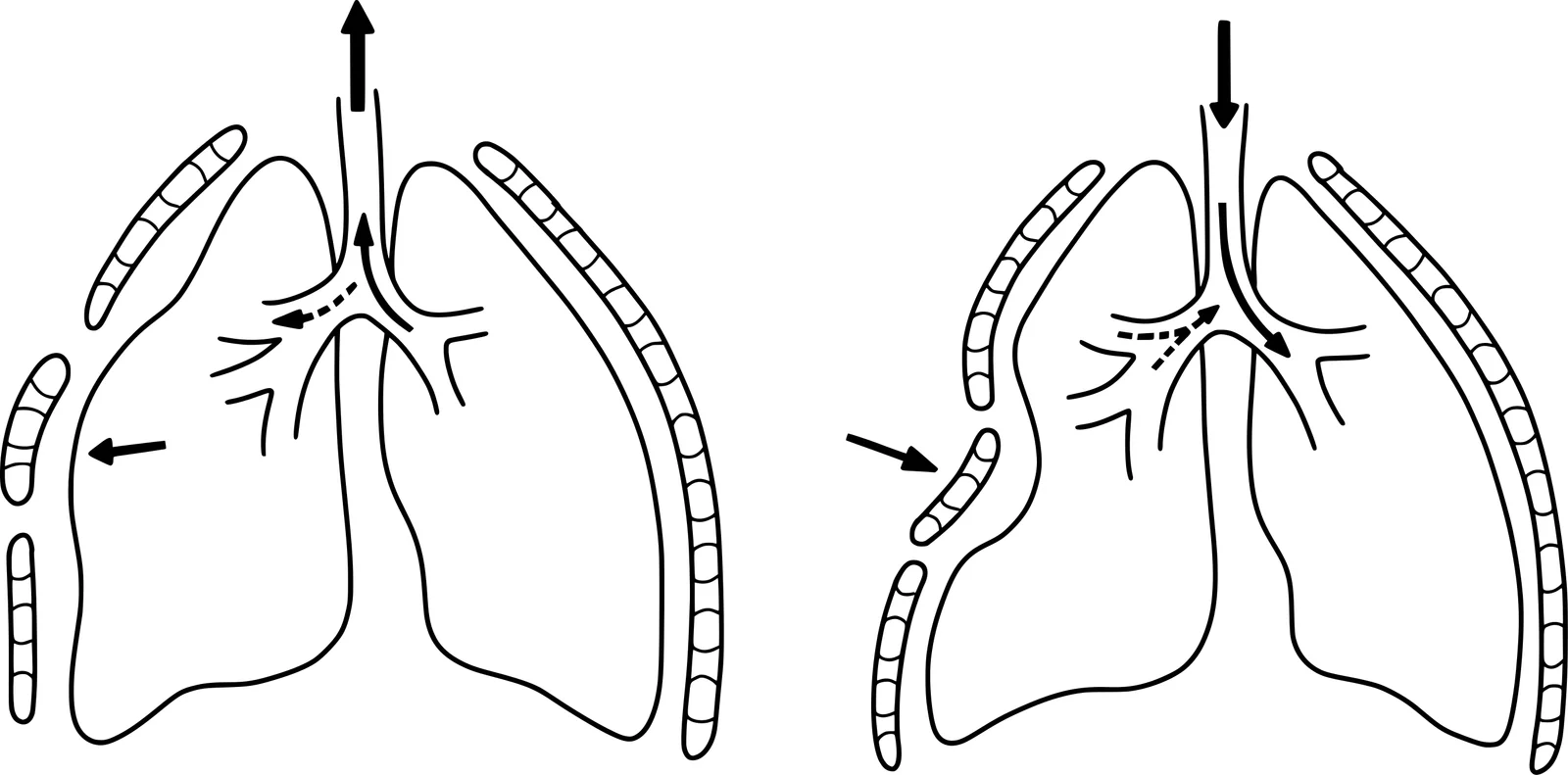
Long description
illustration of paradoxical breathing caused by an unstable chest wall (flail chest).

Pain control is a central part of rib fracture care. Effective analgesia allows deep breathing and coughing, which prevents atelectasis, pneumonia and ARDS.[1] Use a multimodal, opioid-sparing regimen: paracetamol and NSAIDs where safe, regional techniques and, where needed, opioids. Useful regional options include epidural, paravertebral, intercostal, serratus anterior plane and erector spinae plane techniques. [6] Regional blocks can improve pain scores and inspiratory capacity in chest wall injury; ultrasound guidance is required. Sub-dissociative ketamine, topical lidocaine and other adjuvants may be included according to local protocols. [6] For severe pain unresponsive to other treatments, surgical stabilisation may be considered. [1]
Surgical stabilisation of rib fractures is recommended in:
- haemodynamically stable patients with flail chest;
- patients with at least three severely displaced ipsilateral ribs (3rd to 10th), or failure to wean from ventilation; [1]
- within 48 to 72 hours of injury when early intervention is possible, or as soon as possible between 3 and 7 days if delayed. [1]
Traumatic brain injury and pulmonary contusion are not absolute contraindications to fixation. Haemodynamic instability is an absolute contraindication, and advanced age, major comorbidity, active malignancy or chest irradiation are relative contraindications requiring individual assessment. Fractures of the 1st, 2nd, 11th and 12th ribs do not usually affect ventilation unless they are significantly displaced.[1] Fixation improves ventilator, intensive care and pain outcomes in selected patients but should not delay treatment of more urgent injuries. [1]
Figure 3Rippenserienfraktur Röntgen Thorax
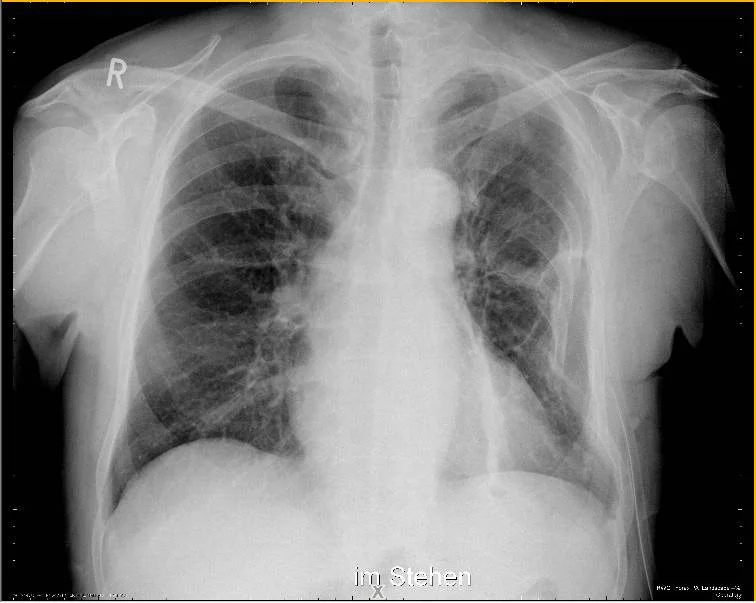
Long description
Chest X-ray showing a serial (multiple) rib fracture on the left involving ribs 2-10, with the dislocated fractures of ribs 5-7 especially conspicuous.

Treatment focuses on oxygen supplementation to maintain adequate oxygenation and on analgesia to facilitate deep breathing and coughing. Non-invasive or mechanical ventilation is used for respiratory failure, with lung-protective strategies. Fluids are given carefully. Routine antibiotic prophylaxis is not indicated in healthy patients without other penetrating injuries requiring chest tube placement. [1]
Figure 4Hemorragic effusion
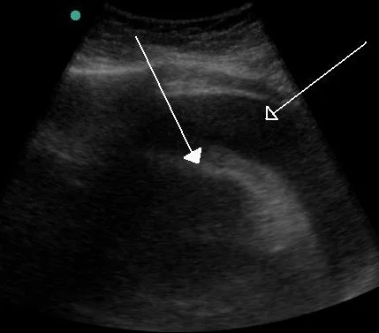
Long description
An echocardiogram/ultrasound of a very large hemorrhagic pericardial effusion; the pericardial effusion article caption identifies closed and open arrows pointing to the heart and the effusion.

Suspect cardiac tamponade in any chest trauma with hypotension, raised jugular venous pressure and muffled heart sounds, and confirm (or exclude) with bedside ultrasound: a pericardial collection with right-heart collapse and exaggerated respiratory variation in flow. Echocardiography also assesses valve, septal and free-wall integrity and myocardial function. [2]
If tamponade is suspected clinically or on ultrasound, pericardiocentesis can be performed; a tamponade from an atrial tear may be managed with a pigtail catheter and periodic drainage until surgical repair. In unstable patients who may not survive transfer, emergency department thoracotomy may be the best treatment, although blunt thoracotomy rarely resuscitates successfully. Severe injuries (ruptured valve, septum or ventricular wall with tamponade) require emergent surgical consultation. [2]
Blunt cardiac injury (BCI) is usually a myocardial contusion. Presentation ranges from none to chest pain, dyspnoea and arrhythmia. The right ventricle is at greatest risk; right bundle branch block is common and left bundle branch block is rare. Histologically there is haemorrhage, necrosis and oedema. Cardiac enzymes such as troponin and creatine kinase rise after injury, but they are markers rather than a standalone test. [2] Diagnosis combines mechanism, clinical findings, ECG, biomarkers and echocardiography, with discharge decisions made according to local protocol. [2]
Figure 5AoDiss ChestXRay
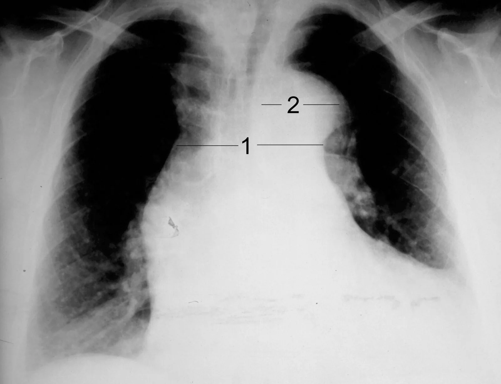
Long description
A chest X-ray of an aortic dissection, Stanford type A.

Mechanism matters more than the chest X-ray. A widened mediastinum (more than 8 cm) appears in only 27.7% of confirmed injuries. [1][3] Gather the mechanism (rapid deceleration, steering-column or seat-belt compression), examine for differential pulses or blood-pressure gradients, and proceed to CTA for any significant deceleration even with a normal film. CTA findings (intimal flap, intramural haematoma, pseudoaneurysm, extravasation) drive management and the Society for Vascular Surgery grade. Grades II to IV are preferably treated with TEVAR; unstable or endovascularly unsuitable cases need open repair. [1][3] Watch for connective tissue aortopathy (Marfan, vascular Ehlers-Danlos), in which fragility permits dissection at small diameters and trauma and unnecessary invasive procedures should be avoided. [4]
The priority is to secure the compromised airway, with careful intubation possibly guided by bronchoscopy. [1] Bronchoscopy provides diagnosis and localisation. Small uncomplicated tears may heal with conservative treatment, close monitoring and infection control. Surgery (minimal debridement, tension-free repair, preservation of blood supply, possible tracheostomy) is required for large ruptures, significant air leaks, injuries with oesophageal or vascular damage, or persistent pneumothorax despite drainage; repair within the first 24 hours is preferred. Prophylactic antibiotics may be considered, especially with surgical repair or delayed drainage of haemothorax. Long-term follow-up with airway endoscopy is needed for late stenosis. [1]
Repair is mandatory as soon as the patient's condition permits, on both sides. Early thoracoscopy or laparoscopy within 24 hours is indicated when the injury is suspected, because missed injuries become post-traumatic hernias. Penetrating trauma makes small tears (63% of diaphragmatic injuries are penetrating); blunt trauma makes larger tears. There is no difference between absorbable and non-absorbable monofilament repair techniques. A chest tube should be placed after repair. Complications include empyema, subphrenic abscess, suture dehiscence and hemidiaphragm paralysis from phrenic nerve injury; mortality is 18 to 40% and morbidity 40 to 60%, mostly from associated injuries. [1][5]
Trauma patient with chest injury
- Primary survey ABCDEAssess airway, breathing, circulation, disability, exposure; give oxygen and monitor
- Control catastrophic haemorrhage and resuscitateFluids, blood products, tranexamic acid, massive transfusion protocol
- Focused chest examinationInspect, palpate, percuss and auscultate both sides; look for open wounds and flail segments
- Question: Haemodynamic or respiratory status?
Unstable or shocked
- Immediate E-FAST +/- chest X-rayDetect pneumothorax, haemothorax, haemopericardium
- Question: Life-threatening thoracic injury found?
Tension pneumothorax
- Needle decompression then chest tube2nd intercostal space midclavicular or 5th midaxillary, then tube in 4-6th space
Massive haemothorax
- Large chest tube immediately>500 ml stable to drain; emergency thoracotomy thresholds apply
Open pneumothorax
- Three-sided occlusive dressing, then chest tubeConvert to closed drainage
Cardiac tamponade
- Bedside pericardiocentesis or emergency thoracotomyUltrasound confirms; severe injuries need surgery
- Damage-control resuscitation and surgeryOR, cath lab or ICU according to injury
Stable
- Contrast-enhanced CT chest +/- CT angiographyDefine all injuries; plan intervention
- Tube thoracostomy for pneumothorax or haemothorax4th to 6th intercostal space mid-axillary line
- Airway assessmentBronchoscopy for suspected tracheobronchial injury
- Vascular assessmentCTA for suspected aortic, great vessel or cardiac injury
- Diaphragm assessmentInspect by CT and consider laparoscopy or thoracoscopy if suspected
- Pain control and chest physiotherapyMultimodal, opioid-sparing analgesia with regional techniques
- Surgical stabilisation of flail or displaced ribsUsually within 48 to 72 hours
- Monitor for complicationsPneumonia, ARDS, empyema, missed injuries
- Pneumonia and ARDS from splinting, retained haemothorax and aspiration.
- Empyema from retained haemothorax (especially more than 300 ml residual) or infection.[1]
- Bronchopleural fistula and persistent air leak.
- Air embolism, a rare catastrophic complication of air in the circulation.
- Missed diaphragmatic hernia presenting weeks to years later with obstruction.
- Delayed aortic rupture or pseudoaneurysm formation after intimal injury.
- Cardiac rupture, arrhythmia and post-contusion heart failure.
- Chronic pain and deformity after multiple rib fractures.
Overall mortality from thoracic trauma is substantial: chest trauma causes 20 to 25% of trauma deaths. [1] Penetrating injuries have higher mortality than blunt. Most patients with blunt chest trauma are managed non-operatively and do well if analgesia and ventilation are optimised. Prognosis is driven by associated brain, abdominal and pelvic injuries and by age and frailty.
Prevention works better than any operation: seat belts, helmets, speed control, fall protection, safer machinery and factory guards, and prompt evacuation. In resource-limited settings simple interventions (good oxygen delivery, early analgesia, tube thoracostomy, basic monitoring) save the most lives.
- Children: the chest wall is more compliant, so significant pulmonary injury can exist with few or no rib fractures. The same primary-survey principles apply; involve paediatric and trauma teams.
- Older adults: frailty, anticoagulation, osteoporosis and reduced reserve worsen outcome; lower thresholds for imaging and admission.
- Pregnancy: resuscitate the mother (left uterine displacement, high-flow oxygen) and involve obstetric, trauma and critical care teams.
- Anticoagulated or bleeding patients: reverse anticoagulation where appropriate and monitor closely.
- Treat tension pneumothorax on clinical grounds with immediate decompression; do not wait for X-ray.
- Tube thoracostomy can be performed with basic sterile equipment; percussion and auscultation remain valuable when imaging is limited.
- Ultrasound (if available) diagnoses free fluid, pneumothorax and haemopericardium.
- Analgesia, oxygen, intravenous fluids and physiotherapy should be prioritised.
- Refer early for thoracotomy, aortic repair or fixation when local expertise is lacking.
East Africa
In many East African settings penetrating trauma is common, prehospital care variable and advanced imaging scarce. Clinical diagnosis, bedside decompression and tube thoracostomy, early referral and simple multimodal analgesia (paracetamol, NSAIDs where safe, nerve blocks when an anaesthetist is available) achieve the best outcomes.
- Treat tension pneumothorax, open pneumothorax, massive haemothorax and cardiac tamponade before any imaging.
- Blunt chest trauma causes most trauma deaths but usually does not need surgery; penetrating trauma more often does.
- A widened mediastinum is present in only 27.7% of confirmed blunt aortic injuries: keep a low threshold for CT angiography after deceleration.
- Chest tube is inserted in the 4th to 6th intercostal space mid-axillary line; needle decompression is at the 2nd intercostal space midclavicular or the 5th midaxillary line.
- Massively bleeding chests drain more than 1,500 ml in 24 hours or 200 ml per hour for 3 hours and need surgery.
- Pain control is a core treatment: it prevents pneumonia and ARDS after rib fractures.
- CT angiography and ultrasound are central; do not rely on chest X-ray to exclude aortic or diaphragmatic injury.
Answer each one in your head first, then open the answer.
A 24-year-old is stabbed in the chest, hypotensive, with decreased breath sounds on the right and bright blood from a chest drain in the first hour. What are the next two critical decisions?
Show answerHide answer
Immediately consider thoracotomy for active intrathoracic bleeding (over 200 ml per hour) and exclude cardiac injury through the pericardium into the right haemothorax; the patient needs massive transfusion and damage-control care in theatre.[1]
A stable polytrauma patient has a chest X-ray that is normal but was in a high-speed deceleration. Why is CT angiography still indicated?
Show answerHide answer
A normal chest X-ray does not exclude blunt aortic injury; a widened mediastinum is present in only 27.7% of confirmed cases, and CT angiography is the preferred test for diagnosis and planning. Rapid deceleration is the predominant mechanism, with shear at the aortic isthmus.
A patient with multiple rib fractures is splinting, tachypnoeic and refusing to cough. What are two important treatments apart from analgesia?
An intubated trauma patient has a non-resolving pneumothorax despite a correctly placed chest drain. List likely causes.
Show answerHide answer
Ongoing air leak from tracheobronchial disruption, blocked or kinked tube, or a large bronchopleural fistula; bronchoscopy confirms airway injury and surgery is indicated for large ruptures or persistent leaks.[1]
A young man hit in the chest has normal vital signs but an ECG shows right bundle branch block and a raised troponin. How should this be interpreted?
Show answerHide answer
It is consistent with blunt cardiac injury, most often a myocardial contusion affecting the right ventricle. Diagnosis is clinical plus ECG, biomarkers and echocardiography; arrhythmias and other complications should be monitored.[2]
A pregnant woman with chest trauma arrives hypotensive. What two adjustments to the primary survey are essential?
Show answerHide answer
Resuscitate the mother first (left uterine displacement, high-flow oxygen, fluids and blood as indicated) and involve obstetric, trauma and critical care teams together.
Primary survey and trauma resuscitation; major haemorrhage and damage control; respiratory failure and mechanical ventilation; blunt abdominal trauma; head injury; spinal injury; pain management and regional anaesthesia; echocardiography and ultrasound in trauma.
- Coccolini F, Cremonini C, Moore EE, et al. Thoracic trauma WSES-AAST guidelines. World J Emerg Surg 2025;20:78. doi:10.1186/s13017-025-00651-1. Accessed .
- Nair L, Winkle B, Senanayake E. Managing blunt cardiac injury. J Cardiothorac Surg 2023;18:71. doi:10.1186/s13019-023-02146-z. Accessed .
- Lee HJ, Rho JY, Kim MY, Kang SR. Aortic Injury and Hemopericardium: Key Findings and Evaluation. J Korean Soc Radiol 2026;87(4):656-672. Accessed .
- Fatehi Hassanabad A, McKenzie MP, Keir M, Fedak PWM. Thoracic aortic disease in young patients: from genetics to management. Front Cardiovasc Med 2026;13:1864645. Accessed .
- Choi JM, Lim S, Keum MA, Lee TY. Chest Wall and Diaphragmatic Injuries: Imaging Evaluation and Clinical Considerations. J Korean Soc Radiol 2026;87(4):641-655. Accessed .
- Hancin E, Patel PP, Egodage T. Regional, multimodal, and opioid-sparing strategies after trauma: a review. Trauma Surg Acute Care Open 2026;11(suppl 1):e002273. Accessed .
Figure credits
- Figure 1: Cupr78up, via Wikimedia Commons, CC BY-SA 3.0, CC BY-SA 3.0.
- Figure 2: Baedr-9439, via Wikimedia Commons, CC0, CC0.
- Figure 3: Sjoehest, via Wikimedia Commons, CC BY-SA 4.0, CC BY-SA 4.0.
- Figure 4: Jmh649, via Wikimedia Commons, CC BY-SA 3.0, CC BY-SA 3.0.
- Figure 5: J. Heuser JHeuser, via Wikimedia Commons, CC BY-SA 3.0, CC BY-SA 3.0. resized.
Educational content for medical students and doctors. It is not a substitute for clinical judgement or local guidelines.