Adrenal insufficiency and adrenal crisis
How the adrenal glands fail, how the patient looks, how to test, and how to treat adrenal crisis and lifelong replacement safely.
- Adrenal insufficiency means the adrenal glands do not make enough of the steroid hormones the body needs to keep blood pressure, salt balance and energy steady. The commonest chronic form is autoimmune destruction of the adrenal cortex, known as Addison disease.
- It divides into primary disease, where the adrenal cortex itself fails; secondary disease, where the pituitary does not make ACTH; and tertiary disease, where the hypothalamus does not make CRH. Glucocorticoid tablets taken for another condition and then withdrawn are the most common cause of the secondary and tertiary forms.
- The everyday picture is tiredness, weight loss, low blood pressure on standing, salt craving, hyponatraemia and often hyperkalaemia; darkening of the skin occurs only when the adrenal (not the pituitary) has failed.
- An adrenal crisis is a medical emergency: severe weakness, fainting, abdominal pain, vomiting, confusion and very low blood pressure. Treat at once with hydrocortisone 100 mg intravenously and rapid saline, before any test result.
- Lifelong treatment replaces what is missing: hydrocortisone daily, fludrocortisone if the mineralocorticoid is missing, together with written sick-day rules, a steroid emergency card and a pre-filled hydrocortisone injection kit.
By the end of this page you should be able to:
- Explain what the adrenal cortex and medulla normally do and how failure of each zone produces symptoms.
- Classify adrenal insufficiency as primary, secondary or tertiary, and name the main causes of each.
- Recognise the clinical features of chronic adrenal insufficiency, including the features that separate primary from secondary disease.
- State the diagnostic pathway: morning cortisol and ACTH, the short Synacthen test, renin, aldosterone and 21-hydroxylase antibodies, with their cut-offs.
- Treat an adrenal crisis without delay and plan lifelong replacement, sick-day dosing and steroid card use.
- Adjust care for pregnancy, children, older adults and low-resource settings.
Adrenal insufficiency is the clinical state produced by a failure of the adrenal glands to secrete enough steroid hormone. It is uncommon, serious and eminently treatable. The adrenal glands are two small glands, one above each kidney. Each has an outer cortex that makes steroid hormones and an inner medulla that acts as part of the sympathetic nervous system. Because cortisol is required for vascular tone, glucose availability and the stress response, its deficiency can kill within hours once a patient is stressed by infection, trauma or surgery. Because aldosterone controls sodium and potassium balance, its deficiency produces salt loss and dangerous hyperkalaemia when the adrenal itself is destroyed.
The topic on this page covers both the chronic disease and its acute presentation, an adrenal crisis. The common form of primary adrenal failure, autoimmune adrenal destruction, is often still called Addison disease after Thomas Addison.
Figure 11818 The Adrenal Glands
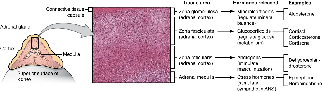
Long description
Both adrenal glands sit atop the kidneys and are composed of an outer cortex and an inner medulla, all surrounded by a connective tissue capsule; the cortex can be subdivided into additional zones, all of which produce different types of hormones.

Each adrenal is a small, flattened gland lying in the retroperitoneum above the kidney. The gland has two parts of different embryological origin.
- Adrenal cortex arises from mesoderm. It makes three classes of steroid hormone from cholesterol:
- the outer zona glomerulosa makes aldosterone, the principal mineralocorticoid;
- the middle zona fasciculata makes cortisol, the principal glucocorticoid;
- the inner zona reticularis makes weak adrenal androgens such as dehydroepiandrosterone (DHEA) and its sulphate (DHEAS).
- Adrenal medulla arises from neural crest. It is essentially a sympathetic ganglion whose cells release catecholamines, mainly adrenaline (epinephrine) and noradrenaline (norepinephrine), and a little dopamine, into the bloodstream.
The cortex is the part that fails in adrenal insufficiency. Adrenal crisis in classical Addison disease is a crisis of cortisol and aldosterone deficiency. The medulla is usually spared, except after bilateral adrenalectomy or massive haemorrhage.
- Cortisol is driven by ACTH (adrenocorticotropic hormone) from the anterior pituitary, which is itself driven by CRH (corticotropin-releasing hormone) from the hypothalamus. Cortisol feeds back negatively on both. The hypothalamus-pituitary-adrenal (HPA) axis is the central stress system. Cortisol has a marked circadian rhythm, peaking in the early morning and reaching its lowest point late at night.
- Aldosterone is driven mainly by the renin-angiotensin system (angiotensin II) and by serum potassium, with a minor contribution from ACTH. Its production is independent of ACTH in the long term, which is why aldosterone is preserved in secondary and tertiary disease.
- Adrenal androgens are ACTH-driven but their loss is clinically important mainly in women, in whom they are the main source of androgen after the menopause.
- Cortisol maintains vascular alpha-adrenergic responsiveness and blood pressure, stimulates gluconeogenesis and protects against hypoglycaemia, raises free-water clearance through effects on the kidney, and has permissive actions on catecholamines. It also suppresses inflammation and the immune response.
- Aldosterone acts on the renal collecting duct to reabsorb sodium and excrete potassium and hydrogen ions. Without it, sodium and water are lost in the urine and potassium accumulates.
- Adrenal androgens contribute to pubic and axillary hair in women and to libido and sense of well-being. Their loss in men is usually unnoticed because the testes produce far more.
A patient with adrenal insufficiency therefore shows the combined effects of these losses: low blood pressure and salt wasting, hypoglycaemia under stress, pigmentation from loss of negative feedback only when ACTH rises, and in women loss of axillary and pubic hair.
Adrenal insufficiency is the inability of the adrenal cortex to produce sufficient glucocorticoids and, variably, mineralocorticoids and androgens. Adrenal crisis is the acute, life-threatening manifestation of that failure.
| Level of failure | Site | ACTH | Aldosterone or renin | Sodium and potassium | Hyperpigmentation |
|---|---|---|---|---|---|
| Primary | Adrenal cortex | High | Low aldosterone, high renin | Low sodium, potassium often high | Present |
| Secondary | Pituitary | Low or inappropriately normal | Normal aldosterone, normal renin | Sodium often low, potassium normal | Absent |
| Tertiary | Hypothalamus | Low or inappropriately normal | Normal aldosterone, normal renin | Sodium often low, potassium normal | Absent |
- Primary adrenal insufficiency (PAI) means the adrenal cortex itself is destroyed or dysfunctional. ACTH rises because feedback is lost; renin rises because aldosterone is low. Pigmentation develops because ACTH and other products of pro-opiomelanocortin stimulate melanocytes.
- Secondary adrenal insufficiency is failure of ACTH secretion from the pituitary (pituitary tumour, apoplexy, surgery, radiation, hypophysitis, or chronic glucocorticoid use).
- Tertiary adrenal insufficiency is failure of CRH from the hypothalamus. In practice the two are managed identically and are usually grouped as central adrenal insufficiency; the distinction matters mainly for research and for understanding the cause.
- Glucocorticoid-induced adrenal insufficiency is the most important form of secondary or tertiary disease. Chronic exogenous glucocorticoid suppresses hypothalamic CRH and pituitary ACTH, and the adrenal cortex atrophies from lack of stimulation. Onset can be insidious and unmask itself only when the drug is tapered or the patient is stressed. Fludrocortisone is not needed because the renin-angiotensin-aldosterone axis is intact. [2]
Adrenal insufficiency can also be described by time course:
- Chronic disease, with weeks to months of non-specific symptoms.
- Acute decompensation on chronic disease, which is the most common cause of adrenal crisis.
- Acute destruction of both adrenals (bilateral haemorrhage, thrombosis or massive infiltration), which can produce crisis as the first event with no prior chronic phase.
Adrenal insufficiency is rare but under-recognised. Reports are mainly from Europe and North America; data from low-resource regions are essentially absent.
- Prevalence of primary adrenal insufficiency ranges from about 15 to 20 per 100,000 in Scandinavia to about 10 per 100,000 in other parts of Europe, and was 0.4 per 100,000 in a national survey from Korea. An earlier Endocrine Society estimate put prevalence at 100 to 140 per million with an incidence of about 4 per million per year in Western societies. [1][3]
- Incidence of primary disease is reported at about 5 new cases per million per year. [3]
- Diagnosis is commonly delayed: in one series fewer than 30% of women and 50% of men were diagnosed within the first 6 months of symptoms, and 20% had been symptomatic for more than 5 years. More than two-thirds saw at least three doctors first and two-thirds were given another diagnosis initially. [3]
- Autoimmune destruction dominates in developed countries, accounting for about 90% of primary adrenal insufficiency in young and middle-aged adults, and 21-hydroxylase autoantibodies are present in more than 90% of newly diagnosed cases. [3] [1]
- Adrenal crisis is common in known disease: in a 24-year Norwegian cohort of 1040 patients with autoimmune primary adrenal insufficiency, 63% had at least one crisis-related hospitalisation and 25% had an overt adrenal crisis after diagnosis, with an incidence of 3.2 overt crises per 100 person-years. Crisis-related in-hospital mortality in that cohort was 0.3%. [4]
- Secondary adrenal insufficiency from long-term glucocorticoid use is far more common than primary disease in absolute terms: at least 1% of the population of the United Kingdom and the United States is on chronic glucocorticoid therapy at any time, and prolonged treatment above a physiologic daily dose equivalent (about 15 to 25 mg hydrocortisone equivalent) suppresses the HPA axis. [2]
The Endocrine Society lists the major causes of primary adrenal insufficiency. [1] They group naturally as destructive, genetic and drug-related.
| Group | Causes |
|---|---|
| Primary, destructive | Autoimmune adrenalitis (isolated or as part of autoimmune polyglandular syndrome type 1 or type 2); tuberculosis and other infections (HIV, cytomegalovirus, candidiasis, histoplasmosis, syphilis, African trypanosomiasis, paracoccidioidomycosis); adrenal haemorrhage (sepsis, anticoagulation, antiphospholipid syndrome, including Waterhouse-Friedrichsen syndrome); metastases (lung, breast, colon, melanoma, lymphoma) and primary adrenal lymphoma; infiltration (haemochromatosis, amyloidosis, sarcoidosis); bilateral adrenalectomy |
| Primary, genetic | Congenital adrenal hyperplasia (commonest cause of primary adrenal insufficiency in children, about 80% of paediatric cases; 21-hydroxylase deficiency is the commonest form); adrenal hypoplasia congenita; X-linked adrenoleukodystrophy in males; ACTH insensitivity syndromes; mitochondrial disease such as Kearns-Sayre syndrome; peroxisomal disorders; familial glucocorticoid deficiency |
| Primary, drug-induced | Enzyme inhibitors (ketoconazole, mitotane, metyrapone, etomidate, aminoglutethimide); drugs that accelerate cortisol metabolism (rifampicin, phenytoin, phenobarbital, carbamazepine); immune check-point inhibitors (nivolumab, pembrolizumab); abiraterone; mifepristone |
| Secondary (pituitary) | Chronic exogenous glucocorticoid use, the commonest overall; pituitary macroadenoma or microadenoma; hypophysectomy or pituitary radiation; pituitary apoplexy; lymphocytic hypophysitis; trauma and empty sella; infiltrative disease |
| Tertiary (hypothalamic) | Chronic exogenous glucocorticoid use; cranial irradiation; hypothalamic tumour or surgery; infiltration; chronic opiate use |
| Neonatal and childhood | Congenital adrenal hyperplasia, adrenal hypoplasia, Allgrove syndrome (triple A), adrenal haemorrhage of the newborn, and maternal glucocorticoid excess |
Some practical points on causes:
- Autoimmune disease. Even when 21-hydroxylase antibodies are positive, other organ-specific autoimmune disease is common: thyroid disease in roughly half of patients, type 1 diabetes in about one in six, vitiligo, pernicious anaemia, coeliac disease and premature ovarian insufficiency. [4] [4]
- Tuberculosis remains an important cause in developing countries and in older migrants, and should be considered where infection is plausible. Bilateral adrenal metastases usually destroy most of the cortex before symptoms appear; a third to a half of patients with bilateral adrenal masses have biochemical insufficiency.
- Congenital adrenal hyperplasia is the commonest cause of primary adrenal insufficiency in children, accounting for about 80% of paediatric cases; 21-hydroxylase deficiency is the commonest form and is associated with hyperandrogenism and, in the salt-wasting form, with neonatal crisis. [1]
- X-linked adrenoleukodystrophy should be considered in males with negative 21-hydroxylase antibodies: measure plasma very long chain fatty acids. [1]
- Glucocorticoid-induced disease is a chronic, avoidable cause. Risk rises with dose and duration: in asthma cohorts biochemical suppression was found in 2.4% on low-dose and 21.5% on high-dose regimens, and rose from 1.4% with treatment under 28 days to 27.4% with treatment over one year; oral, intra-articular and high-dose inhaled routes all carry risk. Treatment for less than 3 to 4 weeks, regardless of dose, rarely suppresses the axis and can be stopped without testing. [2]
In primary adrenal insufficiency the cortex is destroyed (autoimmune, infectious, haemorrhagic or infiltrative) or cannot function (genetic enzyme defects). Cortisol falls first; ACTH rises as feedback is lost. Without cortisol, vascular tone falls, vascular permeability rises and free-water excretion falls. Hyponatraemia follows from loss of free water and, in primary disease, from aldosterone loss. Hyperkalaemia and salt wasting follow from absent aldosterone: urinary sodium loss contracts the extracellular volume, renin rises, and potassium and hydrogen ions accumulate. Hypoglycaemia follows from failed gluconeogenesis, especially during fasting, infection or alcohol. ACTH over-secretion, together with other products of pro-opiomelanocortin such as beta-lipotropin and melanocyte-stimulating hormone, darkens the skin. [1]
A mineralocorticoid deficiency also stimulates salt craving, a useful historical clue. Aldosterone deficiency makes the patient prone to hypotension and volume depletion even before a stress. DHEA loss produces fatigue, low mood and loss of libido in women; in men the testes compensate.
In central disease the adrenal cortex is healthy but unstimulated. ACTH is low, so cortisol is low, but aldosterone and renin remain normal because the renin-angiotensin system is intact. There is no hyperpigmentation and no hyperkalaemia, but salt may be lost if there is co-existing aldosterone problems from severe illness. Volume depletion is less striking than in primary disease. Other pituitary hormone deficits often dominate the picture: secondary hypothyroidism, hypogonadism, growth hormone deficiency or diabetes insipidus.
In glucocorticoid-induced disease, prolonged supraphysiological exogenous steroid suppresses CRH and ACTH and the adrenal cortex atrophies. Recovery after withdrawal is highly variable: the axis may recover in weeks, or may take many months, and sometimes never fully recovers. A long-acting steroid such as dexamethasone carries the highest risk; switching to a shorter-acting steroid when the disease is controlled makes both tapering and recovery easier. [2]
An adrenal crisis occurs when the residual cortisol (or exogenous steroid cover) is inadequate for the stress imposed by infection, trauma, surgical stress, myocardial infarction, pregnancy and delivery, or gastrointestinal illness with vomiting and diarrhoea. The already low blood pressure and intravascular volume collapse, glucose falls, and the patient may die of vasodilatory shock and hypoglycaemia unless cortisol is replaced. A surprisingly large fraction of crises are precipitated by reduction or stopping of the steroid dose rather than by intercurrent illness alone. [2][4]
Adrenal pathology reflects the cause:
- Autoimmune adrenalitis: lymphocytic infiltration of the cortex, most prominent in the zona fasciculata and reticularis, with progressive cortical atrophy; the medulla and capsule are spared.
- Infective adrenalitis: caseating granulomas in tuberculosis, fungal or viral inclusions; in acute meningococcal septicaemia there is bilateral adrenal haemorrhage with cortical necrosis (Waterhouse-Friedrichsen syndrome).
- Haemorrhage and infarction: bilateral adrenal haemorrhage from anticoagulation, sepsis, or thrombotic tendency.
- Metastatic infiltration and primary lymphoma: replacement of cortex by tumour; bilateral enlargement on imaging.
- Genetic disease: hyperplasia of the adrenal cortex (as in congenital adrenal hyperplasia) or hypoplasia (as in DAX1 or SF1 mutations).
A biopsy is rarely needed: the cause is usually clear from antibodies, imaging and the clinical picture.
The Endocrine Society summarises the features in two lists: chronic adrenal insufficiency and adrenal crisis. [1] Many features are non-specific and develop over months, which is why the diagnosis is missed.
Symptoms in the patient's words
- Fatigue, weakness and apathy, worst in the morning and improving as the day goes on.
- Loss of appetite, nausea, abdominal discomfort and (in primary disease) salt craving.
- Weight loss despite a normal or reduced intake.
- Orthostatic dizziness and even syncope on standing.
- Muscle and joint pain.
- Mood change: low mood, anxiety, reduced concentration; sometimes frank depression.
- In women, loss of axillary and pubic hair and reduced libido.
Signs on examination
- Hyperpigmentation in primary disease only: generalised duskiness, but most obvious on sun-exposed skin and areas of friction - knuckles, elbows, palmar creases, gums, areola, old scars and recent scars. In dark-skinned patients the change can be subtle and is best judged against the colour of the patient's own siblings. [3]
- Low blood pressure, with an increased postural drop (a fall of systolic pressure on standing).
- Thin habitus from weight loss.
- In children, failure to thrive.
- Vitiligo and other stigmata of autoimmunity.
Laboratory clues (often the trigger for testing)
- Hyponatraemia is the commonest abnormality and is present in about 85% of patients at diagnosis. [3]
- Hyperkalaemia is less common, present in less than half, partly because vomiting loses potassium. [3]
- Unexplained hyponatraemia is itself an indication to test, and a concomitant hyperkalaemia strengthens the suspicion. [3]
- Hypoglycaemia, more often in children, in diabetics (who become more insulin-sensitive) and in fasting or illness.
- Eosinophilia, lymphocytosis and a mild normocytic anaemia are common; eosinophilia at presentation is a useful pointer, because cortisol normally suppresses eosinophils.
- Raised TSH in about half of patients without known thyroid disease, due to loss of cortisol's permissive effect on TSH secretion. Treating this with thyroxine before the adrenal insufficiency is corrected can precipitate crisis. [3]
- Low DHEAS for age and sex, a sensitive early clue that is often overlooked.
A history of autoimmune disease (type 1 diabetes, autoimmune thyroid disease, vitiligo, pernicious anaemia, coeliac disease, premature ovarian insufficiency) should sharpen suspicion, because most of these patients have or will develop at least a second organ-specific autoimmune condition. [3]
Note that the clinical picture of secondary or tertiary disease differs: no hyperpigmentation (ACTH is not high), no hyperkalaemia (aldosterone is preserved), and volume depletion is less severe. The presenting features are often those of the underlying cause (headache and visual loss from a pituitary mass, drug history, radiation) plus non-specific cortisol deficiency, and often other pituitary hormone deficits such as secondary hypothyroidism, hypogonadism or diabetes insipidus. Glucocorticoid-induced disease may present only when the drug is withdrawn or during stress.
An adrenal crisis is a medical emergency. [4] It may be the first presentation of previously undiagnosed disease (classically in children with congenital adrenal hyperplasia, or with acute bilateral adrenal haemorrhage) or decompensation of known disease during intercurrent illness, surgery or trauma.
Symptoms [1]
- Severe, overwhelming weakness and fatigue.
- Nausea, vomiting and abdominal pain that may mimic an acute surgical abdomen.
- Back or loin pain.
- Confusion, drowsiness, delirium and eventually coma.
Signs [1]
- Hypotension, often profound and unresponsive to fluids until cortisol is given.
- Tachycardia, poor perfusion, cold extremities.
- Abdominal tenderness and guarding, fever.
- Reduced level of consciousness.
Laboratory abnormalities [1]
- Hyponatraemia (almost universal) and often hyperkalaemia.
- Hypoglycaemia, particularly in children.
- Hyperkalaemia may be absent when vomiting has been severe.
- Prerenal uraemia and a raised creatinine from volume depletion; raised urea out of proportion to creatinine.
- Hypercalcaemia occasionally.
Formal criteria used in research combine an acute deterioration with at least one of: systolic blood pressure under 100 mmHg, serum sodium under 130 mmol/L, serum potassium over 5 mmol/L, or blood glucose under 3.5 mmol/L. Incipient crisis is the state of typical symptoms (marked impairment of general health, nausea, vomiting, abdominal pain) without these objective abnormalities, requiring parenteral glucocorticoid. [4] Either way, any sick patient on chronic steroids with these features is treated as an adrenal crisis.
| Condition | How to tell it apart |
|---|---|
| Sepsis or acute abdomen | Crisis mimics both; hyponatraemia plus hyperkalaemia without sepsis points to adrenal failure; ultrasound or CT finds the surgical abdomen; blood cultures and cortisol distinguish sepsis |
| Primary (adrenal) vs secondary or tertiary | Pigmentation, hyperkalaemia, high renin and high ACTH mean primary; their absence means central |
| Dehydration from gastroenteritis | Corrects with fluids alone; no hyperpigmentation; no hyponatraemia-hyperkalaemia combination; cortisol normal |
| Hypothyroidism | Fatigue and low mood overlap; raised TSH and low T4; cortisol may be low-normal; never give thyroxine before cortisol is secure |
| Anorexia nervosa, chronic infection, malignancy | Weight loss and fatigue overlap; check early-morning cortisol and ACTH |
| Congenital adrenal hyperplasia | In a child: virilisation, salt wasting in the neonatal period, and a raised 17-hydroxyprogesterone |
| Adrenal metastasis or lymphoma | Older patient, weight loss, adrenal masses on CT, negative 21-hydroxylase antibodies |
Another trap is to attribute adrenal insufficiency to "depression", "gastroenteritis" or "functional illness"; the combination of hyponatraemia, hyperkalaemia and pigmentation should always prompt cortisol testing.
If the patient is acutely ill or haemodynamically unstable, do not delay treatment to investigate. [1] Draw a baseline blood sample for cortisol (and ACTH if it will not delay anything) and then give hydrocortisone 100 mg intravenously and saline. Diagnostic confirmation can be done later with a Synacthen test after the patient stabilises and steroid is temporarily withheld for 24 to 48 hours. [1]
If the patient is stable, the work-up is usually straightforward.
- Take a simultaneous sample for serum or plasma cortisol and plasma ACTH, ideally at 08:00 to 09:00 because cortisol peaks then; ACTH can be taken at any time if the clinical picture demands.
- A morning (08:00-09:00) cortisol below 100 nmol/L with an ACTH more than two times the upper reference limit is diagnostic of primary adrenal insufficiency. [3] [1]
- The Endocrine Society uses a screening cut-off of morning cortisol below 140 nmol/L (5 microg/dL) together with an ACTH markedly raised, as suggestive of disease until confirmatory testing is available. [1]
- An ACTH value over 66 pmol/L represents a maximal stimulus for cortisol secretion; paired with a low cortisol it confirms primary failure. [1]
- ACTH is low or inappropriately normal in central disease.
- At the same visit take plasma renin and aldosterone together, and DHEAS. High renin with low aldosterone supports mineralocorticoid deficiency; low renin does not, because non-steroidal drugs and age affect renin. [1]
Remember that cortisol is 80% bound to cortisol-binding globulin (CBG) and 10 to 15% to albumin; low CBG (liver disease, nephrotic syndrome, late pregnancy, severe illness) gives artefactually low total cortisol, while high CBG (oestrogen, pregnancy, mitotane) masks deficiency. Where results do not fit, free cortisol can be measured or the test repeated. [1]
The standard short (cosyntropin) test is the gold standard for primary adrenal insufficiency. [1]
- Give cosyntropin (synthetic ACTH 1-24, tetracosactide) 250 microg intravenously (or intramuscularly) as a bolus.
- Draw cortisol before the injection and at 30 and 60 minutes.
- A peak cortisol below 500 nmol/L (18 microg/dL) at 30 or 60 minutes indicates adrenal insufficiency. [1]
- The 30-minute point has been validated against the insulin tolerance test; the 60-minute result is sometimes slightly higher with the high dose and lower with the low-dose test.
- The traditional cut-off has been 500 or 550 nmol/L. With highly specific liquid chromatography tandem mass spectrometry (LC-MS/MS) assays the threshold has fallen to 412 nmol/L at 30 minutes and 485 nmol/L at 60 minutes. [3] Always use the cut-off given by your own laboratory and assay, because different immunoassays show different bias, and quote assay-specific normative data on the laboratory report. [1]
- The test is not affected by the time of day, so it can be done at any hour. [1]
- The 1 microg low-dose test adds no sensitivity or specificity over the 250 microg test for primary disease and is reserved for times of cosyntropin shortage; note that the low-dose test may be more sensitive for secondary disease, but that evidence is indirect. [1]
- Cosyntropin must be made up freshly; it degrades. In some countries porcine ACTH is used, but the evidence base is much thinner. [1]
False reassurance occurs when CBG is low (low total cortisol) or high (masked deficiency). In very recent onset disease (for example, autoimmune adrenalitis with residual function) the test may be normal while ACTH is already high; repeat testing in a few weeks.
All patients with confirmed primary adrenal insufficiency should have an aetiological diagnosis, because it changes treatment and follow-up. [3]
- 21-hydroxylase autoantibody assay in everyone without an obvious cause. A positive result confirms autoimmune aetiology in more than 90% of Western patients; in autoimmune polyglandular syndrome type 1 or type 2 look also for hypothyroidism, type 1 diabetes, premature ovarian insufficiency, pernicious anaemia and candidiasis. [1][3]
- Males with negative antibodies should have plasma very long chain fatty acids to exclude X-linked adrenoleukodystrophy. [1]
- If antibodies are negative, adrenal CT will show atrophy in autoimmune disease, enlargement or masses in infiltration (metastases, lymphoma, granulomatous disease, or haemorrhage), and calcification in tuberculosis and other infections.
- Baseline 17-hydroxyprogesterone above 1000 ng/dL diagnoses 21-hydroxylase deficiency. [1]
- Search for tuberculosis with imaging and culture where relevant; test for HIV and other infections.
- In children, test for congenital adrenal hyperplasia; a salt-wasting 21-hydroxylase deficiency presents in the first two weeks of life and can be fatal if missed.
The backbone is the same: a low morning cortisol with low or inappropriately normal ACTH. Because aldosterone is preserved, hyponatraemia is less often a clue and hyperkalaemia is absent. The short Synacthen test may be normal early, or show a delayed rise, so a low morning cortisol with high pre-test probability is enough to start treatment. Suspect central disease on any of:
- known pituitary or hypothalamic disease, surgery or radiotherapy;
- hypopituitarism (other hormone deficits);
- a recent course of glucocorticoids, inhaled or topical as well as oral;
- abrupt withdrawal of chronic glucocorticoids.
For patients tapering glucocorticoids, do not rely on dynamic testing routinely. Use the morning cortisol as a guide: above 300 nmol/L (10 microg/dL) the HPA axis has recovered and glucocorticoids can be stopped; 150 to 300 nmol/L means continue the physiological dose and repeat in weeks to months; below 150 nmol/L (5 microg/dL) continue and repeat in a few months. [2] Switch long-acting steroids (dexamethasone, betamethasone) to shorter-acting ones (hydrocortisone, prednisone) before tapering. [2]
Approach to suspected adrenal insufficiency
- Suspect from clinical pictureFatigue, weight loss, hypotension, hyponatraemia, hyperpigmentation (primary), hypoglycaemia in children; unexplained hyponatraemia
- Question:
Yes: crisis
- Take baseline cortisol and ACTH if it will not delay care
- Hydrocortisone 100 mg IV now, plus 1 L saline; continue 200 mg per 24 h
- Confirm diagnosis later with a Synacthen test once stable
No: chronic evaluation
- Paired 08:00 cortisol and ACTH; plus renin, aldosterone and DHEAS
- Cortisol under 100 nmol/L plus ACTH more than twice the upper limit of normal is diagnostic
- If uncertain, short Synacthen test: 250 microg IV, cortisol at 0, 30 and 60 min; peak below 500 nmol/L (assay-specific)
- Confirm primary vs centralHigh ACTH and high renin mean primary; low ACTH and normal aldosterone mean central
- 21-hydroxylase antibodies in all primary cases without obvious causePositive confirms autoimmune disease; if negative, CT adrenals and, in males, very long chain fatty acids
- Start replacement and educateHydrocortisone, fludrocortisone if primary, sick-day rules, steroid card, injection kit
- ECG and chest radiograph are non-specific. The ECG may show tall T waves or arrhythmia from hyperkalaemia.
- Blood cultures if infection or infected adrenal is suspected.
- Abdominal CT or MRI defines structure: atrophy suggests autoimmune disease; masses, haemorrhage, calcification or infiltration point to another cause.
- Dual-energy screening for associated autoimmune disease is reasonable and usually annual: thyroid function and antibodies, glucose, B12, coeliac serology, and ovarian reserve where relevant. [1]
There is no validated scoring system; diagnosis rests on biochemistry plus a compatible clinical picture.
Primary adrenal insufficiency is diagnosed when morning cortisol is low with inappropriately high ACTH, or when the Synacthen test fails to raise cortisol adequately, together with a compatible picture and high renin in the absence of other explanations. Central adrenal insufficiency is diagnosed when morning cortisol is low with low or normal ACTH, together with evidence of pituitary or hypothalamic disease or a relevant drug history.
Adrenal crisis has no universally accepted definition, and criteria vary between studies. In practice, treat whenever a patient with adrenal insufficiency (or with recent high-dose glucocorticoid exposure) has acute deterioration together with hypotension (systolic blood pressure under 100 mmHg), hyponatraemia (under 130 mmol/L), hyperkalaemia (over 5 mmol/L) or hypoglycaemia (under 3.5 mmol/L). [4] An incipient crisis is typical symptoms without these objective abnormalities but responding to parenteral glucocorticoid. [4]
It is safer to over-treat than under-treat: guidelines call for a low diagnostic (and therapeutic) threshold in acutely ill patients and in those with predisposing factors, including pregnant women with unexplained persistent nausea, fatigue and hypotension. [1]
Management has three pillars: recognise and treat crisis, replace what is missing, and prepare the patient for the stresses of everyday life.
Start immediately; start before the tests, because delays raise mortality. [1][4]
Steps
- Blood first, then drugs. If possible without delay, take a sample for cortisol, ACTH, urea, electrolytes and glucose. Then proceed.
- Hydrocortisone 100 mg intravenously as soon as you recognise the crisis (50 mg per m2 in children), followed by a further 200 mg over 24 hours (50 to 100 mg per m2 per 24 hours in children), as a continuous infusion or 50 mg every 6 hours intravenously or intramuscularly. [1]
- Fluids. Rapid infusion of 1000 mL of isotonic saline within the first hour (or 5% glucose in isotonic saline), then continue isotonic saline guided by hydration, blood pressure and urinary output; include 5% glucose once hypoglycaemia or sodium is a concern. Children receive 20 mL/kg of normal saline as a rapid bolus, repeatable up to 60 mL/kg within an hour. [1]
- Glucose. If hypoglycaemic, give dextrose 0.5 to 1 g per kg (or 2 to 4 mL per kg of D25W, maximum single dose 25 g) slowly, or 5 to 10 mL per kg of D10W in children under 12 years. [1]
- Monitor. Cardiac monitoring, electrolytes, glucose and urine output; watch for hyperkalaemia resolution and correction of hyponatraemia.
- Treat the trigger (infection, sepsis, myocardial infarction, pregnancy). Send blood cultures before antibiotics.
- Taper and switch to oral as soon as the patient improves. On day 2 the hydrocortisone dose commonly falls to 100 mg per 24 hours, with a planned rapid taper to the oral replacement dose, usually within 2 to 3 days. [1]
- Re-evaluate the cause. If the patient was on chronic glucocorticoids, the crisis may have been precipitated by withdrawal or intercurrent illness; re-test the HPA axis later.
If hydrocortisone is unavailable, use prednisolone; dexamethasone is the least-preferred alternative and should be used only if nothing else is available. [1] If fludrocortisone is given as part of the oral regimen, note that it is contraindicated in glucocorticoid-induced adrenal insufficiency, where the mineralocorticoid axis is intact. [2]
Parenteral cover for patients who cannot take tablets (gastroenteritis, trauma, pre-operative): adults 100 mg of hydrocortisone intramuscularly or subcutaneously; children according to size - 25 mg in infants, 50 mg in school-aged children, 100 mg in adolescents. [1]
Route for emergency treatment by injection kits is usually intramuscular or subcutaneous into the thigh; patients and families need face-to-face training, not just written instructions.
- Exclude or treat infection and dehydration.
- Identify the trigger and correct it.
- Provide written (and preferably pocket-sized) instructions for sick days.
- Ensure the patient carries identification.
Replace what is missing and avoid over-replacement. Chronic replacement doses are lower than crisis doses.
Glucocorticoid (all patients with adrenal insufficiency)
- Hydrocortisone 15 to 25 mg per day, or cortisone acetate 20 to 35 mg per day, divided into two or three oral doses; the highest dose is taken on waking, with the next dose in the early afternoon, and a third in the late afternoon if used. Twice or three times daily dosing mimics the natural cortisol peak better than a single large dose. [1]
- Treatment should be taken with food; missing the morning dose is a common cause of morning fatigue.
- Prednisolone 3 to 5 mg daily, once or twice daily, is an acceptable alternative, especially where compliance with three daily doses is poor; dexamethasone is not recommended for chronic replacement because its long action makes dose titration difficult and causes Cushingoid side effects. [1]
- Monitor by clinical assessment: body weight, postural blood pressure, energy, salt craving, signs of glucocorticoid excess (weight gain, abdominal striae, hyperglycaemia, osteoporosis). Adequate replacement should not need biochemical monitoring; blood cortisol levels do not reflect tissue exposure because of pulses. [1]
- Doses need adjustment during intercurrent illness, surgery, pregnancy and other stress (below).
Mineralocorticoid (primary adrenal insufficiency with aldosterone deficiency)
- Fludrocortisone, starting dose 50 to 100 microg once daily in adults, is recommended for all patients with confirmed aldosterone deficiency; do not restrict salt intake. [1]
- The median dose in treated adults is about 0.1 mg daily, and most patients need between 0.05 and 0.2 mg per day; adjust according to salt craving, postural hypotension, peripheral oedema and blood electrolytes. [1]
- If blood pressure rises on fludrocortisone, reduce the dose; if it remains uncontrolled, treat the hypertension but continue fludrocortisone. [1]
- In children with confirmed aldosterone deficiency, fludrocortisone 100 microg daily plus salt supplementation in infants. [1]
- Children with primary disease: hydrocortisone about 8 mg per m2 of body surface area per day, divided into three or four doses, preferred over longer-acting glucocorticoids. [1]
- Fludrocortisone is not used in glucocorticoid-induced or central adrenal insufficiency: the mineralocorticoid axis is intact. [2]
Adrenal androgen (selected women)
- Consider dehydroepiandrosterone (DHEA) in women with primary adrenal insufficiency who have low libido, depressive symptoms or low energy despite otherwise optimised replacement. Start and review after 6 months; discontinue if there is no sustained benefit. Monitor by morning DHEAS (aim for the mid-normal range for age). Evidence quality is low, so offer as a therapeutic trial rather than a standard of care. [1]
- Minor to moderate surgical or physical stress: hydrocortisone 25 to 75 mg per 24 hours for 1 to 2 days. [1]
- Major surgery, general anaesthesia, trauma or delivery: hydrocortisone 100 mg intravenously, then a continuous infusion of 200 mg per 24 hours (or 50 mg every 6 hours intravenously or intramuscularly), with appropriately weight-based saline containing 5% glucose and isotonic or half-isotonic saline. Taper rapidly and switch to oral therapy according to the clinical state. [1]
- For elective surgery, pre-operative assessment should confirm the diagnosis and adequacy of replacement and record the doses required; the endocrinology team and anaesthetist should plan the steroid cover in advance.
- Minor stress (a febrile illness with temperature over 38 degrees C): double the usual hydrocortisone dose; over 39 degrees C: triple it, usually for 2 to 3 days until recovery, with increased intake of electrolyte-containing fluids as tolerated. [1]
- If the patient cannot absorb oral medication because of gastroenteritis or trauma, give hydrocortisone 100 mg intramuscularly or subcutaneously in adults (25 to 100 mg by age in children) and seek care. [1]
These are the most important practical content of this page.
- When to increase the dose. Double the daily hydrocortisone when feverish (over 38 degrees C), triple when temperature is over 39 degrees C, usually for 2 to 3 days, continuing until the patient is well. [1]
- When you cannot swallow. Vomiting or diarrhoea that prevents tablet absorption means 100 mg of hydrocortisone intramuscularly or subcutaneously at once, followed by medical review; the same rules apply after a larger stress such as surgery. [1]
- Fluids and salt. Take extra water and salt-containing fluids; do not stop added dietary salt.
- Steroid emergency card. Every patient should carry a card informing healthcare staff that they need extra glucocorticoid doses to avert or treat crisis, plus an immediate parenteral steroid if seriously ill, and ideally medical-alert identification. [1]
- Injection kit. Every patient should have a glucocorticoid injection kit for emergency use and be trained to use it, and a named family member or carer should be trained too. An intramuscular injection given by a carer at home prevents many crises. [1]
- Cost and availability. In the United Kingdom and several other countries a free European Emergency Card is available for patients with adrenal insufficiency; its international adoption is uneven, and local card formats differ. [1]
- When to seek help now. Fever, vomiting, diarrhoea, abdominal pain, chest pain, breathlessness, fainting, or any illness requiring bed rest means increasing the dose and contacting the treating team early; if symptoms escalate before help arrives, administer the hydrocortisone injection.
Pregnancy. Pregnancy raises cortisol-binding globulin and total cortisol; treat by clinical assessment rather than biochemical targets. Hydrocortisone is preferred over cortisone acetate, prednisolone or prednisone; dexamethasone is avoided because it is not inactivated in the placenta and suppresses the fetal HPA axis. Hydrocortisone dose is often increased, particularly in the third trimester; fludrocortisone needs may rise. Stress dosing applies during labour and delivery, as for major surgery. Review at least once per trimester with an endocrinologist. [1]
Children. Congenital adrenal hyperplasia is the commonest cause of primary adrenal insufficiency in children (about 80% of paediatric cases). Infants with 21-hydroxylase deficiency can present with a salt-wasting crisis in the second week of life; neonatal screening is standard in many countries. Replacement is hydrocortisone in divided doses (about 8 mg per m2 per day) with fludrocortisone 100 microg per day and salt supplementation up to the age of 12 months. [1] Avoid synthetic long-acting glucocorticoids. Growth, weight, blood pressure and energy guide dose adjustments.
Older adults. Simultaneous autoimmune disease and polypharmacy are common; daily corticosteroid dose is not a predictor of crisis risk, but younger and older age and type 1 diabetes increase overt crisis risk. [4] Trial multiple comorbidities, infection risk and fracture risk from over-replacement.
Immunosuppressed patients and those on immune checkpoint inhibitors may develop central or primary adrenal insufficiency; a morning cortisol should be checked when suggestive symptoms appear. [3]
- See an endocrinologist or an endocrine-experienced clinician at least annually; infants should be seen every 3 to 4 months. [1]
- Review every visit for over- or under-replacement. [1]
- Annual screening for associated autoimmune disease (thyroid, glucose, premature ovarian failure, coeliac disease, B12) in patients whose primary adrenal insufficiency is autoimmune. [1]
- Genetic counselling where a monogenic cause is found. [1]
- Patients should know to increase their steroid dose before intercurrent illness, fever and stress and how to use their injection. [1]
- Crisis is the dominant acute risk. Prevalence of crisis remains high (about two-thirds of patients will be hospitalised with one), but overt crisis incidence is modest (about 3 per 100 person-years) and crisis mortality in a modern cohort was very low (0.3% of admissions); this compares with earlier reports of up to 6% mortality per crisis. Prehospital stress dosing was used in only about half of admissions, highlighting an implementation gap. [4]
- Type 1 diabetes increases crisis risk about two-fold. [4]
- Over-replacement produces iatrogenic Cushing syndrome: weight gain, hypertension, hyperglycaemia, osteoporosis, skin thinning and infection risk.
- Under-replacement leaves the patient fatigued, hypotensive and at risk of crisis.
- Impaired quality of life is well documented even with standard replacement, and is worse where diagnosis was delayed; replacement with extended- or dual-release hydrocortisone preparations may help in selected patients, but evidence is currently very low quality. [1]
- Prognosis is excellent with treatment. Untreated disease is fatal within days to weeks, especially under stress.
This section summarises adjustments for pregnancy, children, older adults and patients in low-resource settings; fuller detail is in Management above.
- Pregnancy: hydrocortisone preferred, often in rising dose across trimesters, and stress dosing in labour. [1]
- Children: hydrocortisone in divided doses and mineralocorticoid with salt supplementation up to the first year; monitor growth velocity. [1]
- Older adults: comorbidity and polypharmacy dominate; crisis risk is U-shaped with age. [4]
- Immunosuppressed: in patients with HIV, tuberculosis or on checkpoint inhibitors, test on suspicion; treat the underlying infection. [1][3]
Most of the natural history and the diagnostic rules come from Europe and North America; reports from developing countries for adrenal insufficiency overall are largely missing. [3] The following are pragmatic steps when laboratories, drugs or specialists are scarce.
- Suspect the diagnosis from the bedside: unexplained fatigue, weight loss, hypotension on standing, salt craving, and the triad of hyponatraemia plus (in younger or male patients without severe vomiting) hyperkalaemia. Skin darkening in a light-skinned patient, or a change in pigmentation in a dark-skinned patient compared with relatives, is a strong clue.
- If a short Synacthen test is not available, a morning cortisol with paired ACTH at any time of day is usually diagnostic (cortisol under 100 nmol/L with ACTH more than twice the upper reference limit). Plasma renin and aldosterone, even if only renin is affordable, support mineralocorticoid deficiency.
- Imaging and specialised tests (CT, antibodies, very long chain fatty acids) may not be available. Treat on clinical grounds, screen for tuberculosis where prevalence is high, and treat empirically. Document the cause as "unknown etiology pending", not as a specific diagnosis.
- Drugs. Hydrocortisone and fludrocortisone are on the WHO Model List of Essential Medicines. Where hydrocortisone is unavailable, prednisolone 3 to 5 mg per day is the specified alternative for chronic replacement; dexamethasone is reserved for when nothing else is available, and even then only with great caution, because it is hard to titrate. [1] The smallest drop-in improvement is usually to provide 100 mg hydrocortisone ampoules for emergencies in remote clinics and to train staff to give them without waiting for senior review.
- Salt and nutrition. Encourage dietary salt and adequate hydration; this partly compensates for missing fludrocortisone but is not a full substitute. Weight and blood pressure guide adequacy.
- Tuberculosis and HIV. Both are common causes of bilateral adrenal destruction in high-burden settings; treat the infection alongside replacement. Patients with sepsis or meningococcaemia may present with Waterhouse-Friedrichsen syndrome; steroid coverage should be started on suspicion.
- Prevention is cheap. A locally printed steroid card, a pre-filled hydrocortisone syringe given to the patient, a one-page sick-day plan taught to the family, and a home visit to check the kit remain the highest-value interventions in resource-limited settings. Patient education about increasing doses during illness and recognising impending crisis is recommended. [1]
- Glucocorticoid-induced disease is the commonest preventable cause: avoid long courses of systemic steroids for trivial indications, set a stop date, taper as soon as the underlying condition is controlled, and never stop long courses abruptly. Never start thyroxine in a patient who might have adrenal insufficiency without first excluding or treating it.
- Pregnancy and childbirth in low-resource settings still require a plan for parenteral hydrocortisone at delivery and immediate post-partum.
NoteRegional guideline differences
The Endocrine Society 2016 guideline was written for primary adrenal insufficiency and largely excludes secondary and critical illness related insufficiency; the joint European/Endocrine Society 2024 guideline covers glucocorticoid-induced disease. The sick-day thresholds used here (over 38 or over 39 degrees C) are the Endocrine Society values. Local formularies and guidelines may differ, and doses should be verified against the current national formulary before prescribing. [1]
- Adrenal insufficiency is failure of the adrenal cortex: cortisol always, aldosterone variably, androgens in women. Primary disease destroys the cortex; secondary disease loses ACTH; tertiary disease loses CRH. Glucocorticoid withdrawal is the usual cause of the latter two.
- Primary disease raises ACTH and (because aldosterone is lost) renin, and produces hyperpigmentation. Central disease does not raise either and does not pigment.
- Fatigue, weight loss, hypotension with postural drop and salt craving dominate; hyponatraemia is present in about 85% and hyperkalaemia in under half. Hypoglycaemia, especially in children, can be the only finding.
- A morning (around 08:00) cortisol with paired ACTH is the diagnostic screen; a cortisol under 100 nmol/L with ACTH over twice the upper reference limit is diagnostic. The short Synacthen test (250 microg cosyntropin, cortisol at 0, 30 and 60 minutes) is the gold standard; a peak under 500 nmol/L means insufficiency. Assay-specific (LC-MS/MS) thresholds are lower. High renin with low aldosterone confirms mineralocorticoid deficiency. [1][3]
- 21-hydroxylase antibodies confirm autoimmune aetiology in most developed-world patients; negative results should trigger a search for tuberculosis, metastasis, haemorrhage or genetic cause (including X-linked adrenoleukodystrophy in men).
- An adrenal crisis must be treated immediately with hydrocortisone 100 mg intravenously and 1 L of saline, before any investigation. Prednisolone or, as a last resort, dexamethasone can be used if hydrocortisone is unavailable.
- Lifelong replacement: hydrocortisone 15 to 25 mg daily in 2 to 3 divided doses, fludrocortisone 50 to 100 microg daily in primary disease with adequate salt intake, and DHEA as a 6-month trial in women with specific symptoms.
- Written sick-day rules: double the dose over 38 degrees C, triple over 39 degrees C; 100 mg hydrocortisone intramuscularly if tablets cannot be absorbed.
- Every patient carries a steroid emergency card and an injection kit, and knows when to use it.
- Screen annually for autoimmune co-morbidity, treat the trigger and the associated conditions, and review at least once a year.
Answer each one in your head first, then open the answer.
A 32-year-old woman has had 6 months of fatigue, weight loss and postural dizziness. She craves salt, her knuckles and gums are darkened and her blood pressure is 92/60 mmHg lying and 74/52 mmHg standing. Sodium is 126 mmol/L and potassium 5.8 mmol/L. Which diagnosis fits best, why, and what is the first investigation to arrange if she is haemodynamically stable?
Show answerHide answer
Primary adrenal insufficiency (Addison disease). Pigmentation plus hyponatraemia plus hyperkalaemia point to adrenal (not pituitary) failure, with loss of both cortisol and aldosterone. The first investigation is a simultaneous 08:00 cortisol and ACTH plus renin and aldosterone; a Synacthen test follows if the result is equivocal. Antibodies to 21-hydroxylase would confirm autoimmune aetiology. The postural drop of 18 mmHg systolic reflects combined volume depletion.
A patient with newly diagnosed autoimmune primary adrenal insufficiency is prescribed hydrocortisone 20 mg daily in two doses. What else must you add, at what starting dose, and what advice on salt?
Show answerHide answer
Add fludrocortisone 50 to 100 microg once daily, because aldosterone is missing in primary disease. Do not restrict salt; encourage adequate dietary salt, especially in hot weather. Monitor by salt craving, postural blood pressure, oedema and electrolytes, rather than by cortisol levels. If blood pressure rises, reduce the fludrocortisone dose rather than stop it abruptly.
A patient with primary adrenal insufficiency develops fever to 39 degrees C with a urinary infection and can keep down tablets. What dose change is required, how long does it continue, and when is parenteral treatment needed instead?
Show answerHide answer
Triple the usual hydrocortisone dose until 24 to 48 hours after the fever resolves (usually 2 to 3 days), and increase intake of electrolyte-containing fluids. If vomiting or diarrhoea prevents absorption, give hydrocortisone 100 mg intramuscularly or subcutaneously immediately and seek medical care; treat the infection promptly.
Why can giving thyroxine to a patient with unrecognised adrenal insufficiency be dangerous?
Show answerHide answer
Thyroxine increases metabolic rate and cortisol demand while the adrenal reserve is already deficient. In addition, severe hypocortisolism itself raises TSH, so the apparent hypothyroidism may be secondary to untreated adrenal failure. Treating with thyroxine before securing cortisol can precipitate an adrenal crisis. Always check cortisol (and ACTH) when TSH is raised with non-specific symptoms, and treat the adrenal axis before thyroid replacement in a sick patient.
A 58-year-old man with severe rheumatoid arthritis has been on prednisolone 10 mg daily for 9 months and now feels generally unwell as it is tapered to 4 mg daily. Morning cortisol is 180 nmol/L. What is your next step and what diagnosis should you exclude?
Show answerHide answer
180 nmol/L lies in the 150 to 300 nmol/L band: continue the current physiological glucocorticoid dose, monitor clinically for adrenal insufficiency, and repeat the morning cortisol in weeks to months. Stop only if a value above 300 nmol/L is reached. Glucocorticoid-induced adrenal insufficiency (secondary or tertiary disease) must be excluded before attributing the symptoms to the underlying arthritis or to non-adrenal illness. Use shorter-acting steroids and avoid dexamethasone, and remember that even 4 to 6 mg of prednisone is a physiological dose equivalent that still suppresses the axis in many patients. [2]
You are on call in a rural hospital and a 60-year-old woman arrives confused, vomiting and hypotensive, with hyponatraemia and hyperkalaemia. Her daughter says she took prednisolone for asthma for 4 months last year. What do you do in the next 5 minutes?
Show answerHide answer
Treat as an adrenal crisis regardless of aetiology or dose history. Take a sample for cortisol and electrolytes if it does not delay treatment, then give hydrocortisone 100 mg intravenously immediately, together with a rapid 1 L infusion of isotonic saline and glucose if hypoglycaemic. Hydrocortisone covers both primary and glucocorticoid-induced disease. Continue 200 mg per 24 hours, treat any infection, and review the steroid history. Confirm with a Synacthen test once she is stable and steroid is briefly withheld if needed. [1]
Addison disease, autoimmune polyglandular syndromes, hypopituitarism, SIADH, hyponatraemia, hyperkalaemia, Cushing's syndrome, congenital adrenal hyperplasia, hyperthyroidism, thyroiditis, hypothyroidism, type 1 diabetes mellitus, osteoporosis, pregnancy, acute kidney injury.
- Bornstein SR, Allolio B, Arlt W, et al. Diagnosis and Treatment of Primary Adrenal Insufficiency: An Endocrine Society Clinical Practice Guideline. J Clin Endocrinol Metab. 2016;101(2):364-389. Accessed .
- Beuschlein F, Else T, Bancos I, et al. European Society of Endocrinology and Endocrine Society Joint Clinical Guideline: Diagnosis and Therapy of Glucocorticoid-induced Adrenal Insufficiency. J Clin Endocrinol Metab. 2024;109(7):1657-1683. Accessed .
- Oksnes M, Husebye ES. Approach to the Patient: Diagnosis of Primary Adrenal Insufficiency in Adults. J Clin Endocrinol Metab. 2023;109(1):269-278. Accessed .
- Stokland AEM, Breivik L, et al. Risk and outcomes of adrenal crisis in primary adrenal insufficiency: Evidence from a 24-year nationwide cohort study. J Intern Med. 2026;300(5):675-688. Accessed .
Figure credits
Educational content for medical students and doctors. It is not a substitute for clinical judgement or local guidelines.