Chronic complications of diabetes
How long-standing high blood glucose damages the eyes, kidneys, nerves, heart, vessels and feet, and how to screen for and reduce that risk.
- Chronic complications of diabetes are the price of years of raised blood glucose acting on small vessels, large vessels and nerves. They are the reason diabetes is a leading cause of adult blindness, kidney failure and non-traumatic amputation.
- The microvascular triad is retinopathy (the leading cause of preventable blindness in working-age adults), nephropathy (now the commonest single cause of end-stage renal disease in many countries) and neuropathy (sensory loss, pain, autonomic failure).
- The macrovascular group is atherosclerosis in its three main beds: coronary, cerebrovascular and peripheral arterial disease, which causes heart attack, stroke and limb ischaemia.
- Complications are not inevitable. Good glucose, blood pressure, lipids and smoking control each cut risk, and the combination cuts it sharply.
- Most complications announce themselves only late. Systematic screening (eyes, kidneys, feet, nerves, cardiovascular risk) is therefore part of treating diabetes, not an optional extra.
- Prevention and screening matter everywhere, but they matter most where clinics, dialysis and laser surgery are hard to reach.
By the end of this page you should be able to:
- Divide the chronic complications of diabetes into microvascular, macrovascular and "other", and name the classic example of each.
- State the stages of diabetic retinopathy and the monitoring interval for each.
- Work up diabetic kidney disease: how to screen (ACR and eGFR), how to stage it, and how to slow it (ACE inhibitor or ARB, blood pressure, SGLT2 inhibitor, finerenone, GLP-1 receptor agonist).
- List the types of diabetic neuropathy and explain the stepwise treatment of painful peripheral neuropathy.
- Recognise and classify a diabetic foot problem, including the difference between neuropathic and ischaemic ulceration and the red flags of Charcot foot and osteomyelitis.
- Explain the risk-reduction package (glycaemic, blood pressure, lipid and statin, antiplatelet criteria) and assemble a complication screening schedule.
- Plan care in a low-resource setting when tests, drugs or specialist care are not available.
DefinitionChronic complications of diabetes
Every long-term condition that develops because of diabetes. They are traditionally grouped as microvascular (retinopathy, nephropathy, neuropathy), macrovascular (coronary, cerebral, peripheral arterial disease) and other (infections, skin disease, musculoskeletal problems, gastroparesis, sexual dysfunction, dental disease). The division is teaching shorthand, not biology: the same process harms every vessel and every tissue.
The rule in one sentence: duration of hyperglycaemia plus cardiovascular risk factors equals complications. Glucose drives the microvascular disease most strongly; lipids, blood pressure, smoking and inflammation drive the large-vessel disease. Because the same person often has both, complications in one system should prompt you to look for the rest.
Small-vessel damage in diabetes is not from glucose alone. Sustained hyperglycaemia activates several pathways at once: advanced glycation end-products accumulate, the polyol and hexosamine pathways run faster, protein kinase C is activated, and reactive oxygen species are generated. The result is basement membrane thickening, endothelial dysfunction, capillary closure and tissue ischaemia. In the retina this produces microaneurysms and neovascularisation; in the kidney, glomerular hyperfiltration then scarring; in nerve, demyelination and axonal loss from both ischaemia and direct glucose toxicity.
Large-vessel damage is accelerated atherosclerosis on top of the usual risk factors. Diabetes converts a slow process into an earlier, more diffuse and more aggressive one, and when a diabetic patient has a myocardial infarction it is more likely to be silent or atypical and more likely to be fatal.
Clinical pearlOne pipe, several leaks
A patient with retinopathy almost certainly has nephropathy and neuropathy to some degree, and carries a greatly raised risk of coronary and cerebral disease. Finding one complication is your signal to screen for all the others.
Figure 1Advanced diabetic retinopathy, fundus photograph
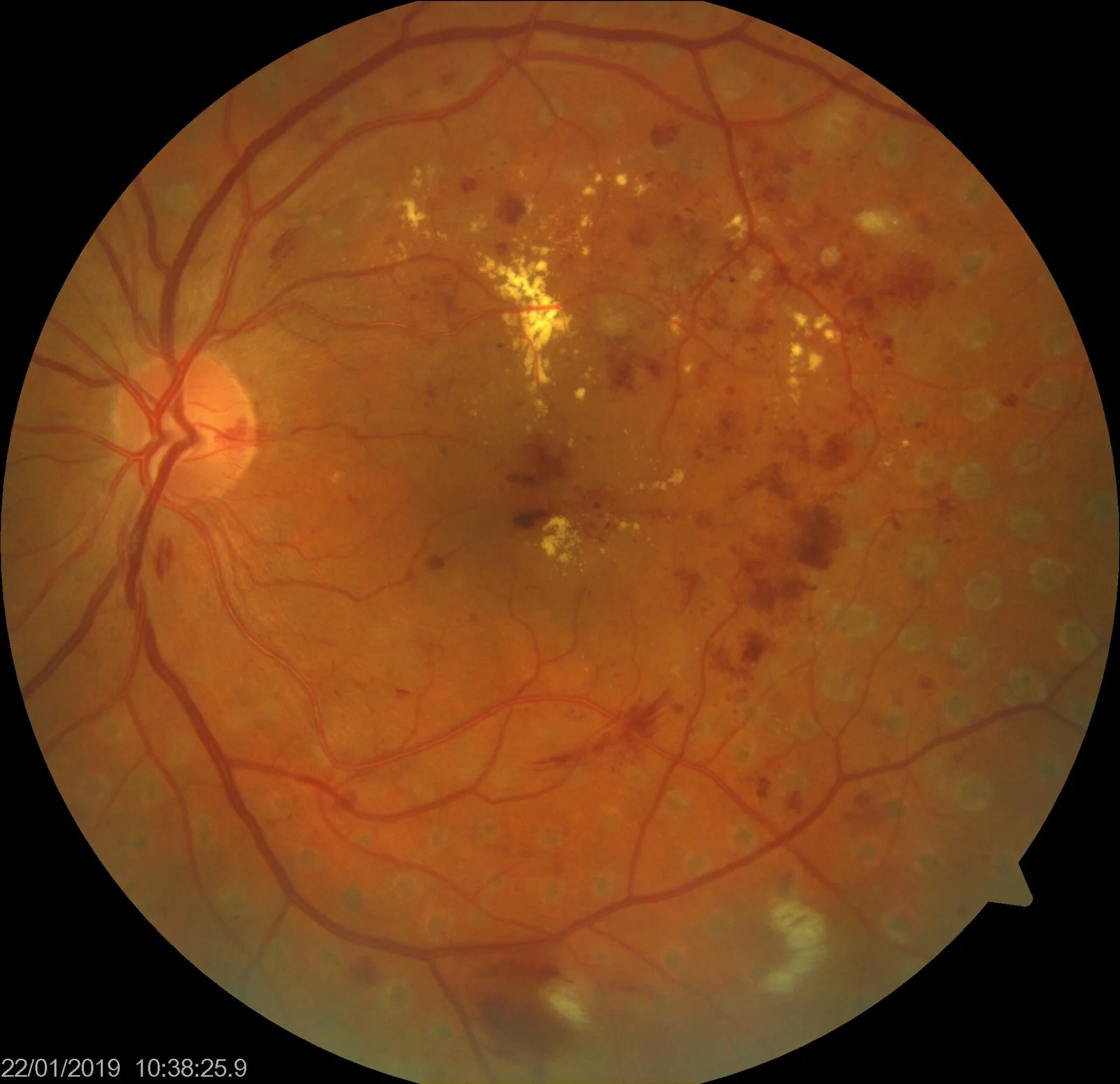
Long description
Fundus photograph in advanced diabetic retinopathy showing hard exudates, retinal haemorrhages and peripheral laser photocoagulation scars.

Figure 2Fundus - diabetic retinopathy
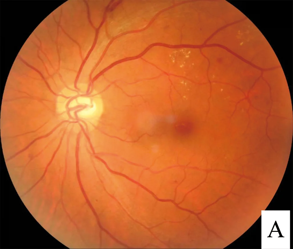
Long description
Fundus photograph in diabetic retinopathy showing scattered hard exudates near the macula.

Diabetic retinopathy is damage to the retinal microcirculation. It is staged so that screening tells you both how far it has gone and how soon it needs treating. The stages, in order of severity, are:
- No retinopathy. Normal retina.
- Mild non-proliferative diabetic retinopathy. Microaneurysms only.
- Moderate non-proliferative diabetic retinopathy. More widespread changes (haemorrhages, exudates, cotton-wool spots) but without the severe changes below.
- Severe and very severe non-proliferative diabetic retinopathy. Extensive haemorrhage, venous beading, intraretinal microvascular abnormalities, approaching the proliferative stage.
- Proliferative diabetic retinopathy. New vessels growing on the disc or retina, with a high risk of vitreous haemorrhage and tractional detachment.
- Diabetic macular oedema. Thickening at the macula, centre-involving or not, which threatens central vision at any stage of retinopathy.
Screening aims to pick up disease before symptoms. In adults with type 2 diabetes, refer at diagnosis to the local diabetic eye screening service. [1] In adults with type 1 diabetes and in children and adolescents, follow the national diabetic eye screening criteria for the start and frequency of screening. Once disease is known, monitoring intervals tighten: moderate non-proliferative disease is reviewed every 6 to 12 months and severe or very severe non-proliferative disease every 3 to 6 months. [4]
Treatment escalates with stage. Background disease is watched and risk factors tightened. Macular oedema that threatens vision is treated with macular laser, intravitreal anti-vascular endothelial growth factor (anti-VEGF) agents or steroids, and proliferative disease with panretinal photocoagulation; anti-VEGF is used where proliferative disease persists after complete photocoagulation, and vitrectomy is used for non-clearing vitreous haemorrhage or retinal detachment. [4]
Red flagRetinal emergencies
Sudden loss of vision, rubeosis iridis, pre-retinal or vitreous haemorrhage, and retinal detachment all need same-day ophthalmology review. Refer anyone with unexplained visual decline promptly, because proliferative disease can be silent until a bleed.
Diabetic kidney disease usually declares itself first as albuminuria in the presence of diabetes, with or without a reduced estimated glomerular filtration rate (eGFR). Patients rarely feel anything until kidney function is badly impaired, so screening is the only way to find it.
Measure urine albumin:creatinine ratio (ACR) and eGFR. An ACR of 3 mg/mmol or more is clinically significant proteinuria, and it should be measured in all adults with diabetes. [2] Track kidney function over time using a combination of GFR and ACR categories; risk of adverse outcomes (CKD progression, cardiovascular events, death) rises with both a lower GFR and a higher ACR, and the two multiply each other. [2] In adults with diabetes, testing is repeated at diagnosis and at least annually thereafter, more often if the category is worse or the clinical situation changes. [2][3]
Albuminuria is divided into:
- A1, normal to mildly increased: ACR under 3 mg/mmol.
- A2, moderately increased: 3 to 30 mg/mmol. [2]
- A3, severely increased: over 30 mg/mmol. [2]
The GFR categories run from G1 (90 ml/min/1.73 m2 or over) down to G5 (under 15, kidney failure). The combination of ACR category A3 and a GFR under 45 puts someone in the very-high-risk group and should trigger referral and intensive management. [2]
The kidney-protective package has five parts:
- Blood pressure control. In adults with chronic kidney disease and an ACR under 70 mg/mmol, target a clinic blood pressure under 140/90 mmHg; if the ACR is 70 mg/mmol or more, target under 130/80 mmHg. [2]
- ACE inhibitor or ARB. Offer an angiotensin-converting enzyme inhibitor or an angiotensin receptor blocker, titrated to the highest licensed dose the patient can tolerate, when the ACR is 3 mg/mmol or more. They must not be combined, and serum potassium and creatinine must be checked before starting and again 1 to 2 weeks after each dose change. [2][6]
- SGLT2 inhibitor. Add an SGLT2 inhibitor (in addition to optimised ACE inhibition or ARB) when the eGFR is 20 to under 45 ml/min/1.73 m2, or between 45 and 90 when the ACR is at least 22.6 mg/mmol, or when the person has type 2 diabetes. [2] [6]
- Finerenone. A non-steroidal mineralocorticoid receptor antagonist, added when there is stage 3 or 4 chronic kidney disease with an ACR of 3 mg/mmol or more associated with type 2 diabetes, on top of optimised ACE inhibition or ARB (and an SGLT2 inhibitor if suitable), provided the eGFR is 25 ml/min/1.73 m2 or more. [2]
- GLP-1 receptor agonist. GLP-1 receptor agonists with demonstrated cardiovascular benefit are recommended in chronic kidney disease and can be considered independent of the current HbA1c or metformin use, because their kidney and cardiovascular effects are not dependent on glucose lowering. [8]
Exam trapTwo common kidney-prescribing traps
Diabetic neuropathy is not one disease but several, and recognising which one you are dealing with determines the investigation and the treatment.
- Chronic distal symmetric polyneuropathy (sensory-motor). The commonest form. Starts in the toes, ascends, and produces "glove and stocking" sensory loss with loss of ankle reflexes. It is usually painless but is the reason people do not feel foot injuries.
- Painful small-fibre neuropathy. Burning, shooting, allodynic pain, often worse at night. Can occur with little objective sensory loss.
- Focal and multifocal neuropathies. Monoeneuropathies (third nerve palsy, carpal tunnel syndrome), radiculoplexopathies ("diabetic amyotrophy"), which can be painful and self-limiting.
- Autonomic neuropathy. Cardiovascular (postural hypotension, resting tachycardia, loss of hypoglycaemia awareness), gastrointestinal (gastroparesis, nocturnal diarrhoea), genitourinary (bladder atony, erectile dysfunction), sudomotor (anhidrosis, gustatory sweating) and pupillary abnormalities. [1]
- Charcot neuroarthropathy. Painless destruction of foot and ankle joints on the basis of severe neuropathy, often triggered by trivial trauma. Treated as a fracture that the patient cannot feel.
Screening is easy to forget but simple: ask annually about pain, numbness or tingling, test light touch and vibration sense and ankle reflexes, and use a 10 g monofilament for foot sensation. [3] Red flags for another cause (B12 deficiency, alcohol, hypothyroidism, drugs, vasculitis) should be worked up rather than assumed diabetic.
Pharmacological treatment follows the standard neuropathic pain ladder: offer a choice of amitriptyline, duloxetine, gabapentin or pregabalin as first-line, titrated to the minimum effective dose, and try a second and third drug if the first is not tolerated or not effective. Tramadol is reserved for acute rescue, and capsaicin cream suits people with localised pain who cannot take oral drugs. [7]
Exam trapNeuropathy traps
- Loss of hypoglycaemia awareness in a person on insulin is itself a clue to autonomic neuropathy; tighten glucose targets, adjust insulin and consider a continuous glucose monitor.
- A warm, swollen, red foot with intact skin in a patient with long-standing diabetes is Charcot until proven otherwise, not cellulitis or gout. Keep it non-weight bearing immediately. [3]
Diabetes multiplies the risk of atherosclerosis in all three main beds. The emphasis is early, aggressive multifactorial risk factor control rather than waiting for symptoms, because diabetic vascular disease presents later and more catastrophically than in people without diabetes.
Silent ischaemia is more common in diabetes, so a strong index of suspicion is needed for atypical presentations: breathlessness, epigastric discomfort, or "indigestion" in an older person. Risk stratify all adults without established cardiovascular disease with QRISK3, which incorporates type 2 diabetes; a score of 10% or more triggers primary prevention. [5] Established coronary disease calls for secondary prevention with high-intensity statin, blood pressure targets and antiplatelet therapy as per the relevant disease guideline. [5] [6]
Diabetes raises the risk of both ischaemic and haemorrhagic stroke, and of silent cerebral small-vessel disease that presents as cognitive decline. Management follows the same multifactorial prevention package: glycaemic, blood pressure, lipid and smoking control, plus antihypertensives and statins as indicated. [5][6]
Peripheral arterial disease is the bridge between the macrovascular and the diabetic foot. Suspect it when pulses are absent, ankle brachial pressure index is abnormal (bearing in mind that calcified tibial vessels in diabetes can falsely elevate it), or there are rest pain, non-healing ulcers or tissue loss. Abnormal results or a strong clinical suspicion should prompt arterial imaging and vascular referral. Manage with the same risk-factor package plus antiplatelet therapy and vascular surgery when indicated. [3]
The diabetic foot is where microvascular neuropathy, macrovascular ischaemia and infection meet, and it is the complication that behaves worst when missed. The clinical task is to find it early (before it becomes a wound) and to act fast once a wound appears.
Assess risk at diagnosis and at least annually: look at pulses, deformity, callus, skin changes and prior ulceration, and test sensation with a 10 g monofilament as part of the foot sensory examination. [3] Risk is then stratified:
- Low risk: no risk factors except callus alone. Assess annually and give foot-care advice.
- Moderate risk: deformity, neuropathy or peripheral arterial disease. Refer to the foot protection service.
- High risk: previous ulcer or amputation, renal replacement therapy, neuropathy plus arterial disease, or neuropathy/arterial disease with callus or deformity. Refer, and reassess from every 1 to 2 months up to every 1 to 2 weeks if there is immediate concern.
- Active diabetic foot problem: any ulcer, infection, chronic limb-threatening ischaemia, gangrene or suspected acute Charcot.
Reassessment intervals as the risk rises: annually for low risk, every 3 to 6 months for moderate risk, every 1 to 2 months (or every 1 to 2 weeks if there is immediate concern) for high risk. [3]
- Neuropathic ulcers occur at pressure points (metatarsal heads, heel) on a warm, well-perfused foot with intact pulses and diminished sensation. They are often painless and surrounded by callus. Offloading is the treatment, plus wound care and infection control.
- Ischaemic ulcers occur on the toes, margins of the foot or pressure areas, on a cool foot with absent pulses, hairless skin and delayed capillary return. They are painful and have a punched-out appearance. They need vascular assessment and revascularisation alongside wound care.
- Neuro-ischaemic ulcers have features of both and are the most common in practice; always look for both components.
Think of osteomyelitis in any person with diabetes who has a local infection, a deep or chronic foot wound, or a swollen, red, non-healing toe. [3] Osteomyelitis may be present despite normal inflammatory markers, X-rays or a negative probe-to-bone test. [3] Send a deep tissue or bone sample for culture after debridement where possible; MRI helps when X-ray is normal. Antibiotic treatment follows severity (mild versus moderate or severe), is started promptly, and is reviewed at 48 hours with a switch to oral agents when safe; courses run from 7 days for soft-tissue infection to up to 6 weeks for osteomyelitis. [3]
Acute Charcot presents as a hot, swollen, red foot with intact skin in someone with severe neuropathy, often after trivial injury. Treat it as an emergency fracture: immobilise in an offloading device, keep the patient non-weight bearing, and refer within one working day to the multidisciplinary foot care service. [3] It is deemed to be resolving when the skin temperature difference between the two feet is sustained at less than 2 degrees and X-rays show no further progression. [3] Bisphosphonates are not used, and radiographs may be normal early on, so MRI is useful when suspicion is high. [3]
Diabetes raises the risk and severity of many infections, partly through hyperglycaemia, partly through vascular insufficiency and partly through immune dysfunction.
- Skin and soft-tissue infection of the feet has been covered above; cellulitis and abscesses recur more readily in diabetes and should trigger a search for an entry portal (tinea, ulcer, cracked heel).
- Urinary tract infection is more common, more often complicated, and more likely to recur, particularly in women; autonomic bladder dysfunction worsens it.
- Mucormycosis and malignant (necrotising) otitis externa are uncommon but classically diabetic and life-threatening; necrotising otitis externa should be suspected in an older diabetic patient with ear pain and discharge that does not respond to drops, and requires imaging and urgent specialist care.
- Infections generally raise blood glucose and can precipitate ketoacidosis or hyperosmolar crisis, so they must be treated promptly and glucose monitored.
- Skin disease. Necrobiosis lipoidica, diabetic dermopathy, granuloma annulare and acanthosis nigricans are more common in diabetes and may be the first clue. Candidal intertrigo and tinea are frequent.
- Musculoskeletal. Limited joint mobility, carpal tunnel syndrome, frozen shoulder, Dupuytren contracture, trigger finger and Cheiroarthropathy occur more often. They are manageable but are easily missed because the patient attributes stiffness to ageing.
- Periodontal disease is bidirectional with glycaemic control, and gum health should be part of the annual review. [1]
Gastroparesis should be considered in adults with type 2 diabetes who have erratic blood glucose control or unexplained gastric bloating or vomiting, after excluding other causes. [1] It worsens glycaemic control (unpredictable absorption), and treatment is dietary (small frequent low-fat meals), glucose control and antiemetics: domperidone, erythromycin or metoclopramide, with domperidone reserved for exceptional circumstances because of its cardiac risk. [1] Autonomic neuropathy should also be thought of when there is nocturnal diarrhoea, unexplained bladder emptying problems, or loss of hypoglycaemia warning symptoms. [1]
Screening for chronic complications is an annual cycle, not a one-off, and its intensity rises with the risk that each system already shows. The table below summarises a practical schedule.
| System | What to do | When |
|---|---|---|
| Eyes | Refer to the local diabetic eye screening service | At diagnosis for type 2 diabetes; for type 1 diabetes follow the national screening criteria |
| Eyes (known disease) | Moderate non-proliferative disease | Every 6 to 12 months |
| Eyes (known disease) | Severe or very severe non-proliferative disease | Every 3 to 6 months |
| Kidneys | ACR and eGFR | At diagnosis and at least annually; more often if ACR or eGFR category is worse |
| Feet | Foot risk assessment and monofilament sensation | At diagnosis and annually if low risk |
| Feet | Reassess according to risk level | Every 3 to 6 months if moderate risk; every 1 to 2 months (or 1 to 2 weeks if immediate concern) if high risk |
| Nerves | Symptom enquiry, reflexes, vibration and light touch | Annually |
| Blood pressure | Measure | Annually if no hypertension; with each visit if treated or raised |
| Lipids | Full lipid profile | With the annual review; after initiating or changing treatment |
| Cardiovascular risk | QRISK3 (for adults 25 to 84 without established disease) | As part of the annual review [5] |
| Other | Skin, oral health, sexual function, mood, cognition | At the annual review |
This is the work that matters most, because it slows or prevents every complication at once.
Lower HbA1c from very high levels gives the largest benefit; long-term tight control in early disease reduces microvascular complications substantially, and the benefit persists for decades. [1] A sensible goal for many adults is an HbA1c under 7.0% (53 mmol/mol) without hypoglycaemia; individualise, and be more lenient where life expectancy is short or treatment harms outweigh benefits. [8] Of note, the newer glucose-lowering agents that carry specific complication benefits should be considered independent of glycaemic goals, so a patient at glucose target may still need one. [8]
In diabetes without chronic kidney disease or established nephropathy, target a clinic blood pressure below 140/90 mmHg (under 150/90 in those 80 or older); with ACR 70 mg/mmol or more, target below 130/80. [6] An ACE inhibitor or an ARB is first-line for adults with type 2 diabetes, with a calcium-channel blocker or diuretic added as needed; never combine two blockers of the renin-angiotensin system. [6] [2]
- Primary prevention: in type 2 diabetes, offer atorvastatin 20 mg when the 10-year QRISK3 score is 10% or more; do not rule it out when the score is lower if risk may be underestimated or the person wishes to take it. In type 1 diabetes, offer statin treatment to adults over 40, or with more than 10 years of diabetes, nephropathy or other cardiovascular risk factors. [5]
- Secondary prevention: offer atorvastatin 80 mg to anyone with established cardiovascular disease, aiming for LDL cholesterol 2.0 mmol/L or less (or non-HDL 2.6 mmol/L or less). [5]
- Chronic kidney disease: offer atorvastatin 20 mg for primary or secondary prevention. [5]
- Do not advise plant stanols or sterols, fibrates, nicotinic acid, bile acid sequestrants or omega-3 compounds for routine cardiovascular prevention in diabetes. [5]
Do not offer aspirin or clopidogrel to adults with type 2 diabetes who do not have cardiovascular disease. [1] In secondary prevention, antiplatelet therapy follows the atherosclerotic disease guideline for the affected bed. [1][5]
The strongest single lesson of diabetic care is that treating all risk factors together is far more effective than treating any one. Combined blood pressure, lipid and glucose control plus stopping smoking and exercise can reduce microvascular and macrovascular events by half or more over time. [1]
Screening for chronic complications of diabetes
- Annual diabetes reviewGlucose control, blood pressure, lipids, weight, smoking, foot and eye screening, symptom enquiry
- Screen each systemRetina (screening service), kidneys (ACR and eGFR), feet (sensation and pulses), nerves, cardiovascular risk, mouth and skin
- Question: Any abnormal finding?
Retinopathy found
- Stage the diseaseNon-proliferative mild, moderate, severe; proliferative; macular oedema
- Tighten risk factors and set the follow-up intervalModerate 6 to 12 months; severe or very severe 3 to 6 months
- Treat proliferative disease or macular oedemaPhotocoagulation, anti-VEGF, macular laser; ophthalmology
Nephropathy found (ACR 3 mg/mmol or more)
- Check eGFR, potassium, blood pressureStage with GFR plus ACR categories
- Start ACE inhibitor or ARB, titrateACR 3 mg/mmol or more; check potassium and creatinine at 1 to 2 weeks
- Add SGLT2 inhibitor, then finerenone if eGFR allowsFinerenone if eGFR 25 or more with ACR 3 or more in type 2 diabetes; add GLP-1 receptor agonist for cardiovascular or kidney benefit
Foot problem
- Classify the footNeuropathic, ischaemic, neuro-ischaemic, infected, Charcot
- Active problem: refer within one working dayUlcer, infection, ischaemia, gangrene, Charcot
- Prevent and protectOffloading, wound care, footwear, revascularisation; risk stratification determines review frequency
No abnormalities
- Reinforce risk factorsGlucose, blood pressure, lipids, smoking, weight, exercise
- Repeat screen next yearAnnually, or more often for higher-risk categories
- Pregnancy. Good pre-conception control matters because diabetic retinopathy can worsen in early pregnancy when glucose is lowered rapidly, so notify ophthalmology before intensive glucose-lowering therapy. [1] [4] Eye screening continues through pregnancy; new gestational diabetes has its own follow-up. [4]
- Children and adolescents. Foot care advice from diagnosis; annual foot assessment after age 12; kidney and eye surveillance from puberty for type 1 diabetes following national guidance; blood pressure and lipids monitored. [1] [3]
- Older adults. Targets should be relaxed where life expectancy is limited or treatment burden high, and hypoglycaemia must be avoided. [8]
- People with chronic kidney disease. Statin and blood pressure targets change; ACE inhibition safety rules apply and need monitoring of potassium and creatinine. [2] [5]
- An annual foot check with a 10 g monofilament, a torch and a look at the pulses costs almost nothing and prevents most amputations. [3]
- Urine dipstick ACR and a serum creatinine with calculated eGFR are far cheaper than in-house nephrology; start ACE inhibition or ARB at the first sign of proteinuria and do not wait for confirmatory tests if supply is reliable.
- If eye screening, laser therapy or dialysis are unavailable, concentrate on what can be done: glucose, blood pressure and smoking; trauma prevention and offloading for feet; and early referral pathways for vitreous haemorrhage or Charcot.
- Amputations can be prevented by separating high-risk feet early and by simple offloading, but once ulceration and infection are present, hospital multidisciplinary care is needed. [3]
- Polypharmacy and monitoring (potassium, renal function) may not be feasible. When they are not, use lower starting doses, longer titration and clinical judgement, and avoid combining nephrotoxic drugs; document what was not done and why.
- Education of patients and family members about daily foot inspection, footwear and when to seek help is the highest-yield intervention in any setting. [3]
- Chronic complications of diabetes divide into microvascular (retinopathy, nephropathy, neuropathy), macrovascular (coronary, cerebral, peripheral arterial disease) and other (infection, skin, musculoskeletal, gastroparesis).
- Retinopathy is staged; refer at diagnosis and tighten monitoring as stage worsens. Treat proliferative disease and macular oedema promptly. [1] [4]
- Diabetic kidney disease is screened with ACR and eGFR; treat with blood pressure control, ACE inhibitor or ARB (ACR 3 mg/mmol or more), then SGLT2 inhibitor, finerenone and GLP-1 receptor agonist as criteria are met. [2] [8]
- Neuropathy is sensory, painful and autonomic; first-line pain drugs are amitriptyline, duloxetine, gabapentin or pregabalin. [7]
- The diabetic foot is preventable: risk-stratify annually and act within one working day on any active problem. [3]
- Macrovascular prevention is package care: QRISK3-led statins, blood pressure targets, smoking advice and specific antiplatelet rules (no routine aspirin in primary prevention). [1] [5]
- Screening is annual but intensifies with severity; do not let a normal screen be the only screen.
- In low-resource settings concentrate on foot checks, urine ACR, blood pressure control and glucose, because these prevent the most disability per unit cost. [3]
Answer each one in your head first, then open the answer.
A person with type 2 diabetes has an eye-screening report of 'moderate non-proliferative diabetic retinopathy' in both eyes, with no macular oedema. He feels well. What is the next step in monitoring, and what general treatment applies?
Show answerHide answer
[4]Monitor under hospital eye services every 6 to 12 months, tightening to every 3 to 6 months if the disease becomes severe or very severe. Tighten risk factors: glucose, blood pressure, lipids and smoking. There is no proliferative or macular disease to treat yet. Arrange screening for nephropathy and neuropathy at the same visit, because one microvascular complication predicts the others.
A patient with type 2 diabetes has moderately increased albuminuria and a reduced eGFR but feels well. Outline the kidney-protective plan.
Show answerHide answer
Check potassium and creatinine, then start an ACE inhibitor or ARB and titrate to the highest tolerated dose, because the ACR is 3 mg/mmol or more. Aim for a clinic blood pressure below 140/90 mmHg with an ACR under 70 mg/mmol, or below 130/80 if the ACR is 70 mg/mmol or more. Add an SGLT2 inhibitor according to the eGFR criteria (20 to 45, or 45 to 90 when the ACR is at least 22.6 mg/mmol, or type 2 diabetes). Add finerenone if stage 3 or 4 CKD with an ACR of 3 mg/mmol or more and type 2 diabetes with an eGFR of 25 or more. Consider a GLP-1 receptor agonist for cardiovascular or kidney benefit independent of HbA1c. Repeat eGFR and potassium 1 to 2 weeks after each change. [2]
Why should domperidone be reserved for exceptional circumstances in diabetic gastroparesis?
Show answerHide answer
Because the evidence for effectiveness is strongest for domperidone, but it carries cardiac risk (QT prolongation and arrhythmia risk) and potential interactions with other medicines. Metoclopramide and erythromycin are considered first, often alternated; domperidone only if it is the only effective treatment and in line with drug safety guidance.
An adult with type 2 diabetes has a QRISK3 score of 10% or more and no history of cardiovascular disease. What statin and what aspirin advice follow?
Show answerHide answer
Offer atorvastatin 20 mg for primary prevention. Do not offer aspirin or clopidogrel routinely for primary prevention of cardiovascular disease in adults with type 2 diabetes without cardiovascular disease. Reinforce smoking cessation, diet, exercise and weight management, and check blood pressure. [1]
A patient with long-standing diabetes presents with a hot, swollen, erythematous right foot but normal inflammatory markers and a normal X-ray. There is no ulcer. What should you suspect and do?
Show answerHide answer
Suspect acute Charcot neuroarthropathy. Keep the foot non-weight bearing, immobilise it in an offloading device, and refer within one working day to the multidisciplinary foot care service. MRI can confirm the diagnosis. It resolves when the temperature difference between the two feet is sustained at under 2 degrees and X-rays show no further progression. Bisphosphonates are not used.
A person with a documented myocardial infarction and type 2 diabetes has an LDL cholesterol above the secondary-prevention target while on atorvastatin 40 mg. What are the lipid targets and next steps?
Show answerHide answer
The secondary prevention target is an LDL cholesterol of 2.0 mmol/L or less, or a non-HDL cholesterol of 2.6 mmol/L or less. Offer atorvastatin 80 mg unless it is contraindicated or not tolerated, discuss adherence and dosing, and consider ezetimibe or other lipid-lowering treatment if maximally tolerated therapy does not reach target.
- Diabetes (type 1, type 2 and the acute complications of diabetes)
- Diabetic ketoacidosis and hyperosmolar hyperglycaemic state
- Hypertension in adults, chronic kidney disease and lipids in primary prevention
- Neuropathic pain, peripheral arterial disease and diabetic foot problems
- Diabetic retinopathy and chronic kidney disease staging
- Nutrition, physical activity and smoking cessation in diabetes
- National Institute for Health and Care Excellence. Type 2 diabetes in adults: management. NICE guideline NG28, last updated 18 February 2026. Accessed .
- National Institute for Health and Care Excellence. Chronic kidney disease: assessment and management. NICE guideline NG203, last updated 24 November 2021. Accessed .
- National Institute for Health and Care Excellence. Diabetic foot problems: prevention and management. NICE guideline NG19, last updated 11 October 2019. Accessed .
- National Institute for Health and Care Excellence. Diabetic retinopathy: management and monitoring. NICE guideline NG242, published 13 August 2024. Accessed .
- National Institute for Health and Care Excellence. Cardiovascular disease: risk assessment and reduction, including lipid modification. NICE guideline NG238, published 14 December 2023. Accessed .
- National Institute for Health and Care Excellence. Hypertension in adults: diagnosis and management. NICE guideline NG136, last updated 26 February 2026. Accessed .
- National Institute for Health and Care Excellence. Neuropathic pain in adults: pharmacological management in non-specialist settings. NICE clinical guideline CG173, last updated 22 September 2020. Accessed .
- American Diabetes Association Professional Practice Committee. 6. Glycemic Goals and Hypoglycemia: Standards of Care in Diabetes—2025. Diabetes Care 2025;48(Suppl 1):S128–S145 (PMC11635034). Accessed .
Figure credits
Educational content for medical students and doctors. It is not a substitute for clinical judgement or local guidelines.