Primary hyperaldosteronism
How excess aldosterone causes hypertension: causes, who to screen, aldosterone-to-renin ratio, confirmatory tests, subtype imaging and treatment.
- Primary hyperaldosteronism (also called primary aldosteronism, and classically Conn syndrome) is the adrenal gland producing aldosterone when it should not: the production is at least partly outside the control of the renin-angiotensin system, and circulating renin is suppressed.
- It is the commonest curable cause of high blood pressure. More than 10% of people with hypertension, and around 20% of those with resistant hypertension, have it. Most are never identified, because fewer than one in ten hypertensive patients is ever screened.
- Aldosterone drives the kidney to retain sodium and water, loses potassium and acid, and raises blood pressure. The result is hypertension that often needs three or more drugs, with or without low potassium.
- A simple blood test, the aldosterone-to-renin ratio, finds most cases. A high ratio with a suppressed renin and an inappropriately high aldosterone is strongly suggestive of the disease.
- If disease is on one side, surgical removal of that adrenal is preferred; if it is on both sides, a mineralocorticoid receptor antagonist such as spironolactone is the treatment. Either way, a treatment that targets the aldosterone itself lowers cardiovascular risk far more than adding another ordinary blood pressure tablet.
By the end of this page you should be able to:
- Explain how autonomous aldosterone from the adrenal produces hypertension and, often, hypokalaemia and metabolic alkalosis.
- Name the main causes of primary hyperaldosteronism and state which are cured surgically.
- State who should be screened, why restricting screening to people with low potassium misses most cases, and what the screening blood tests are.
- Calculate or interpret an aldosterone-to-renin ratio, know the preparation and drug withdrawal, and manage an interfering drug correctly.
- List the confirmatory tests and know when they can be omitted.
- Differentiate unilateral from bilateral disease using adrenal CT and adrenal vein sampling.
- Choose between unilateral adrenalectomy and mineralocorticoid receptor antagonist therapy, and monitor the patient safely.
- Compare primary hyperaldosteronism with secondary hyperaldosteronism and note the practical points for low-resource settings.
DefinitionPrimary hyperaldosteronism
A disorder in which one or both adrenal glands secrete too much aldosterone, at least partly independently of the renin-angiotensin system. Renin is suppressed, plasma aldosterone is inappropriately high for that suppressed renin, and the patient develops hypertension and, in more severe cases, hypokalaemia. The condition is also called primary aldosteronism; the older term Conn syndrome is now used mainly for an aldosterone-producing adenoma.
Primary hyperaldosteronism (PA) is not the same as secondary hyperaldosteronism. In PA the problem starts in the adrenal gland: aldosterone secretion is autonomous and the kidney responds to it. In secondary hyperaldosteronism, the problem starts outside the adrenal - a low effective circulating volume, renal ischaemia, heart failure, cirrhosis, nephrotic syndrome, diuretic use or renovascular disease - which drives the kidney to release renin, and the adrenal simply makes aldosterone in response. Both can raise blood pressure and lower potassium, but renin is suppressed in the first and raised in the second, and that distinction is the first thing to establish in the laboratory.
Secondary hypertension itself is a large topic in cardiology. This page stays on the adrenal endocrine side; any hypertensive patient needs the secondary causes sought as part of the wider work-up.
Aldosterone excess is not benign "high blood pressure with a funny cause". People with PA have a cardiovascular risk that is three times that of people with essential hypertension of the same blood pressure, because aldosterone acts directly on vessels, myocardium and kidney rather than only through pressure. [1] With an estimated prevalence above 10% among all hypertensive patients, and around 20% among those with resistant hypertension, PA is the commonest identifiable cause of elevated blood pressure. [1] Despite this, screening is reported in only 8 to 9% of hypertensive patients, and PA accounts for more than one-third of patients waiting years for a correct diagnosis. [1]
At a tissue level, inappropriately high aldosterone causes endothelial dysfunction, myocardial fibrosis, albuminuria and a fall in glomerular filtration. Glucose metabolism is also impaired, and diabetes and the metabolic syndrome are more common, adding further cardiovascular risk. [1]
PA has one unifying feature: autonomous secretion by the adrenal zona glomerulosa (or, rarely, an ectopic site). The causes differ in how much of the gland is involved, and therefore in whether the patient can be cured with surgery.
| Cause | Approximate share of cases | Typical clinical clue | Treatment |
|---|---|---|---|
| Bilateral adrenal hyperplasia (BAH) | About 60% (estimates 60 to 66%) | Milder, often normokalaemic; no single mass | Lifelong mineralocorticoid receptor antagonist [1] |
| Aldosterone-producing adenoma (APA, classical Conn syndrome) | About 33 to 35% | Younger, marked hypertension, hypokalaemia more common | Unilateral laparoscopic adrenalectomy if unilateral and lateralised [1] |
| Unilateral adrenal hyperplasia | About 2% | Same biochemical pattern as an APA | Usually surgery if lateralised [1] |
| Adrenocortical carcinoma | Less than 1% | Large irregular mass, possible cortisol or androgen co-secretion | Oncological surgery and oncology [1] |
| Ectopic aldosterone secretion (kidney or ovary) | Less than 1% | PA biochemistry without adrenal lateralisation | Remove the source [1] |
| Familial hyperaldosteronism (types I to IV and PASNA) | Rare | Early onset, often severe, family history, children | Type I: low-dose glucocorticoid; others: mineralocorticoid receptor antagonist, with surgery in selected type III disease [1] |
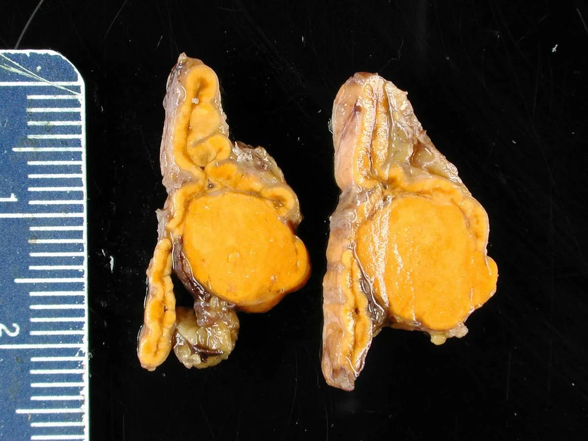
An APA is a benign cortical tumour, usually 1 to 2 cm across, made of cells that release aldosterone without the normal feedback from renin. It produces the most severe biochemical picture, more often with hypokalaemia, and it is the only routine cause removed by surgery. Somatic mutations that keep calcium signalling active are found in the large majority of these tumours; KCNJ5 mutations are the most common and tend to produce larger, clear-cell adenomas, while CACNA1D and ATP1A1 mutations produce smaller, eosinophilic ones. [2]
This is the commonest pattern overall. Aldosterone comes from both glands, usually in the form of multiple small aldosterone-producing nodules or diffuse zona glomerulosa activity rather than one dominant mass. Blood pressure tends to be less severe and potassium is more often normal, and because both adrenals are involved, surgery on one side does not cure the disease. It is treated with a mineralocorticoid receptor antagonist.
These are rare but important, especially in a young hypertensive patient, in a child, or when the family history is positive. The user-facing point is that "essential hypertension in a teenager" should always raise suspicion.
- Type I familial hyperaldosteronism (glucocorticoid-remediable aldosteronism). A chimeric gene places aldosterone synthase under the control of ACTH, so aldosterone is made whenever ACTH rises. Treatment is a low-dose glucocorticoid, ideally dexamethasone 0.125 to 0.25 mg at night, rather than surgery. [1]
- Type II is linked to the CLCN2 gene and presents variably in early life. [1]
- Type III is caused by KCNJ5 mutations and can produce massive bilateral adrenal enlargement with severe early disease. Treatment is usually a mineralocorticoid receptor antagonist; selected patients have surgery on the worse adrenal. [1]
- Type IV (CACNA1H) and PASNA (CACNA1D with seizures and neurological abnormalities) are treated with a mineralocorticoid receptor antagonist, sometimes with a calcium channel blocker. [1]
Germline testing is recommended for all first-degree relatives of a person with familial hyperaldosteronism, and for anyone with PA diagnosed before the age of 20 years. [2]
Conn syndrome originally described a single benign aldosterone-secreting adenoma. Because many APAs are small and non-classical microscopic patterns are common, a CT scan showing a small adrenal nodule does not prove it is the cause. This is exactly why an adenoma seen on CT must be checked for function before it is removed. [1]
PA is common worldwide, and the figures now accepted are higher than older textbooks suggest:
- More than 10% of all people with hypertension, and about 20% of those with resistant hypertension, have PA. [1]
- Using 24-hour urinary aldosterone above 12 microgram/24 h as a measure of overt disease, PA is found in 15.7% of people with stage 1 hypertension, 21.6% with stage 2 hypertension, and 22.0% with resistant hypertension. [1]
- PA is the commonest identifiable cause of curable endocrine hypertension, but it is diagnosed late: it is estimated that only a small minority, roughly 1 to 2% or fewer, of hypertensive patients are ever screened. [1]
There is no known difference in predisposition that would make the disease irrelevant in any country or ethnic group. What does differ is the chance of finding it.
Figure 1Adrenal gland Conn syndrome4
Long description
Gross specimen of a resected aldosterone-producing adrenal adenoma in Conn syndrome

Aldosterone is normally made in the outer layer of the adrenal cortex, the zona glomerulosa, in response to renin, angiotensin II and potassium. In PA that feedback loop is broken: the adrenal cells secrete aldosterone as if the body were always volume-depleted, whatever the salt and volume status actually is.
From there the consequences are straightforward:
- Sodium and water retention. Aldosterone binds the mineralocorticoid receptor in the principal cells of the renal collecting duct and drives ENaC (the epithelial sodium channel) to pull sodium and water back into the blood. Plasma volume expands and blood pressure rises.
- Potassium wasting. Sodium reabsorption is exchanged for potassium secretion, so urinary potassium rises. Only when intake cannot keep up does serum potassium fall, which is why hypokalaemia is a late and inconstant sign, not a rule.
- Hydrogen ion wasting. Acid is lost with potassium, producing a metabolic alkalosis. This shifts potassium into cells and can make hypokalaemia look intermittent.
- Renin suppression. Volume expansion feeds back through the juxtaglomerular apparatus to shut down renin. This suppression is the hallmark of the disease and the reason the aldosterone-to-renin ratio is high.
- Direct organ injury. Apart from the blood pressure, aldosterone itself promotes endothelial dysfunction, myocardial fibrosis, arterial stiffness, microalbuminuria and renal injury. This explains why PA carries higher risk than essential hypertension at the same blood pressure. [1]
The whole mechanism is easier to remember if you think of PA as "the body thinks it is short of salt, so it clings to sodium and throws away potassium, even while the blood pressure is high."
Most people with PA are diagnosed, or not diagnosed, on the basis of resistant hypertension. Many never have a symptom that feels like "an illness". The disease should be suspected particularly in the following patterns:
- Hypertension that needs three or more drugs, or remains above target despite good adherence.
- Hypertension with spontaneous or diuretic-induced hypokalaemia; the presence or absence of hypokalaemia does not rule PA in or out, because only a minority present with low potassium and restricting screening to hypokalaemic patients would miss up to 80% of cases. [1]
- Early onset hypertension, especially before the age of 40, or a stroke before 40.
- An adrenal incidentaloma in a hypertensive person.
- Hypertension with obstructive sleep apnoea.
- Hypertension with unexplained target organ damage out of proportion to the blood pressure: left ventricular hypertrophy, diastolic dysfunction, chronic kidney disease, microalbuminuria. [1]
- Hypertension with unexplained atrial fibrillation.
- A family history of early hypertension or stroke before the age of 40, or a first-degree relative with confirmed PA.
Obstructive sleep apnoea deserves emphasis. OSA is common in PA because of aldosterone-driven fluid retention causing upper airway soft tissue oedema, and PA therapy, whether surgery or a mineralocorticoid receptor antagonist, produces a measurable fall in neck circumference and in the apnoea-hypopnoea index. Conversely, PA is common in OSA through hypoxia-driven increases in endothelin-1 and aldosterone. [1]
- Headache, often from severe blood pressure.
- Muscle weakness, cramps, fatigue and palpitations from hypokalaemia.
- Polyuria and polydipsia in severe hypokalaemia, from nephrogenic diabetes insipidus secondary to the low potassium.
- Worsening renal function.
There is no examination finding specific to PA. Look for:
- Grade of hypertension and retinopathy.
- Evidence of target organ damage: raised jugular venous pressure, basal crackles, displaced apex beat, dyspnoea.
- Volume status: peripheral oedema is uncommon in uncomplicated PA because of the "aldosterone escape" phenomenon, in which the kidney reasserts sodium excretion after a few days even while hypertension persists.
- Secondary causes in general: abdominal bruit for renovascular disease, Cushingoid features, radiofemoral delay, and so on.
The pattern to look for is the combination, not the individual numbers:
- Low or suppressed renin (plasma renin activity under 1 ng/mL/h, or direct renin concentration under 8.2 mU/L). [2]
- Inappropriately high aldosterone for that suppressed renin (10 ng/dL or more, 277 pmol/L, by immunoassay; 7.5 ng/dL, 208 pmol/L, by LC-MS/MS). [2]
- A raised aldosterone-to-renin ratio.
- Hypokalaemia and a metabolic alkalosis in severe cases.
- Normal or borderline aldosterone with suppressed renin still counts as "inappropriately normal", because without PA the aldosterone should be low.
The first branch point is the renin level. A patient with high renin and high aldosterone does not have primary disease; that is secondary hyperaldosteronism.
| Feature | Primary hyperaldosteronism | Secondary hyperaldosteronism |
|---|---|---|
| Problem | Adrenal autonomy | Renin-driven adrenal response |
| Renin | Low or suppressed | Raised |
| Aldosterone | Inappropriately high for renin | High, appropriate to renin |
| Typical causes | Adenoma, bilateral hyperplasia, familial | Renal artery stenosis, diuretics, volume depletion, heart failure, cirrhosis, nephrotic syndrome, oestrogen use |
| Potassium | Often low in severe disease | Often low if hyperaldosteronism is severe |
| Treat the | Adrenal or the aldosterone | Underlying cause |
Within primary disease, the differential is mainly between adenoma and bilateral hyperplasia, because that decides management. The other low-renin mineralocorticoid states, such as Liddle syndrome, apparent mineralocorticoid excess, congenital adrenal hyperplasia due to 11-beta-hydroxylase or 17-alpha-hydroxylase deficiency and deoxycorticosterone-secreting tumours, are rare and usually present in younger patients or with additional features. In older hypertensives, an adrenal nodule seen on CT often turns out to be a non-functional adenoma that is not responsible for the blood pressure, which is the main argument for adrenal vein sampling before surgery.
- Morning aldosterone and renin, with the aldosterone-to-renin ratio, measured in the correct way (see below).
- Serum potassium, aiming to interpret it when corrected to at least above 4 mmol/L if hypokalaemic. [1]
- Basic renal function and bicarbonate (metabolic alkalosis is a clue).
- Optional: urine potassium and sodium when hypokalaemia is present, to confirm renal potassium wasting.
The ARR is simple and reproducible, but it is very dependent on preparation, which is where most errors occur. The Endocrine Society 2025 guideline and current reviews suggest the following. [1][2]
- Timing and posture. Morning sample, taken when the patient has been upright (sitting, standing or walking) for at least 2 hours and then seated for 5 to 15 minutes. [1]
- Potassium. Correct hypokalaemia first and aim for serum potassium above 4 mmol/L, because low potassium suppresses aldosterone and can falsely normalise the test.
- Sodium. Do not restrict sodium in the days before the test; sodium loading above 300 mEq/day should also be avoided. [1]
- Drug withdrawal. Withdraw drugs with a marked effect on the ratio for at least 4 weeks: potassium-sparing diuretics (spironolactone, eplerenone, amiloride, triamterene), potassium-wasting diuretics, and liquorice products. If the result is inconclusive, withdraw drugs with a lesser effect for at least 2 weeks: beta-blockers, clonidine, alpha-methyldopa and NSAIDs. Antihypertensives with the least effect (hydralazine, moxonidine, alpha-1 selective blockers, slow-release verapamil) can be used to control blood pressure meanwhile. [1]
- Interpretation. Beta-blockers, clonidine, alpha-methyldopa and NSAIDs tend to cause false positive ratios; ACE inhibitors, ARBs, diuretics of all kinds and dihydropyridine calcium channel blockers tend to cause false negatives. A normal ratio while on a beta-blocker makes PA unlikely; a raised ratio with suppressed renin while on an ACE inhibitor, ARB or diuretic makes PA very likely.
- Repeat. Between 22 and 25% of the aldosterone concentration and 41 to 42% of the renin concentration vary from one sample to the next, and about 38% of patients have at least one ARR below the diagnostic threshold despite having the disease. Measure it at least twice. [1]
For premenopausal women, sample in the follicular phase, because oral contraceptives and hormone replacement can falsely raise a direct-renin-based ratio. [1]
The Endocrine Society 2025 guideline gives assay-specific guidance, and local laboratory cut-offs should take precedence where they exist. [2]
- Renin is low or suppressed as the hallmark: plasma renin activity 1 ng/mL/h or lower, or direct renin concentration 8.2 mU/L or lower.
- Aldosterone is inappropriately high for that renin: 10 ng/dL (277 pmol/L) or higher by immunoassay, or 7.5 ng/dL (208 pmol/L) or higher by LC-MS/MS.
- The ratio is elevated: aldosterone in ng/dL divided by plasma renin activity in ng/mL/h above 20, or aldosterone in pmol/L divided by direct renin in mU/L above 70 by immunoassay; the corresponding cut-off is about 25% lower when aldosterone is measured by LC-MS/MS. Literature optimal values cited by current reviews are somewhat higher (around 30 with PAC above 10 ng/dL for plasma renin activity, and 2.4 with PAC above 10 ng/dL for direct renin), which is a reminder that no single cut-off is perfect. [1][2]
- Because a very low renin can make any ratio look high, the lowest reported renin is fixed at 0.2 ng/mL/h for plasma renin activity and 2 mU/L for direct renin, and some laboratories require a minimum aldosterone of about 10 to 15 ng/dL (roughly 277 to 416 pmol/L) before calling the screen positive. [1]
- Special handling for potassium sampling matters: blood should be drawn slowly with a syringe and needle, preferably not with a vacuum tube, with the tourniquet released for at least 5 seconds, avoiding fist clenching, and separated from cells within 30 minutes, so that potassium is not falsely raised. [2]
The Endocrine Society 2025 guideline suggests confirmatory tests when the screening result gives an intermediate probability of lateralised disease and the patient would wish to pursue surgery. They can be omitted in:
- Resistant hypertension or hypertension with hypokalaemia, when overt biochemistry is present: plasma renin activity under 0.2 ng/mL/h (or direct renin under 2 mU/L) and plasma aldosterone above 15 ng/dL (416 pmol/L) by LC-MS/MS, or above 20 ng/dL (554 pmol/L) by immunoassay.
- Patients who will not or cannot undergo adrenal vein sampling and adrenalectomy, who can be treated empirically with a mineralocorticoid receptor antagonist based on the screen.
- Familial hyperaldosteronism, where genetic testing is the diagnostic route.
- A low likelihood of lateralised disease, such as a normokalaemic patient with aldosterone below about 11 ng/dL (305 pmol/L) by immunoassay or 8 ng/dL (222 pmol/L) by LC-MS/MS.
When confirmation is needed, the principle is the same in all of them: show that aldosterone is not suppressed by something that should suppress it.
- Fludrocortisone suppression test. The most reliable, but it requires 5 days in hospital. Rarely performed now. [1]
- Saline infusion test. Two litres of 0.9% saline over 4 hours, with aldosterone measured before and after. Performing it seated improves sensitivity with no loss of specificity.
- Oral sodium loading test. High sodium diet, typically with a 24-hour urine for aldosterone.
- Captopril challenge test. Aldosterone is measured before and after captopril; in PA it is not suppressed.
- Losartan suppression and furosemide upright tests exist but are used less.
Volume-expanding confirmatory tests are contraindicated in severe hypertension, renal impairment and cardiac disease. In these situations, captopril challenge may be used, but its discriminatory power is low, and it is often sensible to proceed directly to imaging and adrenal vein sampling, or to a supervised trial of medical treatment. [1]
There is no universal gold standard confirmatory test, and a recent systematic review concluded that the routine use of these tests needs reappraisal because of wide variation in how they are performed and interpreted. [1]
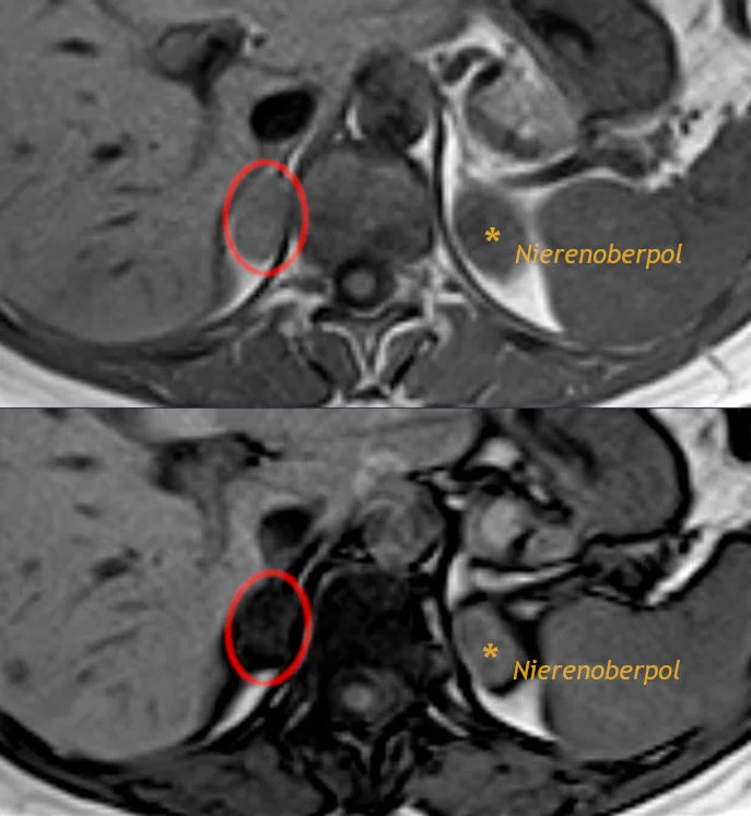
Figure 2Adrenal adenoma on in-phase and out-of-phase T1 MRI
Long description
Axial T1 MRI of an adrenal adenoma (circled): in-phase image above, out-of-phase image below, where the lipid-rich adenoma loses signal. 'Nierenoberpol' on the image means upper pole of the kidney.

All patients with confirmed PA should have adrenal imaging, usually with a thin-slice (3-phase) adrenal CT. CT can show a unilateral or bilateral adenoma, micronodular or macronodular hyperplasia, or, rarely, an adrenocortical carcinoma. CT cannot tell whether a nodule is functional: adrenal adenomas are common in older people and many are incidental. Conversely, in younger patients a unilateral adrenal adenoma above 10 mm is highly likely to be ipsilateral disease. [1]
CT alone is inadequate for deciding on surgery. Concordance between a CT-based and an adrenal-vein-sampling-based strategy is only about 50%: AVS finds unilateral disease in around 22% of CT-negative patients, while CT shows a mass in around 25% of patients whose disease is actually bilateral or contralateral at AVS. Operating on the strength of CT alone would deny surgery to a fifth of eligible patients and expose others to unnecessary adrenalectomy. [1]
AVS is the gold standard for deciding whether aldosterone comes from one adrenal or both, and therefore whether surgery is possible. It is demanding and should be performed by an experienced interventional radiologist; in experienced hands the success rate is above 95%. [1]
- Both adrenal veins are catheterised, usually simultaneously or sequentially, and blood is drawn from both adrenal veins and the inferior vena cava.
- Cortisol correction. The aldosterone from each adrenal is divided by the cortisol from the same side. The adrenal vein-to-inferior-vena-cava cortisol ratio exceeds 3.0 on both sides to confirm that the catheters are in the right place.
- Lateralisation index. The higher cortisol-corrected aldosterone ratio divided by the lower one: a value above 4.0 indicates unilateral aldosterone excess. [1]
- Contralateral suppression index. The lower adrenal cortisol-corrected aldosterone ratio divided by the peripheral ratio: below 0.5 it supports unilateral disease. [1]
- ACTH stimulation. About half of centres use a synthetic ACTH infusion during AVS to reduce sampling variability, deepen the gradients and stimulate aldosterone from aldosterone-producing tumours. [1]
The Endocrine Society 2025 guideline accepts that AVS may be omitted when a unilateral APA is very likely: that is, an individual under 35 years of age with marked PA, hypokalaemia, and a unilateral adrenal adenoma larger than 1 cm on CT. [2] Everyone else who may have surgery should have AVS.
In any patient with PA and an adrenal adenoma, the Endocrine Society recommends a 1-mg overnight dexamethasone suppression test: 1 mg dexamethasone is taken at 23:00 to 24:00 and serum cortisol is measured at 08:00 to 09:00. A cortisol above 1.8 microgram/dL (50 nmol/L) suggests autonomous cortisol secretion, which requires its own management and may affect how the AVS is interpreted. [2]
There is no internationally agreed staging system for PA. Diagnosis is functional and proceeds in stages:
- A compatible clinical picture, plus a positive screen on two occasions (or one in the presence of overt biochemistry).
- Confirmation if the pre-test probability is intermediate.
- Subtyping to unilateral or bilateral disease.
Severity is described by the consequences rather than by a score: resistant or stage 3 hypertension, spontaneous hypokalaemia, and damage such as retinopathy, left ventricular hypertrophy, chronic kidney disease, microalbuminuria and atrial fibrillation. In practice these are the factors that push clinicians toward earlier definitive therapy.
The management of PA hinges on one question: is the excess aldosterone coming from one adrenal or from both? If it is coming from one, unilateral laparoscopic adrenalectomy can cure the hormonal abnormality; if from both, lifelong medical therapy is required. [1][2]
Medical therapy is the treatment for bilateral disease, for patients who are not surgical candidates or decline surgery, and for those with mild disease in whom the probability of unilateral disease is low.
- Mineralocorticoid receptor antagonists (MRAs) are the drugs of choice, and they should be titrated to biochemical effect, not to blood pressure alone. The Endocrine Society 2025 guideline prefers spironolactone because of its low cost and widespread availability, while acknowledging that all MRAs are expected to have similar efficacy when titrated to equivalent potency. MRAs with greater mineralocorticoid receptor specificity, such as eplerenone, produce fewer androgen and progesterone receptor side effects and may be preferred in some patients. [2]
- Start low and titrate. Initial dosing is chosen with the severity of hypertension in mind, and some other antihypertensives can be withdrawn once blood pressure control improves. The dose is then adjusted to blood pressure, potassium, renal function and renin. [2]
- Renin as a target. In patients whose blood pressure stays high and whose renin remains suppressed, therapy should be increased to raise renin from its pre-treatment baseline. Observational data link suppressed renin on treatment to ongoing cardiovascular and renal risk, even when blood pressure looks acceptable. The guideline does not set a specific renin number to aim for. [2]
- Monitor. Potassium, renal function, renin and the blood pressure response should be followed during titration. [2]
- ENaC blockers (amiloride, triamterene) are alternatives when an MRA is contraindicated, such as with hyperkalaemia, advanced renal impairment or pregnancy. The guideline suggests MRAs rather than ENaC inhibitors when both are options. [2]
- Glucocorticoid-remediable disease. Type I familial hyperaldosteronism is treated with low-dose glucocorticoids rather than a mineralocorticoid receptor antagonist. [1]
- Laparoscopic unilateral adrenalectomy is the gold standard for unilateral APA, and it is the only treatment with curative intent. [1][2]
- Total unilateral adrenalectomy is preferred over partial resection, because the disease can be multifocal within the same adrenal. [2]
- Peri-operative care. Blood pressure and potassium must be checked rigorously on the first postoperative day, and a mineralocorticoid receptor antagonist and potassium supplements should be stopped to avoid hypotension and hypokalaemia. [1]
- Cortisol co-secretion. If the dexamethasone test is positive, plan for possible glucocorticoid insufficiency after surgery, including peri-operative steroid cover.
- Results. Biochemical cure, defined by normalisation of aldosterone, renin and potassium after surgery, is achieved in most properly selected patients, because adrenal vein sampling correctly identifies unilateral disease. Blood pressure improvement is common but cure of hypertension is far less frequent: long-standing hypertension, older age, male sex, higher body mass and target organ damage all reduce the chance of stopping all medication. [1]
- Recurrence. Even after a good operation recurrence is possible, because the remaining adrenal still lives in a body with a tendency to aldosterone dysregulation. Non-classical histology (multiple aldosterone-producing micronodules rather than one dominant adenoma) is associated with a much higher rate of biochemical recurrence. Long-term follow-up with clinical assessment and, where useful, aldosterone and potassium is therefore sensible.
A small group of carefully selected patients lateralise on adrenal vein sampling despite evidence of contralateral aldosterone production. Even here, removing the dominant adrenal can help in a minority to a majority of patients, but the result is less reliable and the decision should be made by a multidisciplinary team with the patient fully informed. [1]
A common error is to add antihypertensives indefinitely while never testing for PA. Another is to perform an adrenalectomy on the strength of a CT scan without lateralisation. A third is to stop at "the potassium is normal" and conclude there is no disease.
The whole pathway is summarised in the flowchart. It is a teaching outline, not a replacement for local guidance; follow the Endocrine Society 2025 algorithm where that differs. [2]
Primary hyperaldosteronism: from screening to treatment
- Hypertension in an adultScreen all hypertensive patients when resources allow; prioritise resistant hypertension and the high-probability groups
- Measure morning aldosterone, renin and ARR; measure potassiumSeated sampling after at least 2 hours upright; correct hypokalaemia and handle drugs as described
- Question: Result of the screen
Overt biochemical PA
- Confirm the diagnosis is establishedConfirmatory tests are not required
Intermediate probability
- Confirmatory testFludrocortisone suppression, saline infusion, oral sodium loading or captopril challenge; omit if the patient will not pursue surgery
Negative
- Repeat the screenCorrect potassium, withdraw interfering drugs, retest on a different day
Negative, low probability
- Re-screen later ifResistant hypertension develops, new hypokalaemia appears, or atrial fibrillation appears without heart disease or thyroid disease
- Biochemical PA confirmed: treat the potassium and any metabolic alkalosisStart definitive pathway
- Adrenal CT (thin-slice)Assess for adenoma, hyperplasia, or carcinoma; CT alone does not decide surgery
- Question: Considering surgery?
Yes
- AVSAdrenal vein-to-caval cortisol above 3.0 both sides; lateralisation index above 4.0; contralateral suppression index below 0.5; ACTH stimulation in many centres
- Question: Result of AVS
Unilateral
- Laparoscopic adrenalectomyStop MRA and potassium supplements peri-operatively; monitor blood pressure and potassium on day 1
Bilateral or unclear
- Mineralocorticoid receptor antagonistPreferably spironolactone; titrate to blood pressure and renin
No, or lateralisation unlikely
- Mineralocorticoid receptor antagonistStart low, titrate to blood pressure, potassium, renal function and rising renin; monitor regularly
- In all patients with an adrenal adenoma: 1 mg dexamethasone suppression testCortisol above 1.8 microgram/dL (50 nmol/L) suggests autonomous cortisol secretion
- Follow-upBiochemistry, blood pressure, potassium, renal function and renin; long-term surveillance for recurrence
Untreated PA is a systemic disease, not merely raised pressure. Compared with essential hypertension of the same severity, PA is associated with:
- Higher rates of atrial fibrillation, left ventricular hypertrophy, stroke, coronary disease, heart failure and renal injury. [1]
- Microalbuminuria and chronic kidney disease, from direct glomerular and tubular effects of aldosterone.
- Impaired glucose metabolism, diabetes and the metabolic syndrome. [1]
- Hypokalaemia, sometimes severe enough to cause neuromuscular weakness, metabolic alkalosis and cardiac rhythm disturbances.
Treatment-specific risk matters too. Patients treated medically with a mineralocorticoid receptor antagonist who have persistently suppressed renin carry excess cardiovascular, renal and atrial fibrillation risk compared with those whose renin is unsuppressed, which is the physiological argument for titrating to renin rather than to blood pressure alone. [1]
The long-term outlook is good when the disease is found early and treated specifically, but it worsens with delay: the longer the exposure to aldosterone, the more fixed the hypertension and the vascular damage become.
- Children and young adults. Hypertension under the age of 40 should prompt a secondary-cause work-up, and confirmed PA before age 20 calls for genetic testing and family screening. [2]
- Pregnancy. Spironolactone is generally avoided in pregnancy; mineralocorticoid receptor antagonists are among the contraindications. Medical and obstetric management should be led by a specialist team, and surgery, if considered, is usually deferred.
- Chronic kidney disease. Renin falls as nephrons are lost, while aldosterone may be high, so screening can produce false positives. Care is needed in interpretation and in the use of MRAs, which raise potassium.
- People of African origin. Aldosterone sensitivity may be higher, and PA can present with only modest aldosterone elevations, so different cut-offs may be needed. [1]
- Premenopausal women. Sample aldosterone and renin in the follicular phase to avoid oral contraceptive and luteal-phase effects on direct renin.
- Elderly patients. Non-functional adrenal adenomas are common; do not attribute an incidental adrenal mass to PA without lateralisation or functional proof.
PA can be suspected, screened for and treated cheaply even where advanced testing is unavailable, but the diagnosis is often delayed everywhere, and more so where resources are limited.
- The Endocrine Society recommendation to screen all hypertensives is explicitly conditional on available resources, local expertise and healthcare capacity. [2]
- Resistant hypertension (blood pressure at least 140/90 mmHg despite three drugs, one of them a diuretic) is the pragmatic trigger for investigation; full population screening is rarely feasible in low-resource settings. [1]
- Clinical skills still matter: a resistant hypertensive patient with spontaneous or diuretic-induced hypokalaemia, an adrenal incidentaloma, an early stroke, or a strong family history has a high pre-test probability.
- Adrenal vein sampling is frequently unavailable. Without it, a CT scan alone should not be used to send a patient to surgery, because of the roughly 50% concordance between CT-based and AVS-based decisions. Where lateralisation cannot be done, a supervised trial of a low-cost mineralocorticoid receptor antagonist (spironolactone) is often the safest effective strategy. [1]
- Spironolactone is inexpensive and widely available, which is the main reason guidelines favour it. Monitoring is nonetheless essential: hypokalaemia has to be corrected at initiation, and potassium and renal function must be rechecked after starting or increasing the dose.
- A supervised mineralocorticoid receptor antagonist trial can itself be informative: a clear biochemical and clinical response supports the diagnosis even when confirmatory and localisation tests are impossible.
- Education of primary care clinicians is high-yield, because screening is reported in only 8 to 9% of hypertensive patients globally and unfamiliarity with the tests is one of the main barriers. [1]
- Primary hyperaldosteronism is autonomous aldosterone secretion with suppressed renin; it is the commonest curable cause of hypertension.
- It affects more than 10% of all hypertensive patients and about 20% of those with resistant hypertension; most cases are never found.
- Only a minority present with hypokalaemia, so restricting screening to low potassium misses up to 80% of cases. [1]
- Clinical triggers include resistant hypertension, spontaneous or diuretic-induced hypokalaemia, an adrenal incidentaloma, early-onset hypertension or stroke, obstructive sleep apnoea, unexplained atrial fibrillation and strong target organ damage or family history.
- The aldosterone-to-renin ratio is the workhorse screening test, but preparation - morning sampling after 2 hours upright, potassium above 4 mmol/L, sensible sodium intake, stopping spironolactone and other interfering drugs for 4 weeks and beta-blockers or ACE inhibitors appropriately - decides whether it is trustworthy.
- A positive screen is suppressed renin plus inappropriately high aldosterone, with an elevated ratio, confirmed on at least two occasions because of biological variability.
- Confirmatory tests (fludrocortisone suppression, saline infusion, oral sodium loading, captopril challenge) are not needed when biochemistry is overt or when the patient will not go to surgery.
- Adrenal CT is the first imaging test but cannot establish function; adrenal vein sampling is the gold standard for lateralisation, with a lateralisation index above 4, a contralateral suppression index below 0.5, and bilateral catheter placement confirmed by adrenal vein to inferior vena cava cortisol ratios above 3.
- CT alone is not enough to operate on, because concordance with adrenal vein sampling is only about 50%.
- If disease is unilateral and the patient is fit, laparoscopic adrenalectomy is the treatment of choice and biochemical cure is achieved in most. Blood pressure often improves; cure of hypertension is less common and depends on duration and burden of disease.
- If disease is bilateral or the patient is not a surgical candidate, use a mineralocorticoid receptor antagonist, preferably spironolactone for cost and availability, titrated to blood pressure, potassium, renal function and a rising renin.
- Check for cortisol co-secretion with a 1-mg dexamethasone test in anyone with an adrenal adenoma, and remember glucocorticoid-remediable disease (dexamethasone 0.125 to 0.25 mg at night) in early-onset familial cases. [1]
- In low-resource settings, a supervised mineralocorticoid receptor antagonist trial is often the safest effective option where lateralisation is impossible, and primary care education is a high-yield intervention.
Answer each one in your head first, then open the answer.
A 46-year-old man has blood pressure 168/104 mmHg on amlodipine, ramipril and bendroflumethiazide. His serum potassium is 3.2 mmol/L. His GP checks renin and aldosterone. What result would make primary hyperaldosteronism likely, and what preparation check would you make first?
Show answerHide answer
Primary hyperaldosteronism would be likely if renin is suppressed and aldosterone is inappropriately high, producing a raised aldosterone-to-renin ratio. First check that he has not taken bendroflumethiazide for about 4 weeks or the ramipril for 2 weeks, and that potassium has been corrected to above 4 mmol/L; sample in the morning after at least 2 hours upright. A normal potassium does not exclude the disease, so the screening should not be abandoned.
Why does a patient with primary hyperaldosteronism have suppressed renin, and why is that clinically useful?
Show answerHide answer
Sodium and water retention expand plasma volume, which feeds back to suppress renin release from the juxtaglomerular apparatus. Suppressed renin against an inappropriately high aldosterone is the biochemical hallmark of the disease, and it is what makes the aldosterone-to-renin ratio high. It also explains why primary and secondary hyperaldosteronism separate by renin level.
In whom can confirmatory aldosterone suppression testing be omitted?
Show answerHide answer
Confirmatory testing can be omitted with overt biochemistry (renin under 0.2 ng/mL/h or direct renin under 2 mU/L with aldosterone above 15 ng/dL by LC-MS/MS or above 20 ng/dL by immunoassay) in a patient with resistant hypertension or hypokalaemia; in patients who will not or cannot have adrenal vein sampling or surgery; in familial hyperaldosteronism diagnosed genetically; and when the probability of lateralised disease is very low, such as in a normokalaemic patient with aldosterone below roughly 11 ng/dL by immunoassay.
Adrenal CT shows a 1.4 cm left adrenal nodule in a 52-year-old with confirmed primary hyperaldosteronism. What must happen before adrenalectomy is offered, and why?
Show answerHide answer
Adrenal vein sampling is needed to prove that the nodule is making the excess aldosterone. In adults, concordance between CT-based and AVS-based strategies is only about 50%; about 25% of patients with a CT mass turn out to have bilateral or contralateral disease at sampling, and operating on CT alone would expose some of them to an unnecessary adrenalectomy. [1]
How does adrenal vein sampling prove unilateral disease?
Show answerHide answer
Both adrenal veins and the inferior vena cava are sampled. Cortisol from each adrenal corrects the aldosterone for dilution; bilateral catheter placement is confirmed when the adrenal vein to inferior vena cava cortisol ratio exceeds 3.0 on both sides. A lateralisation index above 4.0 (higher over lower cortisol-corrected aldosterone ratio) indicates unilateral disease, supported by a contralateral suppression index below 0.5. [1]
A patient with bilateral adrenal hyperplasia has well-controlled blood pressure on a mineralocorticoid receptor antagonist. Renin is still suppressed. Why might the clinician increase the dose?
Show answerHide answer
Suppressed renin on treatment reflects residual mineralocorticoid excess and has been linked with ongoing cardiovascular, renal and atrial fibrillation risk, even when blood pressure looks acceptable. The Endocrine Society suggests titrating aldosterone-directed therapy upward until renin rises from its pre-treatment baseline, provided potassium and renal function allow.
A 30-year-old has PA and imaging suggests a unilateral adrenal adenoma, on a background of strong family history. What additional management step is specific to his situation?
Show answerHide answer
Genetic testing should be offered, both to him and, if positive, to first-degree relatives, with appropriate adrenal screening in carriers. Type I familial disease is treated with low-dose glucocorticoids rather than surgery, and young-onset PA diagnosed before age 20 specifically warrants genetic investigation. [2]
Related MedGrasp topics to read next or around this one (plain text, no links yet): Secondary hypertension (Cardiology); Hypokalaemia; Hyperkalaemia; Metabolic alkalosis; Adrenal incidentaloma; Obesity and metabolic syndrome; Chronic kidney disease; Obstructive sleep apnoea; Atrial fibrillation; Left ventricular hypertrophy.
Aldosterone receptor antagonists and other antihypertensives are covered in the drug topics of the pharmacology section.
This page is educational; it is not a substitute for local guidelines or clinical judgement. Thresholds and assays vary by laboratory, and all doses and cut-offs should be checked against your local formulary.
- Fernandez CJ, Nagendra L, Alkhalifah M, Pappachan JM. Endocrine Hypertension: The Urgent Need for Greater Global Awareness. touchREVIEWS in Endocrinology 2023;19(2):31-41. doi:10.17925/EE.2023.19.2.11. Accessed .
- Adler GK, Stowasser M, Correa RR, et al. Primary Aldosteronism: An Endocrine Society Clinical Practice Guideline. Journal of Clinical Endocrinology and Metabolism 2025;110(9):2453-2495. doi:10.1210/clinem/dgaf284. Accessed .
Figure credits
Educational content for medical students and doctors. It is not a substitute for clinical judgement or local guidelines.