Coeliac disease
Immune-mediated small-bowel disease triggered by gluten in HLA-DQ2 or DQ8 carriers, from definition to diagnosis, diet and complications.
- Coeliac disease is a chronic, systemic, immune-mediated disease of the small bowel that occurs in genetically predisposed people after they eat gluten, the protein in wheat, rye and barley.
- It has a broad spectrum: classic malabsorption with diarrhoea and weight loss, "silent" iron deficiency or osteoporosis, or no symptoms at all picked up through screening.
- The key immune players are tissue transglutaminase (TG2), which deamidates gluten peptides, and HLA-DQ2 or HLA-DQ8 molecules, which present them to T cells and drive inflammation, villous atrophy and crypt hyperplasia.
- Diagnosis in most adults needs coeliac serology taken on a gluten-containing diet plus duodenal biopsies showing villous atrophy; in children with a tissue transglutaminase IgA titre at least 10 times the upper limit of normal and a positive endomysial antibody test on a second sample, biopsy may be omitted.
- The only effective treatment is a strict, lifelong gluten-free diet, supervised by a dietitian, with attention to nutrition, vaccination (including pneumococcal vaccine when hyposplenism or autoimmune disease is present) and bone health.
By the end of this page you should be able to:
- Explain why coeliac disease develops only in people carrying HLA-DQ2 or HLA-DQ8, and what tissue transglutaminase does to gliadin.
- Name the main associations and risk groups, and explain why they share susceptibility.
- Recognise the classic, atypical and silent forms, including in children and in adults with iron deficiency, osteoporosis or raised liver enzymes.
- Describe the biopsy findings (Marsh classification) and the no-biopsy diagnostic pathway in children and in selected adults.
- Plan initial and follow-up management of a newly diagnosed patient, including the role of the dietitian, micronutrient checks and bone density.
- Define non-responsive and refractory coeliac disease, including type I and type II, and their link to enteropathy-associated T-cell lymphoma.
Coeliac disease is a chronic, systemic, immune-mediated enteropathy of the small bowel that occurs in genetically predisposed people and is triggered by ingestion of gluten, the storage protein of wheat, rye and barley. It was formerly called coeliac sprue, gluten-sensitive enteropathy or non-tropical sprue. Three facts are needed for the disease to be possible: the person must carry the HLA-DQ2 or HLA-DQ8 haplotype, gluten must be eaten, and the immune system must lose tolerance to gluten.[1][5] Remove any one of these and the disease does not occur, which is the basis of both diagnosis and treatment. [1][5]
Coeliac disease is one of the most common autoimmune diseases, but a large proportion of cases remain undiagnosed. Histologically confirmed prevalence is around 0.7% in western countries, about 1% by serological screening, with pooled estimates 0.75-1.6%. More than 70% of diagnoses are made in adults, and the median age at diagnosis has risen from about 1.9 years before 1990 to 7.6 years since 2000. The female-to-male ratio ranges from about 2:1 to 1.5:1. Prevalence varies by geography: about 1.5-2.4% in Finland, about 0.7% in Italy, 0.3% in Germany; in the USA about 1.01% in white people, 0.2% in Black people, 0.3% in Latino people. Incidence has risen up to 50 cases per 100,000 person-years in Scandinavia, Finland and Spain. Monozygotic twins have about a 70% cumulative probability of disease within five years versus about 9% for dizygotic twins. [1][5][6]
Susceptibility is genetic and necessary; exposure and loss of immune tolerance are also required.
The HLA region contributes roughly 35-40% of genetic risk. 90-95% of people with coeliac disease carry HLA-DQ2.5; 5-10% carry HLA-DQ8 or HLA-DQ2.2; fewer than 1% carry only DQA1*05. Homozygous HLA-DQ2.5 carries the highest risk, up to 30%, versus about 3% in other genotypes. Negative HLA-DQ2/DQ8 typing makes coeliac disease very unlikely (NPV >99%). [1][3]
Testing indicated in all these: first-degree relatives (5-10%; siblings up to about 20%), second-degree (2.3-2.6%), type 1 diabetes (3-10%; pooled ~5.1-6.0%), autoimmune thyroid disease (biopsy-proven coeliac disease ~6.2% in children and 2.7% in adults with thyroid disease), Down and Turner syndromes, autoimmune hepatitis, Addison and Williams syndromes, selective IgA deficiency (2-3% of coeliac patients), unexplained elevated aminotransferases (>95% normalise on diet), osteoporosis and fragility fractures, unexplained ataxia or peripheral neuropathy, recurrent aphthous ulcers and dental enamel defects, infertility, recurrent miscarriage, late menarche, early menopause, hyposplenism/functional asplenia, dermatitis herpetiformis (skin biopsy-proven), psoriasis and other autoimmune disease. [1][3][5]
The disease is a wheat-reactive autoimmune process. Ingested gluten resists digestion; gliadin fragments increase intestinal permeability. TG2 deamidates glutamine residues in gliadin to glutamic acid, allowing high-affinity binding to HLA-DQ2/DQ8 on antigen-presenting cells and activation of gluten-specific CD4+ T cells. A Th1 response (interferon-gamma, IL-15, IL-21) recruits intraepithelial lymphocytes and damages enterocytes and stroma; B cells produce anti-TG2 and anti-endomysial antibodies. Villous atrophy with crypt hyperplasia and at least 25 intraepithelial lymphocytes per 100 enterocytes develops, villous height to crypt depth falling below the normal 3:1 to 5:1 (about 2:1 in the bulb), causing malabsorption of iron, folate, B12, calcium, vitamin D and fat-soluble vitamins. IL-15-driven inflammation triggers enterocyte apoptosis, underlying atrophy and (rarely) malignant transformation. Macroscopic established disease shows scalloping, nodularity, fissuring, mosaic pattern. Microscopic diagnosis: raised intraepithelial lymphocytes, crypt hyperplasia, villous atrophy, graded by modified Marsh (0-4). Marsh 1 alone is common in about 3-4% without coeliac disease and not specific. Histological mimics include tropical sprue/infections, olmesartan/mycophenolate/colchicine/NSAID enteropathy, autoimmune enteropathy/CVID, Crohn disease, and lymphoma. [3][5]
Children: classic failure-to-thrive with diarrhoea, distension, poor appetite, often neuropsychiatric symptoms and secondary lactose intolerance; other presentations include iron deficiency without GI symptoms, short stature, dental enamel defects, delayed puberty. Adults: classical diarrhoea, steatorrhoea, bloating, pain, weight loss; atypical/silent commoner. Extraintestinal clues include iron deficiency anaemia (often the adult presentation), folate and B12 deficiency, osteoporosis/osteopenia in up to half (osteoporosis 11-38%), unexplained raised aminotransferases (>95% normalise on diet), infertility, recurrent miscarriage, late menarche, early menopause, neurological disease in 10-22% (gluten ataxia, peripheral neuropathy, headache), recurrent aphthous ulcers, dental enamel defects, fatigue and low mood. [1][3][5]
Figure 1Dermatitis herpetiformis
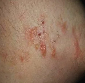
Long description
The Commons description states that this is dermatitis herpetiformis on the elbow of a 37-year-old Caucasian woman.

Dermatitis herpetiformis is the cutaneous manifestation of coeliac disease, intensely pruritic grouped papulovesicles on extensor surfaces, with granular IgA deposits in the dermal papillae (pathognomonic). Mostly adults, slightly more men, mean onset about 50 years. Three-quarters have villous atrophy. Treatment: strict gluten-free diet (skin clears over months to years) plus dapsone symptomatic control, 25-50 mg daily rising to 100-200 mg daily, tapered over about two years; monitoring and G6PD exclusion required because of haemolysis and methaemoglobinaemia. Dapsone has no effect on enteropathy or lymphoma risk. [6]
Villous atrophy and raised lymphocytes are not specific to coeliac disease.
| Condition | Pattern | Feature that separates it from coeliac disease |
|---|---|---|
| Coeliac disease | Villous atrophy, crypt hyperplasia, IEL at least 25 per 100, positive TG2 IgA/EMA | Positive serology on gluten; HLA-DQ2/DQ8 positive |
| Infections | Villous atrophy, malabsorption | Negative coeliac serology; organism on testing |
| Drug-induced enteropathy | Villous atrophy or IEL rise | Drug history; improvement on withdrawal |
| Autoimmune enteropathy/CVID | Villous atrophy; no plasma cells in CVID | Negative TTG; hypogammaglobulinaemia |
| Crohn's disease | Patchy inflammation, granulomas | Skip lesions, strictures or fistulae; negative TTG |
| H. pylori gastritis | Marsh 1 picture | Gastric biopsy positive; IEL count normalises after eradication |
| Functional bowel disease | Normal mucosa | Normal biopsy and serology |
All serology must be done on a gluten-containing diet: IgA anti-TG2 plus total IgA; IgG-based tests if IgA deficient (2-3% of patients); EMA as confirmatory; anti-DGP IgA/IgG as adjunct. Biopsy in adults: at least 4 specimens from the second part of duodenum plus 2 from the bulb. Baseline tests: FBC, ferritin, folate, B12, calcium, vitamin D, fat-soluble vitamins, LFTs, renal and thyroid function. Video capsule for complicated or slow-responding disease. Adult diagnosis standard: positive serology with villous atrophy on a gluten diet; a conditional no-biopsy pathway is allowed in adults under 45 with IgA anti-TG2 at least 10 times ULN, confirmed on a second sample, no alarm features. In children, no biopsy is needed if initial IgA anti-TG2 exceeds 10 times ULN with positive EMA on a second sample, regardless of symptoms, by a paediatric gastroenterologist with consent; lower titres need 4 distal plus 1 bulb biopsy. IgA-deficient serology-positive children should be biopsied. HLA-DQ2/DQ8 typing excludes coeliac disease when negative and helps in uncertainty (equivocal, already gluten-free, potential/seronegative disease). [1][3][4]
Coeliac disease has no other curative therapy. Strict lifelong gluten-free diet (GFD) is the recommendation, with strong, evidence-based support. Symptom improvement is usually expected within 4 weeks to 4-5 months; anti-TG2 titres decline from 2-4 weeks and normalise in most within about 12 months; mucosal healing is generally expected around 1 year, although full histological recovery occurs in only 50-83% of adults after 1-5 years on the diet. Normal levels of IgA anti-TG2 do not guarantee healed mucosa. [2]
Safe gluten intake: generally accepted threshold no more than 10 mg of gluten per day; foods labelled gluten-free contain no more than 20 mg per kg (20 ppm). Certified gluten-free oats can be included from diagnosis; standard oats are often contaminated and unsafe. A well-balanced GFD should emphasise naturally gluten-free foods, with attention to label reading, dairy or fortified alternatives, adequacy of macronutrients and micronutrients. [2]
A dietitian experienced in coeliac disease should provide education on the lifelong GFD at diagnosis, perform a baseline nutritional assessment, address deficiencies and plan follow-up. Dietetic review is central to detecting inadvertent gluten exposure and to evaluating persistent or recurrent symptoms, and detects ongoing villous atrophy more reliably than questionnaires alone. Gluten immunogenic peptides (GIP) in stool or urine can objectively detect recent intake. Adherence varies, and is reduced by younger age, lower socioeconomic status, eating outside the home, absence of symptoms and limited knowledge.
Correct deficiencies identified at diagnosis and during follow-up: iron, folate, vitamin B12, calcium, vitamin D, magnesium, zinc, and fat-soluble vitamins. Bone mineral density assessment as below. Temporary lactose restriction may help until villi heal. In persistent GI symptoms after histological healing, a low-FODMAP diet may be considered after excluding other causes. Psychosocial support and patient support groups improve adherence and quality of life.
Pneumococcal vaccination is recommended for adults with coeliac disease who have evidence of functional asplenia, concurrent autoimmune disorders or refractory coeliac disease type II, as well as patients over 65 years. Because hyposplenism and infection risk (including pneumonia) are increased, influenza and other routine vaccines should also be kept up to date. Screening for associated autoimmune conditions (for example thyroid function at diagnosis) is advised. Vitamin D and calcium repletion are part of prevention.
Adults with coeliac disease are at increased risk of reduced bone mineral density and fractures. A baseline DXA scan is recommended after 1 year on a gluten-free diet for adults with additional risk factors (delayed diagnosis, severe malabsorption or marked weight loss at presentation, prior fragility fracture, other osteoporosis risk factors, Down syndrome). For adults without these, routine DXA is not mandatory but may be considered approaching midlife (over 35-40 years); FRAX can support individualised assessment. [2]
Non-responsive coeliac disease is defined as persistent or recurrent symptoms despite being on a gluten-free diet. It requires a systematic work-up, especially for unintentional gluten contamination.
Non-responsive coeliac disease evaluation
- Persistent or recurrent symptoms on a GFD
- Re-verify the original diagnosisSerology and biopsy review; exclude seronegative villous atrophy
- Assess adherence and cross-contaminationDietitian review; consider gluten immunogenic peptides (GIP)
- Question: Adherence confirmed?
No
- Correct the diet; reassess in 3-6 months
Yes
- Repeat duodenal biopsy
- Question: Villous atrophy persists after at least 12 months strict GFD?
No
- Seek alternative/overlapping conditionsIBS, SIBO, microscopic colitis, pancreatic insufficiency, lactose intolerance, IBD
Yes
- Evaluate for refractory coeliac disease and exclude EATLFlow cytometry for aberrant T cells, TCR clonality, imaging, enteroscopy
Refractory coeliac disease (RCD) is defined as persistence or recurrence of symptoms and villous atrophy after at least 12 months on a strict gluten-free diet, in the absence of other causes. It can be primary or secondary and is subdivided by the proportion of aberrant T cells into RCD-I and RCD-II. RCD accounts for about 0.04-1.5% of all coeliac disease cases, mainly in people diagnosed over 50. There are no evidence-based treatments from controlled trials; management is based on expert consensus. For RCD-I, open-capsule budesonide is considered first-line, with conventional immunosuppressants such as azathioprine in selected cases, re-evaluating azathioprine for discontinuation after 2-3 years of stability. For RCD-II, mild-to-moderate disease may be treated with open-capsule budesonide; in selected patients cladribine (or fludarabine), with or without autologous haematopoietic stem-cell transplantation, or JAK inhibitors may be considered individually. Accurate subtyping requires comprehensive immunophenotyping of small bowel lymphocytes; flow cytometric analysis is superior to immunohistochemistry for detecting aberrant lymphocytes. [2][5]
- Malignancy: in non-refractory coeliac disease the risk of malignant complications is only slightly increased in absolute terms and decreases with long-term strict gluten-free diet; routine screening beyond general population recommendations is not indicated without clinical suspicion. Enteropathy-associated T-cell lymphoma (EATL) and small-bowel adenocarcinoma are the most feared. RCD-II carries a substantial risk of progression to EATL (up to half of affected patients), which dominates its markedly reduced 5-year survival. [2]
- Osteoporosis/osteopenia from calcium and vitamin D malabsorption, with fracture risk, improve with diet and repletion.
- Hyposplenism/functional asplenia predisposing to infections (hence pneumococcal vaccination).
- Nutritional deficiencies (iron, folate, B12, fat-soluble vitamins), anaemia, short stature in children, infertility and adverse pregnancy outcomes, neurological sequelae, dental enamel defects, secondary lactose intolerance.
- Other reported comorbidities: type 1 diabetes, autoimmune thyroid disease, and neuropsychiatric disease.
Long-term follow-up is recommended to monitor adherence, detect complications and comorbidities, and provide nutritional and psychosocial support, in hospital-based specialist clinics where feasible, with primary-care follow-up acceptable for stable patients. Both fixed-interval and individualised strategies are used; follow-up should be patient-centred and based on disease activity, adherence, comorbidities and individual needs. Anti-TG2 is useful for detecting gluten exposure but a negative result neither confirms strict adherence nor excludes villous atrophy. Follow-up biopsy is not routine; it is considered for persistent or worsening symptoms, severe disease at diagnosis or inadequate response. Serologic surveillance commonly checks antibodies every 3-6 months in the first year, then every 1-2 years. Healing timelines: symptoms 4 weeks to 4-5 months, serology in the majority within about 12 months, mucosa around 1 year (50-83% full histological recovery after 1-5 years). First-degree seronegative relatives can be re-tested every 4-5 years. A "coeliac passport" can support transition from paediatric to adult care. [2][5]
- Children: presentation often failure to thrive; growth and development are goals of management; screen children of patients with coeliac disease from infancy/childhood with HLA typing and serology; annual thyroid etc.
- Older adults: non-classical symptoms, delayed diagnosis, slower response to diet but substantial benefit; higher osteoporosis and malignancy risk.
- Pregnancy: preconception and perinatal nutritional optimisation; continue GFD; postpartum support.
- Associated exocrine pancreatic insufficiency may require pancreatic enzyme replacement.
- Potential (latent) coeliac disease: monitor.
Diagnosis may have to rely on case-finding with whatever tests are available: total IgA plus IgA anti-TG2 where obtainable, endomysial confirmation if possible; where endoscopy and biopsy are unavailable, diagnosis should still follow guideline criteria with serology on a gluten-containing diet and involve a specialist where possible. Where gluten-free foods are scarce or expensive, patients should base the diet on naturally gluten-free staples (rice, maize, potatoes, cassava, millet, sorghum, vegetables, fruit, legumes, eggs, meat, fish) and avoid cross-contamination in shared kitchens, processing and serving. Cross-contamination is common even in products carrying gluten-free labels, and gluten-free products are often less nutritious and more expensive, affecting adherence and nutritional status. Oats should only be eaten if certified gluten-free. Education, family involvement and patient-support networks are particularly important when specialist dietetic support is limited; telemedicine can extend follow-up. A gluten challenge (minimum 3 g gluten daily for 6 weeks) supports diagnosis when a patient has already started a gluten-free diet before testing, after confirming HLA-DQ2/DQ8. [1]
- Coeliac disease is an HLA-DQ2/DQ8-dependent, immune-mediated small-bowel enteropathy triggered by gluten in genetically predisposed people.
- The disease process depends on tissue transglutaminase deamidation of gliadin, high-affinity binding to HLA-DQ2/DQ8, T-cell-driven inflammation and villous atrophy.
- Prevalence of about 0.6-0.7% histologically, 1% by screening, with a diagnostic iceberg.
- Strong associations: first-degree relatives (5-10%), type 1 diabetes, autoimmune thyroid disease, Down and Turner syndromes, IgA deficiency, hyposplenism, unexplained anaemia, osteoporosis, raised transaminases, ataxia, infertility, dermatitis herpetiformis.
- Histology shows villous atrophy, crypt hyperplasia and raised intraepithelial lymphocytes (at least 25 per 100), graded by the modified Marsh classification.
- Serology is IgA anti-TG2 with concurrent total IgA; IgG-based tests when IgA-deficient; all on a gluten-containing diet.
- Adults need multiple duodenal biopsies; a conditional no-biopsy pathway exists in adults under 45 with very high titres, and in children with titres at least 10 times ULN plus positive EMA on a second sample.
- HLA-DQ2/DQ8 negativity makes coeliac disease very unlikely and can exclude it.
- Treatment is a strict lifelong gluten-free diet, with a limit of about 10 mg gluten daily and 20 ppm in labelled foods, supervised by a dietitian.
- Most patients respond clinically within 4 weeks to 4-5 months; serology normalises within about 12 months in most; only 50-83% of adults achieve full histological healing after 1-5 years.
- Refractory coeliac disease is villous atrophy persisting or recurring after 12 months on a strict gluten-free diet: RCD-I treated with open-capsule budesonide, RCD-II with budesonide and, in selected cases, cladribine/fludarabine or JAK inhibitors.
- RCD-II progresses to enteropathy-associated T-cell lymphoma in up to half of patients.
- Non-refractory coeliac disease carries only a slight absolute increase in malignancy, reduced by strict diet; no extra screening required.
- Bone density monitoring, correction of micronutrient deficiencies, pneumococcal vaccination (for functional asplenia, autoimmune disease, RCD-II or over 65 years) and cardiovascular/osteoporosis prevention are part of long-term care. [2]
- Low-resource care relies on case-finding, naturally gluten-free foods and avoiding cross-contamination.
Answer each one in your head first, then open the answer.
Why does coeliac disease only develop in people who carry HLA-DQ2 or HLA-DQ8?
Show answerHide answer
HLA-DQ2/DQ8 molecules are the only antigen-presenting molecules that can present deamidated gliadin fragments to CD4+ T cells with high affinity. Tissue transglutaminase deamidates gliadin glutamine to glutamic acid, which is the binding step; without the haplotype the adaptive immune response cannot be triggered.
A 16-year-old has fatigue and an isolated iron deficiency anaemia with no GI symptoms. Coeliac serology (IgA TG2 12 times the upper limit of normal) is positive on a gluten-containing diet; total IgA is normal and EMA is positive on a second sample. What is the next step?
Show answerHide answer
In a child, biopsy can be omitted when initial IgA TG2 exceeds 10 times the upper limit of normal with positive EMA on a second sample; the diagnosis is confirmed by a paediatric gastroenterologist with family consent. Full blood count, ferritin, folate, B12 and thyroid function should be checked and deficiencies corrected, with growth and development monitored.
A man has duodenal villous atrophy but coeliac serology is negative. What possibilities should you consider before labelling him as 'seronegative coeliac disease'?
Show answerHide answer
Check for IgA deficiency first (then use IgG-based tests). Consider other causes of villous atrophy: tropical sprue/infections, drug-induced enteropathy (olmesartan, mycophenolate, colchicine, NSAIDs), autoimmune enteropathy, CVID, Crohn's disease, enteropathy-associated T-cell lymphoma. Seronegative coeliac disease is a diagnosis of exclusion requiring HLA-DQ2/DQ8 and response to a gluten-free diet.
Which patients with coeliac disease should receive pneumococcal vaccine, and why?
Show answerHide answer
Pneumococcal vaccination is recommended for adults with coeliac disease who have evidence of functional asplenia, concurrent autoimmune disorders or refractory coeliac disease type II, and for patients over 65 years. Hyposplenism and impaired immunity raise the risk of serious infection (including pneumonia), and vaccination addresses that excess risk.
A treated coeliac patient reports bloating and loose stools 3 years after diagnosis, serology negative, biopsy normal. What is the most likely explanation and how do you manage it?
Show answerHide answer
With healed mucosa and negative serology, coeliac disease is controlled; symptoms are more likely an overlapping functional disorder (IBS, non-coeliac gluten sensitivity) or secondary lactose intolerance. Consider a low-FODMAP diet trial for persistent GI symptoms, review the diet with a dietitian, and exclude other conditions such as SIBO, microscopic colitis, pancreatic insufficiency or bile acid malabsorption.
Describe the difference between RCD-I and RCD-II in one or two sentences, and say why the distinction matters.
Show answerHide answer
RCD-I is villous atrophy persisting after 12 months on a strict gluten-free diet without an excess of aberrant T cells and is treated with open-capsule budesonide (plus azathioprine if needed). RCD-II shows at least 20% aberrant intraepithelial T cells and has a much higher risk of progression to enteropathy-associated T-cell lymphoma, requiring specialist multidisciplinary care.
Type 1 diabetes mellitus; autoimmune thyroid disease; osteoporosis; small-bowel lymphoma; hyposplenism; dermatitis herpetiformis. Related test: HLA-DQ2/DQ8 typing. (No live links: topics listed as plain text; link when verified topics are available.)
All references are listed in the front matter and cited inline with Ref tags throughout the text.
- Al-Toma A, Zingone F, Branchi F, et al. European Society for the Study of Coeliac Disease 2025 Updated Guidelines on the Diagnosis and Management of Coeliac Disease in Adults. Part 1: Diagnostic Approach. United European Gastroenterology Journal 2025;13:1855-1886. doi:10.1002/ueg2.70119. Accessed .
- Al-Toma A, Branchi F, Zingone F, et al. European Society for the Study of Coeliac Disease 2025 Updated Guidelines on the Diagnosis and Management of Coeliac Disease in Adults. Part 2: Management, Follow-Up, and Complex Disease Courses. United European Gastroenterology Journal 2026;14:e70195. doi:10.1002/ueg2.70195. Accessed .
- Al-Toma A, Volta U, Auricchio R, et al. European Society for the Study of Coeliac Disease (ESsCD) guideline for coeliac disease and other gluten-related disorders. United European Gastroenterology Journal 2019;7:583-613. doi:10.1177/2050640619844125. Accessed .
- Riznik P, Levstik P, Kukovic M, Dolinsek J. Exploring Tissue Transglutaminase Antibodies as a Confirmatory Tool for Diagnosing Coeliac Disease in Children. Diagnostics 2026;16:1216. doi:10.3390/diagnostics16081216. Accessed .
- Kowalski MK, Domżał-Magrowska D, Małecka-Wojciesko E. Celiac Disease - Narrative Review on Progress in Celiac Disease. Foods 2025;14:959. doi:10.3390/foods14060959. Accessed .
- Nguyen CN, Kim S-J. Dermatitis Herpetiformis: An Update on Diagnosis, Disease Monitoring, and Management. Medicina 2021;57:843. doi:10.3390/medicina57080843. Accessed .
Figure credits
Educational content for medical students and doctors. It is not a substitute for clinical judgement or local guidelines.