Colorectal cancer
Colorectal cancer from polyp to metastasis: epidemiology, risk factors, site-specific presentation, staging, surgery, chemotherapy and screening.
- Colorectal cancer is a malignancy of the colonic or rectal mucosa, almost always an adenocarcinoma arising from a pre-existing adenoma or, less often, a serrated polyp.
- It is the third most common cancer worldwide and second only to lung cancer among cancers in the numbers it kills in the United States, where 2025 projections run to 154,270 new cases and 52,900 deaths.[3]
- Overall incidence is falling slightly in older adults, but colorectal cancer is rising in younger adults: 2.4% per year in those younger than 55 (NCI Screening PDQ, 2012 to 2021) and 2.9% per year in those under 50 (ACS, 2013 to 2022).[3][7]
- Surgery is the only curative treatment for localized disease; chemotherapy, targeted therapy and immunotherapy add benefit by stage.
- Screening from age 45 by colonoscopy, FIT or other validated strategies can both detect early cancer and prevent it by removing premalignant polyps.[5]
By the end of this page you should be able to:
- Explain how adenomas and serrated polyps become invasive cancer, and how family history and hereditary syndromes change risk.
- Recognise the different clinical pictures produced by right-sided, left-sided and rectal tumours.
- Choose and interpret the investigations that diagnose and stage colorectal cancer.
- Stage the disease with TNM and Dukes terminology.
- Outline the surgical, adjuvant and metastatic treatment pathways and the role of rectal total mesorectal excision.
- Plan appropriate screening and surveillance for average-risk adults, for families, and for patients with inflammatory bowel disease.
DefinitionColorectal cancer
Colorectal cancer (CRC) is a malignant neoplasm of the large intestine, arising from the mucosal epithelium of the colon or rectum. In routine clinical use, colon and rectal cancers are managed as one disease with important anatomical differences.
Nearly all colorectal cancers are adenocarcinomas: more than 95% of colorectal cancers are carcinomas and about 95% of these are adenocarcinomas.[4] The remainder include mucinous and signet-ring subtypes, squamous and neuroendocrine tumours. Anatomically the disease is divided into right-colon (caecum to splenic flexure), left-colon (descending and sigmoid), and rectal tumours, because site strongly influences symptoms, endoscopic approach and prognosis.
CRC is the third most commonly diagnosed cancer worldwide and second only to lung cancer among cancers in the numbers it kills in the United States.[3] In 2022 there were an estimated 1.93 million new cases of colorectal cancer and 903,859 deaths worldwide.[1] In the United States in 2025 an estimated 107,320 new colon cancers, 46,950 new rectal cancers and 52,900 combined deaths are projected, and about 4% of Americans will develop the disease in their lifetime.[1][3] Lifetime risk of dying from CRC is about 1.4%.[3]
Age is the dominant risk factor: 90% of all CRCs are diagnosed after age 50 and the median case is in the seventh decade. Incidence is higher in men. Between 2012 and 2021, incidence fell by about 1% per year overall, but this improvement is confined to adults 65 and older. In adults younger than 55, incidence rose 2.4% per year according to NCI Screening PDQ (2012 to 2021),[3] whereas ACS statistics report a 2.9% per year rise in adults under 50 during 2013 to 2022.[7] Mortality has fallen about 1.7% per year in both sexes over the last decade. About 77% of cases occur in people 55 or older, about 10.5% of new CRC cases occur in people under 50, and a further 15% of cases occur between ages 45 and 54.[3]
Of the total, about 75% of colorectal cancers are sporadic, 10% to 30% show a family history suggesting hereditary contribution or shared exposures, and 5% to 6% are due to high-penetrance inherited syndromes.[4]
Risk accumulates from age, diet, body habitus, inflammation and inheritance. The strongest everyday factors are advancing age, a first-degree relative with CRC (risk roughly doubled, and roughly triples to quadruples if more than one relative or if the relative was diagnosed before 45), a personal history of advanced adenoma, long-standing ulcerative colitis or Crohn colitis, alcohol and smoking, obesity, type 2 diabetes and race (African American and American Indian/Alaska Native populations).[1][3][4][7]
| Factor | Magnitude or detail | Mechanism in brief |
|---|---|---|
| Age | 90% diagnosed after 50; lifetime risk ~4% | Time for the adenoma-carcinoma sequence to run |
| Family history | One FDR ~2-fold; more than one FDR ~4-fold; FDR diagnosed under 45 ~4-fold | Shared genes and environment |
| Hereditary syndromes | 5% to 6% of all CRC | Germline variants in APC, mismatch-repair or other genes |
| Adenomatous polyps | Advanced: at least 10 mm, villous histology, or high-grade dysplasia[3] | The lesion itself is the precursor |
| Inflammatory bowel disease | UC more than Crohn; risk relates to extent and duration | Chronic inflammation, dysplasia |
| Diet | High-fat, low-fibre; processed and red meat | Alters bile acids and microbiota; nitrosamine exposure |
| Obesity / type 2 diabetes | Increased risk | Insulin and IGF-1 signalling, inflammation |
| Smoking, alcohol | Excess alcohol and long-term smoking | Carcinogen exposure, folate depletion |
| African American and Indigenous ethnicity | Higher incidence and mortality | A mix of biology, access and screening |
Hereditary syndromes deserve special emphasis because management changes completely.[4]
- Familial adenomatous polyposis (FAP): caused by pathogenic variants in APC. Hundreds to thousands of adenomas develop, often from the early teens, and carriers almost all develop colorectal cancer by the early 40s (American Cancer Society: near 100% in classic FAP, average diagnosis at 39), whereas NCI Genetics PDQ cites 90% by age 45, unless the colon is removed. FAP accounts for about 1% of CRC. Carriers also have duodenal, gastric, thyroid and desmoid risk.[4][7]
- Lynch syndrome: caused by germline variants in the mismatch-repair genes MLH1, MSH2, MSH6, PMS2 or EPCAM. It is the most common hereditary syndrome (NCI Genetics PDQ: 2% to 3% of CRC; American Cancer Society: about 2% to 4%) and is dominantly inherited. Colorectal risk runs from about 10% to 56% by age 75 depending on the gene, with faster adenoma-to-carcinoma progression than in sporadic disease. Carriers also have substantial risk of endometrial, ovarian, urothelial, gastric and other cancers.[4][7]
- MUTYH-associated polyposis: recessively inherited variants in MUTYH produce an attenuated polyposis; lifetime colorectal risk about 35% to 53%.[4]
- Peutz-Jeghers syndrome: STK11 mutation, hamartomatous polyps with high risk of colorectal, small-bowel, pancreatic and breast cancers.[4]
- Juvenile polyposis: BMPR1A or SMAD4 mutation, colorectal cancer risk 17% to 68% by age 60.[4]
- Serrated polyposis: multiple serrated lesions with familial clustering; genetic basis still being defined.[4]
Without intervention, most patients with FAP develop colorectal cancer by the early 40s (NCI Genetics PDQ: 90% by age 45; American Cancer Society near 100% by about 39 in classic FAP).[4][7]
The adenoma-carcinoma and serrated pathways explain almost all of the disease.[4]
- Chromosomal instability pathway: inactivation of APC, activating KRAS variants, then TP53 loss. This is the classic adenoma-to-carcinoma sequence and accounts for most CRC.
- Microsatellite instability pathway: failure of mismatch repair, either inherited (Lynch) or sporadic through MLH1 promoter hypermethylation. MSI tumours are mucin-rich, with lymphocytic infiltration and a better stage-adjusted prognosis; their faster progression explains interval cancers after a recent colonoscopy.
- CpG island methylator phenotype (CIMP) and serrated pathway: sessile serrated and traditional serrated adenomas of the proximal colon, often with BRAF variants and MSI. These lesions are flat, easily missed and may progress quickly.
Polyp morphology matters: adenomas are graded tubular, tubulovillous and villous, in order of increasing malignant potential, with larger size, villous histology and high-grade dysplasia predicting transformation.[4] Villous and sessile lesions carry more risk than tubular or pedunculated ones.
Over 95% of colorectal cancers are adenocarcinomas.[4] Grossly the lesions range from subtle mucosal plaques in the right colon to annular, napkin-ring strictures in the sigmoid and rectum. Microscopically look for irregular glands, desmoplasia, mucin and, in MSI tumours, marked lymphocytic infiltration.
History and examination are guided by where the tumour sits.
Symptoms are often late and non-specific. Ask about:
- change in bowel habit, alternating constipation and diarrhoea, tenesmus or urgency (left and rectal disease);
- blood: bright red rectal bleeding suggests a distal source, while occult bleeding presents as iron-deficiency anaemia;
- abdominal pain or a palpable lump;
- weight loss, anorexia and fatigue;
- family history of CRC or polyps, age at diagnosis of relatives, and personal history of IBD, polyps or ovarian cancer.
- Right-sided (proximal) tumours grow in a wide lumen and bleed slowly and silently: iron-deficiency anaemia, occult positive stool testing, weight loss and a palpable mass are typical, while obstruction is rare.[3][8]
- Left-sided tumours narrow the lumen and produce a change in bowel habit, ribbon-like or narrow stools, alternating constipation and diarrhoea, lower abdominal pain and blood-streaked stool. Obstruction and a short, annular "apple-core" narrowing are classic.[1]
- Rectal tumours present with bright red rectal bleeding, tenesmus, urgency, mucus discharge and a feeling of incomplete emptying; pain on defaecation suggests invasion of the sphincter or surrounding structures.
Unexplained iron-deficiency anaemia in an adult man or postmenopausal woman, or new iron deficiency in any older adult, should be treated as colorectal cancer until proven otherwise.[8]
Two special associations are worth remembering: Streptococcus gallolyticus (formerly Streptococcus bovis) bacteraemia or endocarditis is strongly associated with colonic neoplasia and is an indication for colonoscopy.[9] Clostridium septicum infection, including non-traumatic gas gangrene or unexplained sepsis, is also linked to underlying colorectal malignancy.[10]
Look for pallor, cachexia and weight loss; a right-sided mass in the right iliac fossa; hepatomegaly from metastasis; and signs of intestinal obstruction. A digital rectal examination can detect distal rectal tumours, and in women a pelvic examination may reveal a posterior mass. Test for occult blood in stool[3], and take blood for full blood count, ferritin, liver and renal function, and CEA.[1]
The picture depends on site and whether presentation is with bleeding, obstruction or a mass.
- Obstruction: diverticular stricture, ischaemic colitis, Crohn disease, incarcerated hernia, volvulus.
- Bleeding: haemorrhoids, anal fissure, diverticular disease, angiodysplasia, ischaemic or infective colitis, peptic ulcer (melaena).
- Mass or pain: appendicitis, ovarian or uterine tumour, sarcoma, metastatic disease.
- Systemic upset: IBD, TB, lymphoma, connective-tissue disease (especially with a right-sided tumour masquerading as inflammation).
A mobile, easily friable, exophytic necrotic mass with iron-deficiency anaemia in an older adult is cancer until the biopsy says otherwise.
Figure 1Colonic carcinoma of rectosigmoid portion
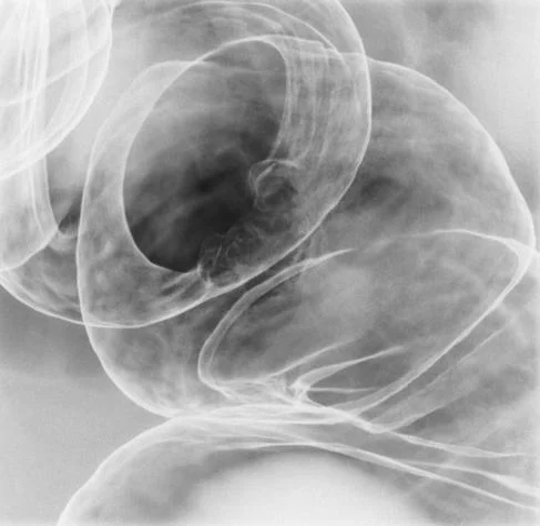
Long description
A barium-enema X-ray of a colorectal carcinoma (described as a type 2 tumor) in the rectosigmoid portion.

- Full blood count and iron studies; iron deficiency mandates bowel investigation.[8]
- CEA at baseline to aid follow-up (it is not a screening test).[1]
- Colonoscopy with biopsy is the most accurate diagnostic test: it visualises the whole colon, obtains tissue, and allows polypectomy. When passable, examine the proximal colon even if the tumour is thought to be distal.[1]
- CT of chest, abdomen and pelvis to detect metastases (liver, lung, peritoneum) and assess surgical resectability.[1]
- Pelvic MRI for rectal cancer: T stage, nodal status, mesorectal fascia and sphincter relation. This determines resectability and the need for neoadjuvant therapy.[2]
- Flexible sigmoidoscopy may be used when colonoscopy cannot be completed, and CT colonography can be useful when colonoscopy is unsafe or incomplete.[3]
- MMR/MSI testing on the tumour is recommended in all new cases (universal screening is standard in many centres): it identifies Lynch syndrome, predicts prognosis and selects patients for immunotherapy.[4]
- In metastatic disease, test RAS (KRAS/NRAS) and BRAF (especially V600E) on tumour tissue, and HER2 status where available, because these determine eligibility for anti-EGFR, anti-BRAF and HER2-directed therapy.[2][11]
There is no standalone diagnostic score; diagnosis rests on histology from an endoscopic biopsy or operative specimen. Staging is by the AJCC TNM 8th edition for both clinical and pathological stage. Dukes staging (A to D) is the older clinical shorthand and is still widely quoted.
| Stage | TNM grouping in outline | Meaning of the group |
|---|---|---|
| 0 | Tis, N0, M0 | Carcinoma in situ, confined to mucosa |
| I | T1-T2, N0, M0 | Invades submucosa or muscle, no nodes |
| II | T3-T4, N0, M0 | Through bowel wall, no nodes; split A/B/C by depth and serosal involvement |
| III | Any T, N1-N2, M0 | Regional nodes involved; A/B/C by depth and number of nodes |
| IV | Any T, Any N, M1 | Distant metastasis (IVA single organ, IVB multiple, IVC peritoneal) |
| Dukes | Extent |
|---|---|
| A | Confined to bowel wall |
| B | Through bowel wall (B1/B2 by depth) |
| C | Regional lymph nodes involved |
| D | Distant metastasis |
At least 12 lymph nodes should be examined in the resection specimen to confirm a negative nodal status.[1] Microsatellite-instable and mismatch-repair-deficient tumours have a somewhat better stage-adjusted prognosis and may be less responsive to fluoropyrimidine chemotherapy, particularly MMR-deficient tumours in the adjuvant setting.[2]
Surgical resection with lymphadenectomy is the best initial treatment for localized disease. The standard is a segmental colectomy (right or left hemicolectomy, sigmoid colectomy, or anterior resection) with clear margins and harvest of at least 12 regional nodes. Sphincter-preserving surgery is preferred where oncologically safe. For very early cancers confined to the mucosa (T1) and selected favourable polyps, endoscopic removal or local (transanal) excision can be curative. For obstructed patients, a Hartmann procedure or, in selected low-risk cases, a one-stage resection may be used.
Rectal cancer changes the surgical plan: total mesorectal excision (TME) to the pelvic floor is the standard operation and sharply reduces local recurrence.[2] If the anal sphincter cannot be preserved safely, an abdominoperineal excision with a permanent colostomy is required. Minimally invasive, robotic and transanal TME are used in specialist centres.
- Rectal cancer: preoperative (neoadjuvant) long-course chemoradiotherapy (commonly 50.4 to 54 Gy with capecitabine or 5-fluorouracil) or short-course radiotherapy (25 Gy in five fractions) before TME is standard for locally advanced disease.[2]
- Total neoadjuvant therapy (TNT): all radiotherapy and chemotherapy given before surgery improves adherence and response, and currently is the preferred approach for most locally advanced rectal cancers without distant metastasis. Complete clinical responders may be managed with a non-operative "watch and wait" strategy under intensive surveillance.[2]
- Adjuvant chemotherapy: for stage III disease and for selected high-risk stage II disease (T4, perforation/obstruction, inadequate nodal harvest, high-grade histology, lymphovascular invasion), an oxaliplatin-based regimen such as mFOLFOX6 or CAPOX is usual, typically for about 6 months. Adjuvant therapy has not shown benefit in MMR-deficient stage II disease, where surgery alone is often adequate.[1][2] FOLFOX and CAPOX dosing details follow local and NCCN/ESMO protocols; check the local formulary.
- Rectal cancer adjuvant: if neoadjuvant therapy was incomplete or omitted, adjuvant chemoradiation and postoperative chemotherapy are used, adapted to final histology.
About 50% of patients with colon cancer are diagnosed with hepatic metastases at presentation or later. In selected patients, resection of liver (and, more rarely, lung) metastases is curative, with 5-year survival around 25% to 40% after a negative-margin resection. Ablation, chemoembolization and hepaticartery-directed therapy are options when resection is not possible.[1]
Systemic therapy is tailored to biology and site:
-
Cytotoxic backbone: FOLFIRI (irinotecan with 5-FU and leucovorin) or FOLFOX/CAPOX, with duration and dose per protocol. FOLFIRI: irinotecan 180 mg per m2 with leucovorin and 5-FU every 2 weeks.[2]
-
Anti-angiogenic therapy: bevacizumab with chemotherapy in selected metastatic patients.
-
Anti-EGFR therapy: cetuximab or panitumumab for RAS wild-type (preferably left-sided) metastatic disease.[11]
-
Immunotherapy: pembrolizumab for mismatch-repair-deficient/MSI-high disease; about 4% of stage IV cases are MSI-high.
-
BRAF V600E (about 10% of metastatic CRC): encorafenib with cetuximab.
-
HER2-amplified tumours: HER2-directed combinations.
- Local progression, obstruction, perforation, bleeding.
- Metastasis to liver, lung, peritoneum, bone.
- Treatment complications: anastomotic leak, chemoradiotherapy toxicity, oxaliplatin neuropathy.
- Extracolonic cancers in Lynch syndrome.
Localized disease: surgery cures roughly 50% of patients.[1] After negative-margin liver metastasis resection, 5-year survival is about 25% to 40%.[1] MMR-deficient tumours have better stage-adjusted prognosis but poorer adjuvant fluoropyrimidine monotherapy response.[2]
Screening begins at 45 for average-risk adults.[5][6]
| Modality | Interval |
|---|---|
| FIT or high-sensitivity gFOBT | Every year |
| Multitarget stool DNA | Every 1 to 3 years |
| CT colonography | Every 5 years |
| Flexible sigmoidoscopy | Every 5 years (or every 10 with annual FIT) |
| Colonoscopy | Every 10 years |
| Risk category | Approach |
|---|---|
| Average risk | Screening begins at 45; choose one of the options above.[5][6] |
| First-degree relative with colorectal cancer | Colonoscopy at age 40, or 10 years before the relative's diagnosis.[6] |
| Ulcerative colitis or Crohn colitis | Surveillance colonoscopy from about 8 years after onset, then every 1 to 3 years.[6] |
| Familial adenomatous polyposis (FAP) or Lynch syndrome | Specialist endoscopic surveillance from youth.[4] |
- Pregnancy: defer unless red flags; multidisciplinary care.
- Older adults: shared decision-making over 75.
- IBD: high-risk surveillance.
Late presentation is common. Use symptom recognition, FIT programmes where feasible, train endoscopy/surgery, and refer early for curative resection.
- CRC is preventable and curable when found early.
- Site determines the presentation.
- Iron-deficiency anaemia in older adults is CRC until proven otherwise.
- S. gallolyticus and C. septicum infection are red flags for colonic neoplasia.
- Staging uses TNM (Dukes as shorthand).
- Surgery is first treatment; chemotherapy and targeted agents added by stage/biology.
- Screening starts at 45.
Answer each one in your head first, then open the answer.
A 62-year-old man has changed bowel habit and ribbon-like stools. Which part of the colon is most likely affected?
Show answerHide answer
Left-sided colon (narrowing and altered bowel habit; obstruction classic).
Why is unexplained iron-deficiency anaemia a red flag?
Show answerHide answer
It is colorectal cancer until proven otherwise, particularly in adults over 50 or with right-sided (silent) disease.
What is the standard rectal cancer operation?
Show answerHide answer
Total mesorectal excision, after neoadjuvant chemoradiotherapy or radiotherapy where indicated.
When is immunotherapy first line?
Show answerHide answer
In metastatic mismatch-repair-deficient or MSI-high disease.
- Ulcerative colitis and inflammatory bowel disease overview
- Crohn disease
- Gastrointestinal bleeding
- PDQ Adult Treatment Editorial Board, National Cancer Institute. Colon Cancer Treatment (PDQ) - Health Professional Version. National Cancer Institute PDQ (updated 12 Feb 2025). Accessed .
- PDQ Adult Treatment Editorial Board, National Cancer Institute. Rectal Cancer Treatment (PDQ) - Health Professional Version. National Cancer Institute PDQ (updated 12 Feb 2025). Accessed .
- PDQ Screening and Prevention Editorial Board, National Cancer Institute. Colorectal Cancer Screening (PDQ) - Health Professional Version. National Cancer Institute PDQ (updated 10 Apr 2025). Accessed .
- PDQ Cancer Genetics Editorial Board, National Cancer Institute. Genetics of Colorectal Cancer (PDQ) - Health Professional Version. National Cancer Institute PDQ (updated 21 Mar 2025). Accessed .
- US Preventive Services Task Force. Colorectal Cancer: Screening. Final recommendation statement (18 May 2021). Accessed .
- American Cancer Society. Colorectal Cancer Guideline: How Often to Have Screening Tests. ACS guideline recommendation page (accessed 2026). Accessed .
- American Cancer Society. Colorectal Cancer Risk Factors. Cancer.org risk factors (updated 2025). Accessed .
- Kolarš B, Mijatović Jovin V, Živanović N, et al. Iron Deficiency and Iron Deficiency Anemia: A Comprehensive Overview. Pharmaceuticals 2025;18(7):1012. Accessed .
- Starosta RT, Rivero R, de Oliveira FH, et al. Misdiagnosis of Streptococcus gallolyticus endocarditis. Autopsy and Case Reports 2016;6(3). Accessed .
- Abraham AT, Padam S. Clostridium septicum Bacteremia As the Presenting Sign of Colon Cancer. Cureus 2023;15(9):e45343. Accessed .
- Airoldi M, Bartolini M, Fazio R, et al. First-Line Therapy in Metastatic, RAS Wild-Type, Left-Sided Colorectal Cancer. Current Oncology Reports 2024;26:1489-1501. Accessed .
Figure credits
Educational content for medical students and doctors. It is not a substitute for clinical judgement or local guidelines.