Peptic ulcer disease
Peptic ulcer disease: causes, symptoms, endoscopic diagnosis, eradication of H. pylori, and management of bleeding, perforation and obstruction.
- A peptic ulcer is a break in the lining of the stomach or duodenum that goes through the mucosa into the wall, conventionally 5 mm or more with depth (some series and reviews quote 3 to 5 mm).
- Most duodenal ulcers and many gastric ulcers are caused by Helicobacter pylori; non-steroidal anti-inflammatory drugs (NSAIDs) and aspirin are the other major cause.
- The classic pain is epigastric and tied to meals: duodenal ulcer pain tends to come 2 to 3 hours after a meal and at night and ease with food, while gastric ulcer pain starts 15 to 30 minutes after eating.
- Upper endoscopy (oesophagogastroduodenoscopy) with biopsy confirms the diagnosis; every gastric ulcer is biopsied to exclude cancer.
- Most patients heal with a proton pump inhibitor (PPI) for 4 to 8 weeks, plus eradication of H. pylori when present, or withdrawal of NSAIDs.
By the end of this page you should be able to:
- Define a peptic ulcer and distinguish gastric from duodenal ulcer in terms of symptoms and risk factors.
- Explain the roles of H. pylori, NSAIDs, aspirin, and rare causes in pathogenesis.
- Recognise alarm features and complications (bleeding, perforation, gastric outlet obstruction, penetration).
- Plan the investigation of dyspepsia and suspected ulcer, including when to biopsy and when to repeat endoscopy.
- Choose initial pharmacological treatment, H. pylori eradication, and prevention strategies for patients who must continue NSAIDs.
- Describe the acute management of upper gastrointestinal bleeding, perforation, and obstruction.
DefinitionPeptic ulcer disease
Peptic ulcer disease (PUD) is a chronic, relapsing condition in which there is a break in the inner lining of the upper gastrointestinal tract, usually the stomach or proximal duodenum, that extends through the mucosa into the deeper wall; by convention this is a mucosal defect of 5 mm or more with depth (some authors quote 3 to 5 mm), as opposed to the superficial defect of an erosion. The term covers gastric ulcers and duodenal ulcers, and occasionally ulcers of the lower oesophagus, distal duodenum or jejunum.
Ulcers may be acute or chronic, active or healing, and single or multiple. The two sites differ in behaviour and risk profile, so they are best considered together but managed with these differences in mind.
Peptic ulcer disease is common worldwide. A review of complicated ulcers puts lifetime prevalence at 5 to 10% of the population, with an incidence of about 0.1 to 0.3% per year. [6] Complications (haemorrhage or perforation) occur in 10 to 20% of patients with ulcers. [6]
Peptic ulcer bleeding remains the most common cause of upper gastrointestinal bleeding and is a common emergency, with mortality from an upper gastrointestinal bleed of 2 to 10%. [4] Incidence has fallen in many regions with H. pylori eradication and PPI use, but patients presenting now are older, sicker, and more often take antiplatelet or anticoagulant drugs, which offsets much of that gain. [4]
Helicobacter pylori is responsible for most duodenal ulcers and for many gastric ulcers, while NSAIDs and aspirin are the other major cause. [7] Ulcers can also be stress-related, drug-induced, ischaemic, or idiopathic. [1]
- H. pylori. Present in the majority of patients with duodenal ulcer and a large proportion of those with gastric ulcer. Eradication removes this driver and prevents recurrence. [7]
- NSAIDs and aspirin (including low-dose aspirin). These drugs suppress protective prostaglandins. Risk rises with dose, age over 65 years, prior ulcer, concomitant anticoagulants, corticosteroids or other NSAIDs, and H. pylori infection. [1]
- Rare causes. Zollinger-Ellison syndrome (gastrinoma causing extreme acid secretion), Crohn disease, cytomegalovirus infection, malignancy (gastric cancer, lymphoma), stress, smoking, and (in some regions) heavy alcohol use. Idiopathic ulcers occur in patients without H. pylori and without NSAID use, especially in older adults. [1]
- Other risk factors. Smoking impairs healing and promotes relapse; alcohol in excess and severe physiological stress (intensive care, major burns, head injury) increase risk. [7]
The normal stomach tolerates its own acid through a mucosal barrier: a surface layer of mucus and bicarbonate, intact epithelium with rapid restitution, good blood flow, and prostaglandin-mediated protection. Ulceration occurs when aggressive factors (acid, pepsin) overpower these defences, or when defence is weakened.
- Acid-peptic injury. Gastric acid and pepsin digest the mucosa once the barrier fails. The ulcer therefore penetrates the muscularis mucosae and reaches the submucosa or deeper, which is what distinguishes it from an erosion. [7]
- Antral stasis and hyperacidity in duodenal ulcer. H. pylori-driven antral inflammation suppresses somatostatin, so gastrin and acid output rise; duodenal defences are then overwhelmed by the acid load. [7]
- NSAID injury. Systemic inhibition of cyclo-oxygenase-1 reduces mucosal prostaglandins, weakening mucus-bicarbonate secretion, blood flow and epithelial repair. Topical irritation by some NSAIDs adds a local effect. [1]
- Stress ulceration. In critical illness, burns or head injury, mucosal ischaemia and acid back-diffusion produce ulcers that can bleed (see Curling and Cushing ulcers below). [6]
Patients report a burning or gnawing epigastric discomfort, often described as hunger pain or indigestion. The timing relative to meals is the classic clue. [7]
- Duodenal ulcer. Pain tends to occur 2 to 3 hours after a meal and at night, and is relieved by food or antacids. [7]
- Gastric ulcer. Pain usually starts 15 to 30 minutes after eating. Food may not relieve it, and food-related pain tends to reduce intake. [7]
- Weight loss favours a gastric ulcer rather than a duodenal one, and should always raise suspicion of malignancy in an older patient. [1]
- Alarm (red-flag) symptoms. Haematemesis, coffee-ground vomiting, melaena, dysphagia, early satiety, persistent vomiting, unexplained weight loss, anaemia or an abdominal mass require prompt endoscopy. [1]
- Medication and risk history. NSAID and aspirin use, anticoagulants, corticosteroids, previous ulcer or bleed, smoking, alcohol, and any family history of gastric cancer or H. pylori treatment.
The examination is often normal between flares. Look for epigastric tenderness in the midline and signs of anaemia or occult bleeding. In gastric outlet obstruction there may be a succussion splash and visible gastric peristalsis. [1] Signs of peritonism (guarding, rigidity, rebound) indicate perforation. Vital signs and a shock assessment come first in any suspected complication.
Epigastric pain has many causes. The table lists the main competitors and the feature that separates each.
| Condition | Features that point away from uncomplicated PUD |
|---|---|
| Gastro-oesophageal reflux disease | Burning retrosternal pain and acid regurgitation; symptoms worse when lying flat or after meals; endoscopy may show oesophagitis, not ulcer. [1] |
| Functional dyspepsia | Chronic upper abdominal symptoms with a normal endoscopy and no structural disease. [1] |
| Gastritis or duodenitis | Endoscopic inflammation without a deep mucosal defect; symptoms overlap closely with ulcer. [7] |
| Gallstone disease / biliary colic | Right upper quadrant pain, often after fatty meals, with normal endoscopy and abnormal biliary imaging. [1] |
| Pancreatitis | Severe constant epigastric pain radiating to the back, raised amylase or lipase, and CT changes. [6] |
| Gastric or pancreatic cancer | Older age, alarm symptoms, weight loss, anaemia or an abdominal mass; mandates endoscopy and biopsy. [1] |
| Angina or cardiac ischaemia | Exertional chest or epigastric discomfort with cardiac risk factors; check ECG and troponin. [4] |
| Medication-induced dyspepsia | Symptoms linked to iron, antibiotics or other drugs, usually normal endoscopy. [1] |
A patient without alarm features who is younger can be tested for H. pylori non-invasively and treated if positive. Anyone with alarm features, older age, or a complicated course needs endoscopy. [1]
Oesophagogastroduodenoscopy is the diagnostic test of choice when endoscopy is indicated. It identifies the site, size and bleeding stigmata of an ulcer, allows biopsy, and permits treatment of high-risk stigmata in the same session. [1][4]
- Take biopsies of every gastric ulcer to exclude malignancy; do not simply assume a benign ulcer. [1]
- In regions of high gastric cancer incidence, also take biopsies from the antrum and corpus for H. pylori and to look for atrophy or intestinal metaplasia. [1]
- Repeat endoscopy at 8 to 12 weeks when a gastric ulcer is not healed, or the original histology was suspicious, to confirm benignancy and healing. [8]
- Test for H. pylori in all ulcer patients, by biopsy-based tests (rapid urease, histology or PCR) when endoscopy is done, or by urea breath test or stool antigen test when it is not. [7]
- Full blood count for anaemia; ferritin in suspected chronic blood loss.
- Urea and creatinine. A disproportionately raised urea is common in upper gastrointestinal bleeding. [4]
- Coagulation screen and platelets, especially if bleeding is suspected or the patient takes anticoagulants.
- Serum gastrin when ulcers are recurrent, multiple, large or resistant, to exclude Zollinger-Ellison syndrome. [1]
- Erect chest radiograph (or cross-sectional imaging) to look for free air under the diaphragm when perforation is suspected. [6]
- Contrast CT when the diagnosis is unclear, when perforation is not visible on plain film, or for complications such as penetration or abscess. [6]
There is no symptom-based gold standard; a peptic ulcer is confirmed at endoscopy. Staging of an ulcer episode is therefore clinical and complication-based. For bleeding risk stratification use a pre-endoscopic score.
- Glasgow-Blatchford score (GBS). A score of 0 to 1 identifies patients at very low risk who can be managed as outpatients without early endoscopy. [5] Higher scores predict need for hospital intervention, transfusion or endoscopic therapy. [4] Cut-offs vary by source: a GBS of 12 or more has been used to select patients for the earliest endoscopy, and 7 or more to predict the need for intervention. [4]
- Forrest classification. Describes the haemostatic stigmata seen at endoscopy and guides treatment and rebleeding risk (see table below). [4]
- Post-endoscopic scores (full Rockall, Cedars-Sinai, PNEID) and AIMS65 are used for prognostication; none predicts rebleeding well. [4]
Approach to suspected peptic ulcer disease
- Patient with dyspepsia or alarm featuresAssess red flags, NSAID and aspirin use, H. pylori risk, age
- Question: Any alarm features, age over about 50 to 60 (guidelines vary), or complicated course?
Yes
- Urgent upper endoscopyBiopsy all gastric ulcers; test for H. pylori
- Treat according to findingsEradicate H. pylori if positive; stop NSAIDs; PPI 4 to 8 weeks
No
- Test for H. pylori (breath or stool, or serology)Eradicate if positive; PPI if needed
- Question: Symptoms persist or H. pylori negative?
Yes
- Upper endoscopyBiopsy gastric ulcers; consider non-ulcer causes
No
- Discharge with follow-up
Any suspected complication is managed as an emergency: airway, breathing, circulation, two large-bore cannulae, intravenous crystalloid to a systolic pressure around 90 to 100 mmHg, analgesia, and venous bloods (full count, renal function, clotting). [4] Do not transfuse to a normal haemoglobin; use a restrictive strategy (see below).
- Stop the offending NSAID or aspirin where it is safe to do so; healing is fastest once the drug is withdrawn. [1]
- Give a PPI for 4 to 8 weeks (most ulcers heal within 8 weeks on a PPI). [1]
- When a PPI cannot be used, H2-receptor antagonists, or (less effectively) sucralfate, misoprostol or mucosal protective agents are alternatives. [1]
- P-CABs (such as vonoprazan) are at least as effective as PPIs for acid suppression and ulcer healing. [1]
- Sucralfate protects the ulcer base and has a useful role at sites of mucosal injury and in stress-ulcer prophylaxis; it coats the ulcer and promotes local defence. [1]
- Eradicate H. pylori whenever it is present (next section).
Eradicate in every patient with a peptic ulcer and documented H. pylori, including patients taking NSAIDs (in whom eradication reduces recurrence). [1] Confirm success with a urea breath or stool antigen test at least 4 weeks after treatment, ideally after stopping acid suppression for the appropriate washout period, because acid suppression suppresses the test. [2] The choice of regimen follows local resistance patterns and availability (the American College of Gastroenterology 2024 guideline and other current guidelines stress this). [3]
Common evidence-based empirical regimens, of which 14-day bismuth quadruple is a widely recommended first-line option in regions with clarithromycin resistance over 15%: [2]
- Bismuth-based quadruple therapy, 10 to 14 days: standard-dose PPI twice daily, metronidazole 500 mg three times daily, bismuth 120 mg and tetracycline 500 mg four times daily. [2]
- Concomitant (non-bismuth quadruple) therapy, 10 days: standard-dose PPI, clarithromycin 500 mg, amoxicillin 1 g and metronidazole 500 mg, all twice daily. [2]
- Clarithromycin-based triple therapy, 7 to 14 days (only where local clarithromycin resistance is under 15% and no susceptibility testing is available): standard-dose PPI, amoxicillin 1 g and clarithromycin 500 mg, all twice daily. [2]
- Tailored therapy based on clarithromycin resistance testing (PCR): 7-day clarithromycin triple when susceptible, 10-day bismuth quadruple when resistant. [2]
- Salvage after failure: 10 to 14 days of bismuth quadruple therapy, then rifabutin- or other susceptibility-guided regimens in specialist centres. [2]
Supporting principles from current guidelines: [1][2][3]
- Avoid empirical clarithromycin triple therapy when resistance is likely to be above 15% or there is past macrolide exposure.
- vonoprazan combined with antibiotics is recommended as first-line by the Japanese guideline, with PPI- or vonoprazan-based therapy as second line.
- A second-line or salvage regimen should be chosen that does not reuse antibiotics already given.
- Serious adverse events with bismuth quadruple therapy are more common (about 40% report some adverse event), so adherence and tolerability should be discussed.
Where stopping the NSAID is not possible, or the patient needs antiplatelet protection, acid suppression reduces recurrence and bleeding. [1]
- Use a PPI as first line when NSAIDs must be continued. [1]
- For high-risk patients with a history of ulcer or bleeding ulcer who need NSAIDs, a COX-2 selective inhibitor plus a PPI is recommended for prevention of recurrent bleeding. [1]
- In patients at high thrombotic risk on aspirin, continue aspirin when indicated; continuing aspirin after a bleed has better cardiovascular outcomes and, with PPI co-therapy, does not increase rebleeding rates. [1]
- For low-dose aspirin users at ulcer risk, a PPI (or a potent acid blocker) is recommended; for secondary prevention after a bleed, PPI co-therapy is recommended. [1]
- H. pylori should be eradicated in NSAID-naive patients before starting NSAIDs for ulcer prevention, although after an ulcer is established PPIs outperform eradication alone. [1]
Surgery is reserved for complications that cannot be managed endoscopically or radiologically: uncontrolled bleeding, perforation (especially with generalised peritonitis), gastric outlet obstruction not relieved endoscopically, and (rarely) suspected malignancy or giant ulcers. [1][6] After surgery that involves the stomach, test and eradicate H. pylori when positive. [1]
Peptic ulcer bleeding is the leading cause of upper gastrointestinal bleeding. Management follows resuscitation, risk scoring, endoscopy and PPI therapy, with escalation for rebleeding.
- Resuscitation. Restore intravascular volume with crystalloid, target a systolic pressure around 90 to 100 mmHg, and transfuse using a restrictive threshold: a target haemoglobin of about 7 to 9 g/dL (guidelines differ on the exact figure), and higher (about 8 g/dL or more) with significant cardiovascular disease or ischaemia. [5][4]
- Intravenous PPI. Give high-dose intravenous PPI while awaiting and after endoscopy: an 80 mg bolus followed by an 8 mg per hour infusion. [5][4]
- Timing of endoscopy. Upper endoscopy within 24 hours of presentation for admitted patients; in high-risk patients endoscopy within 6 hours has not been shown to improve mortality or rebleeding, so timing is guided by resuscitation, haemodynamics and comorbidity after adequate resuscitation. [4]
- Endoscopic haemostasis for high-risk stigmata (Forrest Ia, Ib, IIa, and IIb after clot removal). Techniques include injection (adrenaline plus a second method), thermal coagulation, clips or over-the-scope clips; adrenaline alone is inadequate. [4]
- Low-risk lesions (Forrest IIc and III) need no endoscopic therapy and may be discharged on standard PPI. [4]
| Forrest class | Appearance at endoscopy | Rebleeding risk | Endoscopic treatment |
|---|---|---|---|
| Ia | Spurting arterial bleeding | Very high | Yes (plus second method) |
| Ib | Oozing bleeding | High | Yes |
| IIa | Non-bleeding visible vessel | High | Yes |
| IIb | Adherent clot over the ulcer | Moderate | Consider removing the clot and treating underlying vessel |
| IIc | Flat pigmented spot | Low | No |
| III | Clean ulcer base | Low | No |
- Rebleeding is recognised by fresh haematemesis, melaena, a fall of more than 20 g/L in haemoglobin after stabilisation, or new tachycardia and hypotension; manage with repeat endoscopy and haemostasis. [4]
- Escalation. If endoscopic control fails, use interventional radiology embolisation or surgery. [4][6]
- Tranexamic acid is not recommended in gastrointestinal bleeding outside clinical trials; the HALT-IT trial showed no mortality benefit and an increased risk of venous thromboembolism. [4]
- Aspirin for secondary prevention should be restarted promptly (immediately for low-risk stigmata; otherwise guidelines give anything from as soon as haemostasis is secure to 3 to 5 days) once haemostasis is adequate. [4]
- Test and eradicate H. pylori in anyone who bleeds from an ulcer. [5]
- Transfusion threshold. A restrictive strategy improves outcomes in upper gastrointestinal bleeding; patients who are exsanguinating need resuscitation rather than threshold-driven care. [5][4]
Figure 1Free air under the diaphragm from a perforated viscus
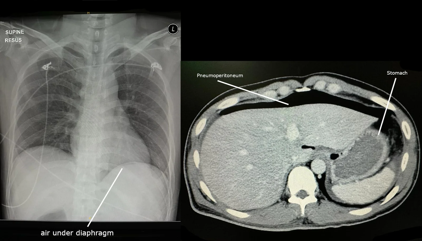
Long description
Left: chest radiograph with a crescent of free air labelled under the diaphragm. Right: axial CT of the upper abdomen with free intraperitoneal air labelled pneumoperitoneum, anterior to the liver and stomach. A perforated peptic ulcer lets gas escape from the stomach or duodenum into the peritoneal cavity; on an upright chest film it collects as a crescent under the diaphragm, and CT is more sensitive for small amounts.

Perforation presents with sudden severe generalised abdominal pain, a rigid abdomen, air under the diaphragm on an erect chest radiograph, and shock. Management is resuscitation, broad-spectrum antibiotics, and surgery; small or contained perforations may occasionally be managed non-operatively by experienced teams. [6]
- Surgical repair is usually an omental patch (Graham patch) repair of the perforation, with peritoneal lavage. [6]
- Larger perforations, delayed presentation, or failed non-operative management require resection or more definitive repair. [6]
- Test for and eradicate H. pylori afterwards. [1]
Obstruction presents with vomiting of undigested food, early satiety, weight loss and a succussion splash; it arises from ulcer oedema, scarring or both. [1]
- Initial management is nasogastric decompression, intravenous fluids and a PPI.
- Endoscopic balloon dilatation can be tried for a narrowed pylorus or duodenal bulb; refractory or malignant obstruction needs surgery. [6]
- Eradicate H. pylori to prevent relapse. [1]
Penetration into an adjacent organ (pancreas, liver, lesser sac) produces deep, persistent pain often radiating to the back, without free perforation. It is managed with intensive acid suppression and eradication of H. pylori; surgery is reserved for refractory or complicated cases. [6]
These are acute stress ulcers linked to severe physiological stress rather than H. pylori or NSAIDs, and they remind us that mucosal ischaemia and acid together cause ulceration. [6]
- Curling ulcer develops after major burns (classically duodenal, especially the first part of the duodenum), due to mucosal ischaemia and acid hypersecretion. [6]
- Cushing ulcer follows raised intracranial pressure or head injury; classically there is gastric acid hypersecretion and ulcers may occur in the stomach, duodenum or oesophagus, with a higher risk of bleeding. [6]
- Management is the same as for other ulcers: resuscitate, correct the underlying stressor, give a PPI, and eradicate H. pylori if present. Prophylaxis with a PPI (or H2 blocker) is usual in critically ill patients at high risk; this is low-grade evidence from stress-ulcer prophylaxis trials, mainly for ventilated or coagulopathic patients. [1]
- Pregnancy. PPIs (especially those with established safety profiles) and H. pylori testing can be used when indicated; avoid unnecessary endoscopy in the first trimester unless alarm features are present, and manage by a gastroenterology and obstetric team.
- Older adults. NSAID use, anticoagulants, and comorbidity dominate; the threshold for endoscopy is lower because malignancy must be excluded.
- Children. Peptic ulcer is uncommon; when present, H. pylori, NSAIDs and Crohn disease are the usual causes, and specialist referral is needed.
- H. pylori can be eradicated empirically with a 14-day regimen suited to local resistance; if resistance testing is unavailable and clarithromycin resistance is likely to be high, prefer bismuth quadruple therapy and avoid clarithromycin triple therapy. [2]
- Where endoscopy is not available, treat clinically and refer anyone with alarm features, anaemia, weight loss, or a poor response.
- Transfusion depends on local blood supply; a restrictive strategy and crystalloid resuscitation still apply. [5]
- Surgery for perforation or obstruction may need referral, but a simple omental patch can be performed in district hospitals with anaesthesia and aseptic technique. [6]
- H. pylori test-and-treat at the community level is cost-effective where test availability allows; otherwise treat based on a positive urease test at endoscopy.
- Reduce harm by prescribing the lowest effective NSAID dose, co-prescribing a PPI, and warning about red-flag symptoms.
- A peptic ulcer is a deep mucosal break (conventionally 5 mm or more) in the stomach or duodenum, usually from H. pylori or NSAIDs and aspirin.
- Duodenal ulcers typically cause nocturnal or late post-prandial pain relieved by food; gastric ulcers cause early post-prandial pain, weight loss and less relief from food.
- H. pylori is found in most duodenal and many gastric ulcers; rare causes include Zollinger-Ellison syndrome, Crohn disease, CMV, malignancy, stress and smoking.
- Always biopsy gastric ulcers to exclude cancer, and repeat endoscopy at 8 to 12 weeks if an ulcer has not healed.
- Uncomplicated disease is treated by stopping the NSAID where possible and giving a PPI for 4 to 8 weeks.
- Eradicate H. pylori after confirming the infection; choice of regimen should follow local clarithromycin resistance (bismuth quadruple therapy where resistance is over 15%).
- For patients who must continue NSAIDs, use a PPI, or a COX-2 inhibitor plus a PPI in high-risk patients; continue aspirin for secondary prevention.
- Upper gastrointestinal bleeding: resuscitate, give intravenous PPI as an 80 mg bolus then 8 mg per hour, perform endoscopy within 24 hours, and use a restrictive transfusion threshold (about 7 to 9 g/dL, higher with cardiovascular disease); tranexamic acid is not recommended.
- Bleeding ulcers with high-risk stigmata need endoscopic haemostasis; rebleeding is managed with repeat endoscopy, then embolisation or surgery.
- Perforation needs an erect chest film looking for free air, resuscitation, antibiotics and usually an omental patch repair.
- Gastric outlet obstruction presents with vomiting, weight loss and a succussion splash; manage with decompression, PPI and (if needed) dilatation or surgery.
- Curling ulcers follow major burns and Cushing ulcers follow head injury or raised intracranial pressure; both are stress ulcers from mucosal ischaemia and acid.
Answer each one in your head first, then open the answer.
What is the main difference between a peptic ulcer and an erosion?
Show answerHide answer
An ulcer is a mucosal defect that extends through the mucosa into the deeper wall (5 mm or more with depth), whereas an erosion is superficial and does not reach the muscularis mucosae. [7]
A 45-year-old man has burning epigastric pain that wakes him at night and is relieved by a midnight snack. Where is the ulcer most likely and why?
Show answerHide answer
Duodenal: duodenal ulcer pain tends to occur 2 to 3 hours after a meal and at night and is relieved by food, reflecting acid load on the duodenal mucosa in the fasting state. [7]
A 60-year-old woman on ibuprofen for arthritis has a haematemesis. Endoscopy shows a bleeding gastric ulcer. What are the first three steps in management?
Which bleeding stigmata at endoscopy require haemostasis, and which do not?
Show answerHide answer
Treat Forrest Ia, Ib, IIa (and IIb after clot removal); lesions with a pigmented spot (IIc) or clean base (III) have low rebleeding risk and do not need haemostasis. [4]
A patient needs long-term NSAIDs for arthritis and has a history of peptic ulcer bleeding. What preventive strategy is recommended?
Show answerHide answer
A COX-2 selective inhibitor together with a PPI; eradicate H. pylori before starting if the patient is NSAID-naive, and continue low-dose aspirin for cardioprotection with PPI cover when indicated. [1]
How do you confirm that H. pylori eradication has worked?
Show answerHide answer
Test with a urea breath test or stool antigen test at least 4 weeks after treatment, after stopping any PPI for the washout period. [2]
A patient with a perforated duodenal ulcer has free air under the diaphragm. What is the standard operation?
Show answerHide answer
Omental (Graham) patch repair with peritoneal lavage, combined with PPI and H. pylori eradication. [6]
A burn patient develops haematemesis. Which stress ulcer should you consider?
Show answerHide answer
A Curling ulcer, which follows major burns (classically duodenal) from mucosal ischaemia and acid hypersecretion. [6]
- Gastro-oesophageal reflux disease
- Upper gastrointestinal bleeding
- Gastric cancer
- Zollinger-Ellison syndrome
- Stress ulcer prophylaxis
- NSAID safety in the older adult
- Kamada T, Satoh K, Itoh T, Ito M, Iwamoto J, Okimoto T, Kanno T, Sugimoto M, Chiba T, Nomura S, Mieda M, Hiraishi H, Yoshino J, Takagi A, Watanabe S, Koike K. Evidence-based clinical practice guidelines for peptic ulcer disease 2020. Journal of Gastroenterology 2021;56(4):303-322. Accessed .
- Bang CS, Kang SJ, Lim H, Kim SH, Lee MW, Nam SY, Tae CH, Kim SE, Kim SY, Jung HK, Kim BJ, Choi M, Jung HY, Kim BW, Korean College of Helicobacter and Upper Gastrointestinal Research. Evidence-Based Guidelines for the Diagnosis and Treatment of Helicobacter pylori Infection in Korea: 2025 Revised Edition. Helicobacter 2026;31(4):e70149. Accessed .
- Chey WD, Howden CW, Moss SF, Morgan DR, Greer KB, Grover S, Shah SC. ACG Clinical Guideline: Treatment of Helicobacter pylori Infection. American Journal of Gastroenterology 2024;119(9):1730-1753. Accessed .
- Orpen-Palmer J, Stanley AJ. Update on the management of upper gastrointestinal bleeding. BMJ Medicine 2022;1:e000202. Accessed .
- Gralnek IM, Dumonceau JM, Kuipers EJ, Lanas A, Sanders DS, Kurien M, Rotondano G, Hucl T, Dinis-Ribeiro M, Marmo R, Racz I, Arezzo A, Hoffmann RT, Lesur G, de Franchis R, Aabakken L, Veitch A, Radaelli F, Salgueiro P, Cardoso R, Maia L, Zullo A, Cipolletta L, Hassan C. Diagnosis and management of nonvariceal upper gastrointestinal hemorrhage: European Society of Gastrointestinal Endoscopy (ESGE) Guideline. Endoscopy 2015;47(10):a1-46. Accessed .
- Tarasconi A, Coccolini F, Biffl WL, Tomasoni M, Ansaloni L, Picetti E, et al. Perforated and bleeding peptic ulcer: WSES guidelines. World Journal of Emergency Surgery 2020;15:3. Accessed .
- Malik TF, Gnanapandithan K, Singh K. Peptic Ulcer Disease. StatPearls Publishing, Treasure Island (FL) 2023. Accessed .
- Maioli ME, Frange RFN, Grion CMC, Delfino VDA. Helicobacter pylori eradication in renal transplant candidates. Jornal Brasileiro de Nefrologia 2022;44(2):215-223. Accessed .
Figure credits
Educational content for medical students and doctors. It is not a substitute for clinical judgement or local guidelines.