Small bowel obstruction
Mechanical blockage of the small intestine: causes, pathophysiology, diagnosis by CT and water-soluble contrast, and when to operate or observe.
- Small bowel obstruction means the lumen of the small intestine is blocked, so gas, fluid and food cannot pass downstream. It is one of the commonest surgical emergencies in adults.
- Postoperative adhesions are by far the commonest cause in adults; hernias and cancer follow. The first task is to separate a simple obstruction from a strangulated or closed-loop one, because the latter needs surgery without delay.
- Small bowel obstruction accounts for roughly 12-20% of patients presenting with an acute abdomen and about 12-16% of hospital admissions in the United States, with an average stay of about 8 days per episode and about 3% in-hospital mortality per episode.[1][4]
- The clinical picture is colicky central pain, early vomiting, variable distension and failure to open the bowels; pain later becomes continuous when the bowel is ischaemic.
- A plain abdominal X-ray can suggest the diagnosis, but a contrast-enhanced computed tomography (CT) scan is the investigation of choice because it shows the level, the cause, the transition point and signs of ischaemia with about 91% sensitivity and 89% specificity for the diagnosis.[4]
- Most adhesive small bowel obstructions, about 70-90%, resolve with bowel rest, a nasogastric tube and intravenous fluids and electrolytes within 72 hours; surgery is reserved for strangulation, peritonitis, closed loop, or failure to improve.[1]
By the end of this page you should be able to:
- Define mechanical small bowel obstruction and classify it as partial or complete, simple or closed loop, and uncomplicated or strangulated.
- List the causes of small bowel obstruction by frequency in adults and children, from adhesions to gallstone ileus.
- Explain how a proximal obstruction dehydrates and endangers the patient, and why closed-loop obstruction is the most dangerous form.
- Elicit the symptoms, examine every hernia orifice and scar, and recognise the signs of strangulation.
- Choose and interpret abdominal X-ray, CT and water-soluble contrast studies, including the Gastrografin challenge.
- Manage an uncomplicated adhesive small bowel obstruction conservatively for up to 72 hours, and state the criteria that mandate emergency surgery.
- Outline the principles of laparoscopic and open adhesiolysis, special situations (Crohn disease, malignancy, early postoperative obstruction, children), and adhesion prevention.
- Adapt care when CT, contrast or surgical services are unavailable.
DefinitionSmall bowel obstruction
Mechanical small bowel obstruction is a partial or complete blockage of the passage of intestinal contents through the small intestine. It is caused by a physical barrier in or around the bowel, as opposed to a paralytic ileus, in which the bowel is open but not moving.
A useful classification follows the severity of the mechanical problem rather than its cause.
- Partial (incomplete) obstruction. Some luminal contents still pass. The patient may still pass flatus and even watery diarrhoea, especially early, and symptoms are less severe. Many partial adhesive obstructions resolve without surgery.
- Complete obstruction. No contents pass beyond the point of blockage. Vomiting becomes more frequent and feculent, and the bowel dilates more.
- Simple (single-point) obstruction. One point of blockage with a free proximal and patent distal bowel. This is the usual adhesive type and the form most likely to resolve with conservative treatment.
- Closed-loop obstruction. A segment of bowel is obstructed at two points, sealing a loop that is tethered, twisted or incarcerated. Gas and fluid accumulate in the loop with no escape either way, so pressure rises rapidly, the blood supply is compromised early, and the mortality of the condition is high, up to 10-35% if strangulation follows.[3]
- Strangulated obstruction. The blood supply of the obstructed bowel is compromised, leading to ischaemia and infarction. It is a surgical emergency with a mortality that rises from about 3% to about 25% when ischaemia is present with delayed surgery.[1][4]
- Obstruction by a partial (rather than fixed) obstruction can be intermittent and mistaken for gastroenteritis, especially in older patients in whom pain is less prominent.
The small bowel normally contains gas and fluid; on its own that content acts as contrast, which is one reason a CT without oral contrast is already diagnostic in many patients.[2]
- Global burden. Bowel obstruction as a whole causes about 15% of hospital admissions for acute abdominal pain in the United States and about 20% of cases needing acute surgical care.[2]
- Small bowel versus large bowel. Small bowel obstruction is the commonest indication for surgery on the small intestine, and obstruction overall causes about 15% of hospital admissions for acute abdominal pain and about 20% of cases needing acute surgical care.[2]
- Adhesive disease. Postoperative adhesions are the leading cause, accounting for 60% of small bowel obstructions and about 55-75% in other series.[1][2] About 1 in 10 patients develops at least one episode of adhesive small bowel obstruction within 3 years of colectomy, and reoperation for adhesive obstruction occurs in 3.2% of colorectal patients and in 4.2-12.6% of paediatric surgical patients.[1]
- Recurrence. After an adhesive obstruction, about 12% of non-operatively treated patients are readmitted within a year, rising to 20% after 5 years; after operative treatment the figures are about 8% and 16% respectively.[1][2]
- Morbidity. Adhesive small bowel obstruction alone causes about 8 days of hospitalisation on average, with an in-hospital mortality of about 3% per episode; mortality rises sharply with ischaemia and delayed surgery.[1][4]
- Age and sex. Adults of any age are affected. Women and men are both affected by adhesive disease in proportion to their prior surgical history; volvulus and cancer become more prominent causes in older adults and in populations with untreated hernias.
The incidence of adhesive small bowel obstruction is somewhat lower after laparoscopic colorectal surgery than after open surgery, although the difference was not statistically significant in the randomised trials reviewed by the World Society of Emergency Surgery (WSES) guideline (odds ratio 0.50, 95% confidence interval 0.20 to 1.2).[1]
The older a patient, the more likely cancer or volvulus is; in younger adults with prior surgery, adhesions dominate.
| Cause of small bowel obstruction | Approximate share | Notes |
|---|---|---|
| Adhesions | 55-75% | Single most common cause in adults with prior abdominal surgery.[1][2] |
| Hernias (external and internal) | 15-25% | Commoner where hernias are not repaired early; femoral hernias in particular strangulate.[2] |
| Malignancy (small bowel, colon, metastatic or peritoneal disease) | 5-10% | Cancer accounts for about 60% of large bowel obstructions.[2] |
| Crohn disease stricture | Part of "other" | Fibrotic stricture after years of disease; usually affects the terminal ileum. |
| Volvulus | Uncommon in small bowel | Midgut volvulus and small bowel volvulus; closed loop and high risk of strangulation. |
| Intussusception | Uncommon in adults | Usually has a lead point such as a polyp or tumour. |
| Gallstone ileus | Uncommon | Gallstone erodes into bowel (usually terminal ileum). |
| Foreign body or bezoar | Uncommon | Dense vegetable matter, hair, or swallowed objects. |
| Ischaemic or radiation stricture | Uncommon | Late sequela of mesenteric ischaemia or abdominal radiotherapy. |
| Other (carcinomatosis, endometriosis, TB, post-anastomotic stricture) | about 10-15% in total | Collectively the rarer causes.[2] |
The causes of small bowel obstruction are not the causes of large bowel obstruction. A separate page deals with large bowel obstruction, but the quick comparison below is worth learning, because the two are confused clinically and radiologically.
| Feature | Small bowel obstruction | Large bowel obstruction |
|---|---|---|
| Commonest cause | Adhesions | Colorectal cancer |
| Pain | Central, colicky early, later continuous | Lower abdomen, more constant |
| Vomiting | Early, frequent, may become feculent | Late or absent |
| Distension | Moderate, periumbilical / central | Marked, lower abdomen |
| Bowel sounds | High-pitched early, absent later | Tinkling, visible peristalsis sometimes |
| Plain film | Dilated small bowel loops, central air-fluid levels, little colonic gas | Dilated colon with haustra, peripheral air-fluid levels |
| Typical emergency test | Contrast-enhanced CT abdomen and pelvis | Contrast-enhanced CT abdomen and pelvis (with rectal contrast if needed) |
| Surgery | Adhesiolysis or resection, often through midline laparotomy | Resection, stenting or decompression depending on cause |
- Previous abdominal or pelvic surgery is the strongest risk factor for adhesions: a history of surgery has about 85% sensitivity and 78% specificity for predicting an adhesive obstruction.[2]
- The risk is highest after colorectal, oncological gynaecological and paediatric surgery; about 1 in 10 patients has an adhesive obstruction within 3 years of colectomy.[1]
- Other adhesion causes: radiotherapy, endometriosis, chronic inflammation, tuberculosis and tumour-related local reaction.[1]
- Untreated or recurrent hernias, particularly femoral hernias in older women, are the other major preventable cause.
- Crohn disease, previous radiotherapy and peritoneal malignancy create strictures and can obstruct.
The harm in small bowel obstruction comes less from the blockage itself than from what accumulates above it and what happens to the bowel wall.
1. Proximal accumulation. The bowel proximal to the obstruction continues to secrete and to swallow air. Gas and fluid collect; the small bowel dilates. A small bowel loop is considered dilated when it is more than about 3 cm across, which is the threshold used in most radiological criteria.[5]
2. Fluid sequestration. The distended bowel wall becomes oedematous and leaks plasma into the lumen and mesentery (third spacing). Vomiting adds to the loss. The patient dehydrates rapidly and may become hypovolaemic: tachycardia, oliguria, dry mucous membranes, and pre-renal acute kidney injury are common.
3. Electrolyte loss. Gastric acid and pancreatic secretions are lost; hypokalaemia is particularly frequent and drives the ileus further. Hyponatraemia and a metabolic alkalosis are typical, but severe sepsis shifts this towards acidosis. Bicarbonate, arterial pH, lactate, amylase and full blood count help detect ischaemia.[2]
4. Bacterial translocation. The dilated, oedematous bowel loses its mucosal barrier. Luminal bacteria and endotoxin cross into the mesenteric lymphatics and blood, fuelling systemic inflammation and sepsis. Fever and leucocytosis in an obstruction are therefore warning signs rather than expected features.
5. Ischaemia and strangulation. Bowel wall tension rises with distension. Venous outflow is obstructed first, then arterial inflow. The mucosa dies first (it is most metabolically active), then the full thickness. Bacterial translocation accelerates; if perforation follows, faecal peritonitis carries a high mortality. The strongest predictor on imaging is decreased enhancement of the bowel wall after intravenous contrast, with a specificity around 95% for ischaemia; mesenteric fluid, free peritoneal fluid, pneumatosis and portal venous gas are other warning signs.[2][3][4]
6. Closed-loop catastrophe. In a closed loop both entry and exit are blocked, so the segment acts like a closed pressure vessel; ischaemia occurs early and progresses to infarction, which is why closed-loop obstruction is the form most feared and the one with the highest mortality, up to 10-35% if strangulation supervenes.[3]
The site of obstruction determines which symptoms dominate.
- Pain. Intermittent, colicky, poorly localised central or periumbilical pain that comes in waves seconds to a minute long, reflecting vigorous peristalsis against a fixed point. Early in a proximal obstruction the patient is comfortable between bouts. Pain that becomes constant, worsening and not relieved by vomiting suggests ischaemia, not simply more obstruction.[2]
- Vomiting. Earlier and more frequent with a proximal obstruction (vomiting within an hour of a high block) than a distal one. Vomit is initially gastric content, then bilious, then feculent in distal small bowel obstruction; feculent vomiting is characteristic of a distal small bowel or a large bowel obstruction.[2]
- Distension. Variable: it may be absent in a very proximal or very recent obstruction, and marked in a distal one. Sudden distension suggests a volvulus; progressive distension suggests an indolent stricture or tumour.[2]
- Bowels. Failure to pass stool or flatus is the hallmark of complete obstruction, but patients with a high obstruction admitted early, and those with a partial obstruction, may still pass stool or flatus, including watery diarrhoea from bacterial overgrowth. Do not be reassured by a bowel movement.[1]
- Associated features. Previous operations (ask exactly what and when: a hernia repair, appendicectomy, gynaecological or colorectal surgery), a history of Crohn disease, radiotherapy, known cancer, or a hernia the patient already knows about. Anorexia and weight loss point to malignancy.
- General. Look for shock: tachycardia, hypotension, dry mucous membranes, reduced skin turgor, oliguria, cool peripheries. Fever is a red flag.
- Abdomen. Inspect for scars (every prior incision, laparoscopic ports, stomas), stomas and hernias, visible peristalsis, and distension. Palpate gently for focal tenderness, guarding, rigidity and localised peritonism. Test for rebound without excess. Bowel sounds change with time: hyperactive, high-pitched tinkling sounds early as the bowel fights the obstruction, becoming quieter and finally silent as ischaemia or exhaustion sets in.
- Hernia orifices. Examine every hernia orifice: umbilical, inguinal, femoral (do not forget the femoral canal in women), any incisional hernia, and the obturator and trocar sites where relevant. A patient with a small strangulated femoral hernia can have deceptively mild abdominal findings.[2]
- Scars. Palpate every surgical scar; an incisional hernia is a common obstructing cause and may be the site of a Richter hernia, in which only part of the bowel wall strangulates.
- Rectal examination. Digital rectal examination (and proctoscopy where available) can show blood, a mass, faecal loading or a pelvic collection, and tells you whether the rectum is empty and collapsed as expected in a complete obstruction.[2]
- Other. Check for a gastric succussion splash, abdominal aortic pulsation and, in women, a pelvic mass.
The classical signs of strangulated obstruction are fever, tachycardia, continuous (rather than colicky) abdominal pain, peritoneal signs, leukocytosis and metabolic acidosis. Each alone has low sensitivity and specificity; combinations matter more, and imaging plus serial examination decide.[3]
Red flagStrangulated small bowel obstruction
Constant pain with fever, tachycardia, peritonism, a rising white cell count or C-reactive protein, or rising lactate means the bowel may be ischaemic. Treat as a surgical emergency: do not wait for the results of a water-soluble contrast study.
The main confusable conditions are other causes of acute abdomen, and paralytic ileus in particular, because giving laxatives to an ileus worsens it.
- Paralytic (adynamic) ileus. Follows surgery, peritonitis, sepsis, trauma, electrolyte disturbance, opioids and many medical conditions. The abdomen is diffusely tympanitic, with absent or uniformly decreased bowel sounds; vomiting is not as projectile and feculent; and pain is not colicky. Imaging shows dilated bowel without a transition point. Management is treat the cause, correct electrolytes and wait; surgery is not indicated.
- Acute colonic pseudo-obstruction (Ogilvie syndrome). Marked colonic dilatation without a mechanical cause, usually in a hospitalised, immobilised, medically ill patient. CT excludes a mechanical cause.
- Gastroenteritis. Watery diarrhoea, self-limiting; may mimic partial obstruction. A low threshold for imaging in the elderly avoids missing an obstruction.[1]
- Mesenteric ischaemia. Pain out of proportion, high lactate, often in a patient with atrial fibrillation or vascular disease; imaging and lactate are decisive.
- Rarer mimics. Abdominal aortic aneurysm, ureteric colic, pelvic inflammatory disease, diabetic ketoacidosis, and (in the young) an intussusception or an internal hernia.
These quantify the insult and guide resuscitation as much as they diagnose.
- Full blood count, urea and electrolytes, creatinine, liver enzymes, amylase, C-reactive protein, lactate, and a venous or arterial blood gas.
- Dehydration shows raised urea, creatinine and haematocrit; hypokalaemia is common and should be corrected.[1]
- Leucocytosis and a C-reactive protein above 75 mg per litre, or a white cell count above 10,000 per mm3, support peritonitis or strangulation but have modest sensitivity and specificity, so they are trended rather than trusted in isolation.[1]
- A raised lactate, metabolic acidosis, low bicarbonate and hyperamylasaemia are the biochemical markers of ischaemia.[2]
- A coagulation screen is needed because emergency surgery is a real possibility.[2]
Figure 1Lateral decubitus abdominal radiograph: air-fluid levels
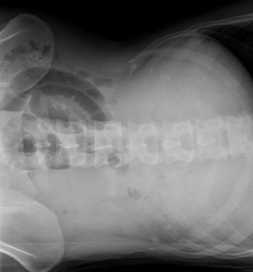
Long description
Abdominal radiograph taken with the patient lying on their side (the image is turned on its side) showing dilated, air-distended small bowel loops with air-fluid levels, the typical stepladder pattern of small bowel obstruction.

Figure 2Supine abdominal radiograph: small bowel obstruction
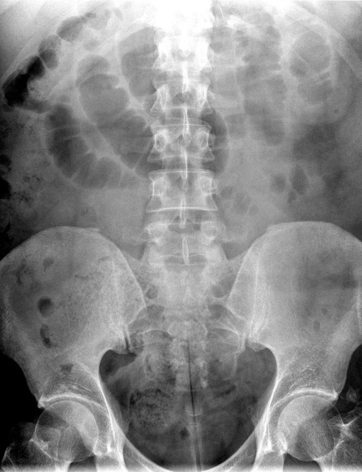
Long description
Supine abdominal film showing dilated, gas-filled loops of small bowel across the central abdomen with relatively little gas in the colon.

A plain supine and erect abdominal film, plus an erect chest film, is the traditional first imaging study. In high-grade obstruction the triad of multiple air-fluid levels, dilated small bowel loops and absent colonic gas is almost diagnostic of small bowel obstruction. In practice, however, plain films are diagnostic in only about 50-60% of cases, inconclusive in 20-30% and misleading in 10-20%, and they miss the cause and early ischaemia entirely.[2] A large pneumoperitoneum from a perforation shows best on an erect chest film.
Sensitivity of plain abdominal radiography for small bowel obstruction is about 69% with specificity about 57% in one series, although another review reported 63-77% sensitivity and 50-78% specificity.[3] Plain film is therefore a triage tool: if the picture is clear and there are no red flags it supports the diagnosis, but an abnormal or equivocal film always needs a CT, and so does any patient with red flags regardless of the X-ray.
Ultrasound can show dilated small bowel loops above 2.5 cm with absent peristalsis, with reported sensitivity 90% and specificity 96% for small bowel obstruction, and works well at the bedside and in low-resource settings, but it is operator dependent and poorer at giving the cause than CT.[2]
This is the investigation of choice for small bowel obstruction. Modern helical CT has about 91% sensitivity and 89% specificity for the diagnosis, with a 95% confidence interval of 84-95% and 81-94% respectively in a meta-analysis of 19 studies covering more than 1,200 patients.[4] Intravenous contrast is preferred when ischaemia is a concern; oral contrast is not needed for diagnosis because the fluid and gas already in the obstructed bowel act as contrast, and it can make the patient vomit. Rectal water-soluble contrast can be added if large bowel obstruction is suspected.
On the scan, follow the loops of bowel proximally from the rectum and look for the transition point where dilated bowel becomes collapsed. A transition point is identified with about 92% sensitivity and 87% specificity on CT in pooled data (95% confidence intervals 87-95% and 74-95%).[4] The cause is read from the anatomy at that point:
- Adhesions give a simple obstruction with an abrupt transition from dilated to collapsed bowel, often a single band, and no mass. CT accuracy for identifying adhesions as the cause is about 95%.
- Hernia shows bowel entering a sac (inginal, femoral, obturator, incisional, internal); accuracy for the cause is lower, about 70%.
- Tumour shows a mass, wall thickening or a stricture, with accuracy about 82%.
- Volvulus or closed loop shows a U- or C-shaped dilated loop, a whirl sign of twisted mesentery and converging folds, with a beak-like taper at the twist point and, if the loop is long and perpendicular to the imaging plane, a clump of loops.
- Gallstone ileus shows Rigler's triad: small bowel obstruction, a gallstone in the bowel and air in the biliary tree (pneumobilia), usually at the terminal ileum.
- Crohn disease shows a thickened terminal ileum with fat wrapping and fibrofatty proliferation.
The CT accuracy for ischaemia is about 82% sensitivity and 92% specificity in pooled data (area under the curve 0.94). Decreased mural enhancement after intravenous contrast is the most specific sign, around 95%; other supporting signs include bowel-wall thickening, mesenteric oedema and fat stranding, mesenteric vascular congestion, ascites, pneumatosis intestinalis and portal venous gas. A recent meta-analysis reported that the absence of mesenteric fluid had about 89% sensitivity for excluding strangulation, and peritoneal fluid among CT findings the highest sensitivity for a complicated course.[4]
Plain film sensitivity 46-69% and specificity 57-67% by comparison, which is why CT, not X-ray, is the default for adults with suspected obstruction.[4]
When contrast-enhanced CT changes management. The Bologna guidelines describe CT as the preferred imaging when the diagnosis is doubtful or urgent surgery may be needed; the workgroup considers it the preferred technique if there is any doubt about adhesive small bowel obstruction, to assess the need for urgent surgery, and to differentiate a complete from a partial obstruction.[1] On the scan, signs of a closed loop, ischaemia or free fluid suggest surgery without delay, and a complete obstruction guides the decision between a trial of conservative management and an operation.[1]
This is both a diagnostic test and, for some patients, a therapeutic one. A dose of 50-150 mL of a water-soluble iodinated contrast, most commonly diatrizoate (Gastrografin), is given by mouth or down a nasogastric tube once the stomach has been decompressed and the patient rehydrated.[2][5] A plain abdominal film is taken at 6 hours and again at 24 hours. Contrast that reaches the caecum or colon within 24 hours predicts successful non-operative management with a resolution in about two thirds to three quarters of patients treated with a Gastrografin protocol; failure of the contrast to reach the colon at 24 hours is highly indicative of failure of conservative management and the need for surgery.[1][2][5] A higher dose (100 mL versus 50 mL) does not appear to improve resolution rates.[5]
Safety points. The osmolarity of diatrizoate is about six times that of plasma, so it draws fluid into the bowel and can worsen dehydration, especially in children and older adults; rehydration first is mandatory. Aspiration pneumonia and pulmonary oedema are life-threatening complications if the contrast is given while the stomach is not adequately decompressed. Rare anaphylactoid reactions are reported, and caution is warranted in patients at risk of gastropathy.[2]
- Magnetic resonance imaging avoids ionising radiation and is a valid alternative in children and pregnant women, with reported sensitivity 95% and specificity 100% in one prospective study.[2] It takes longer and is less good at resolving the cause or assessing bowel viability.
- Endoscopy has no role in small bowel obstruction beyond decompression of a volvulus and assessment of malignant large bowel obstruction; it is not used to prop open a small bowel obstruction.
There is no single scoring system that dictates operation in adhesive small bowel obstruction, and management is clinical. Two derived tools are used to estimate the risk of requiring surgery:
- Zielinski score. Mesenteric oedema, absence of the small-bowel faeces sign on CT, and obstipation identified patients at higher risk of needing surgical exploration, with a concordance index of 0.77 in the validation study.[1]
- Baghdadi score. Radiological findings, sepsis criteria and a comorbidity index predict the need for surgery with an area under the curve of 0.80 in a validation cohort of 351 cases, but it is somewhat complex to apply at the bedside.[1]
Both are decision aids, not protocols. The 70% of malpractice claims in adhesive small bowel obstruction that relate to delayed or missed initial diagnosis underline how much the diagnosis depends on keeping a high index of suspicion and on early imaging.[1]
A related concept is the contrast prediction of outcome: contrast that reaches the colon within 24 hours, resolution of pain, reduction in vomiting, softening of the abdomen, return of bowel sounds and falling nasogastric aspirate all indicate resolution; persistent high nasogastric output with ongoing pain or distension suggests obstruction persists.
- Nil by mouth and a wide-bore nasogastric tube on free drainage, with the output measured and subtracted from fluid balance.
- Intravenous crystalloid and electrolyte replacement, matching losses, with potassium added once urine output is established; balanced crystalloids are preferred for resuscitation, and a urinary catheter monitors output in the sick or elderly.
- Analgesia, ideally opioids in adequate dose: treating pain does not mask deterioration and improves assessment of the abdomen by settling guarding.
- Anti-emetic as needed.
- Investigations as above, including a lactate and cross-match.
- Broad-spectrum antibiotics are not routine in simple adhesive obstruction; they are reserved for strangulation, ischaemia, perforation or a suspected source.
- Communication with a senior surgeon early, and a clear plan reviewed on every shift.
Non-operative management is the standard first strategy in adhesive small bowel obstruction without signs of peritonitis, strangulation or ischaemia.[1] It succeeds in about 70-90% of patients, and the WSES guideline recommends a trial continued safely for 72 hours.[1] Failure rates for a trial of management quoted beyond 3 days are associated with increased morbidity, so the decision to operate should be made deliberately, usually between 24 and 72 hours rather than allowed to drift.[6]
What conservative management actually consists of is the measures above plus the water-soluble contrast challenge at the first safe opportunity, monitoring for passage of contrast to the colon, serial clinical examination, and daily bloods including lactate. Partial (incomplete) adhesive obstruction is the form most likely to settle this way. A faeculent nasogastric aspirate at admission supports a distal small bowel obstruction but is not a reason on its own to operate.
The WSES two-tier approach captured in the Bologna recommendations is: if the diagnosis is certain and there are no signs that immediate surgery might be warranted, a water-soluble contrast study alone is sufficient; if the diagnosis is uncertain, or red flags are possible, CT with oral water-soluble contrast is the preferred technique.
Operate without a completed contrast study if any of the following are present:
- Peritonism or generalized guarding and rebound.
- Strangulation features: fever, tachycardia, continuous pain, leukocytosis, rising C-reactive protein or rising lactate.
- Imaging evidence of a closed loop, strangulation signs, bowel ischaemia or perforation (free gas, free fluid, pneumatosis, portal venous gas).
- An irreducible hernia with obstructive symptoms; attempt controlled manual reduction first in uncomplicated hernia but proceed to surgery if it fails.
- Complete obstruction with a clear transition point from a non-adhesive cause (tumour, gallstone, bezoar, internal hernia).
- No improvement after 48-72 hours of non-operative management, or clinical deterioration at any point.
- Patient factors: immunosuppression, frailty or a closed loop already established.
Approach to a patient with suspected small bowel obstruction
- Patient presents with acute abdomen and suspected obstructionHistory, examination including every hernia orifice and scar, bloods
- Red flags for strangulation or peritonitis?Fever, tachycardia, constant pain, peritonism, rising lactate, leukocytosis
- Question: Red flags present?
Yes
- Resuscitate and operateCross-match, senior surgeon, laparotomy or laparoscopy as local practice dictates
No
- Resuscitate: nil by mouth, nasogastric tube, IV fluids and electrolytes, analgesia, urinary catheter
- Imaging: CT abdomen and pelvis with IV contrast if doubt or red flags not yet clear; plain film for high-grade obstruction
- Question: Cause and severity?
Adhesive, uncomplicated, partial or simple
- Non-operative management with water-soluble contrast challengeNil by mouth, nasogastric tube, fluids and electrolytes, start contrast when rehydrated
- Reassess at 24 and 48 hoursContrast should reach the colon by 24 hours; stop and operate if pain, lactate or aspirate worsen
Closed loop, strangulation, complete non-adhesive or failure to improve
- OperateLaparoscopic or open adhesiolysis and bowel resection as needed
Surgery is required in about 20-30% of adhesive obstructions and in most non-adhesive ones.[1]
- Laparoscopic adhesiolysis is increasingly used for simple adhesive obstruction: less postoperative pain, fewer adhesions re-formed, earlier bowel function and shorter stay in carefully selected patients. Predictors of a successful laparoscopic approach include at most two previous laparotomies, appendicectomy as the index operation, no previous median laparotomy and a single adhesive band.[1] The risk of inadvertent bowel injury is higher than with open surgery (reported at 6.3-26.9% in case series), mostly in patients with multiple complex adhesions or very distended bowel, and the WSES guideline still regards laparotomy as the standard for emergency exploration with laparoscopy as an option in selected simple cases.[1]
- Open adhesiolysis through a midline laparotomy remains the standard for emergency and complex cases. The surgeon enters slowly, using sharp dissection, releases the single band that is usually responsible (most adhesive obstructions are caused by one band) and inspects the whole bowel for non-viable segments, serosal injuries and a second point of obstruction. Avoid seromuscular injuries progressing to enterotomy or delayed perforation.
- Bowel resection is required for non-viable bowel, tumour, a Crohn stricture that cannot be safely stricturoplastied, and a gallstone that cannot be milked proximally. Where the residual bowel is at risk of short gut, resection should be as conservative as possible; stomas are fashioned if viability or contamination is a concern.
- Other causes have their own treatment: incarcerated hernia is reduced and repaired, sometimes with mesh (primary repair without mesh if the field is contaminated); internal hernia is reduced and the defect closed; malignant obstruction is treated according to the primary cancer (resection, stent or bypass); gallstone ileus is relieved by enterotomy and stone removal, with cholecystectomy delayed.
- Adhesion barriers (hyaluronate carboxymethylcellulose, oxidised regenerated cellulose, icodextrin, polyethylene glycol) are placed at operation to reduce reformation of adhesions. Hyaluronate carboxymethylcellulose reduced reoperations for adhesive obstruction in three colorectal trials (relative risk 0.49, 95% confidence interval 0.28-0.88) and is the barrier with the best evidence for prevention.[1]
In sporadic adhesive obstruction, recurrence occurs in about 12% of non-operatively treated patients within a year, and 20% within 5 years; after operation the figures are about 8% and 16%.[1][2] Age, number of bands and prior surgery predict recurrence. Older and frailer patients benefit most from a conservative approach and are more likely to fail it and to end up in surgery.[5]
- Ischaemia and infarction of the obstructed or closed-loop segment, the commonest immediate life-threatening complication.
- Perforation with faecal peritonitis, especially after closed-loop volvulus or prolonged high-grade obstruction.
- Aspiration pneumonia from uncontrolled vomiting in the inadequately decompressed patient.
- Third spacing, dehydration, renal failure and electrolyte collapse from fluid sequestration and vomiting.
- Sepsis from bacterial translocation before or after perforation.
- Enterotomy or delayed perforation from operative adhesiolysis, especially laparoscopic.
- Short bowel syndrome after extensive resection, particularly in Crohn disease or with multiple loops.
- Recurrent obstruction early (residual band) or late (re-formation of adhesions).
- Stoma complications when resection and anastomosis are judged unsafe.
Simple adhesive obstruction treated non-operatively has a low mortality and resolves in most patients within 24-72 hours. Each operative episode, and particularly one with resection, raises the risk of future adhesive obstruction and of a stoma. Strangulated obstruction carries a sharply higher mortality, especially when surgery is delayed, and rises to about 25% if ischaemia is present with delayed operation.[4] In the small bowel itself, obstruction from cancer and in the very frail carries the worst outlook because of the underlying disease rather than the blockage.
Prevention is worth discussing with anyone who operates on the abdomen, because adhesions are the price of access.
- Technique: minimise peritoneal injury with gentle handling, frequent irrigation, meticulous haemostasis and avoidance of foreign bodies (starch, excessive suture, meshes where avoidable). Bipolar or ultrasonic energy injures less than monopolar diathermy in animal models.[1]
- Approach: laparoscopy reduces adhesion formation more than open surgery, although the reduction in reoperation for adhesive obstruction in randomised colorectal trials did not reach significance (odds ratio 0.50, 95% confidence interval 0.20 to 1.2).[1]
- Bars and barriers: hyaluronate carboxymethylcellulose reduced reoperations for adhesive obstruction in three colorectal trials (relative risk 0.49, 95% confidence interval 0.28-0.88); liquid (icodextrin) and gel (polyethylene glycol) barriers are easier to use laparoscopically; oxidised regenerated cellulose is mostly used in gynaecological operations. In secondary prevention after an operation for adhesive obstruction, icodextrin reduced recurrence from about 11% to about 2% in one trial at a mean follow-up of about 41 months.[1]
- Younger patients have the highest lifetime risk of adhesion-related complications and therefore the greatest potential benefit from barrier use at both primary and secondary prevention, including in children, where a 12.6% incidence of adhesive obstruction at a median follow-up of 14.7 years after paediatric surgery has been reported.[1]
Incarcerated hernia is one of the commonest non-adhesive causes and a common cause of emergency laparotomy. Try controlled manual reduction with analgesia and, in selected cases, sedation; if it succeeds, admit for same-day or early elective repair. If it fails, operate. At operation reduce the hernia, assess viability and resect compromised bowel, and repair the defect, with mesh only if the field is clean. Local (spigelian, obturator) and internal hernias can be elusive on clinical examination and are best found on CT.
Malignancy accounts for roughly 5-10% of small bowel obstructions, and cancer for about 60% of large bowel obstructions. Small bowel tumours (adenocarcinoma, neuroendocrine tumour, GIST, lymphoma) are resected with anastomosis if fit. Obstructing left-sided colorectal cancer is managed according to the patient: stenting can palliate or bridge to elective surgery with fewer stomas, but the oncological long-term evidence is still insufficient and stents are reserved for selected patients in experienced centres.[2] Extraperitoneal rectal cancers are usually diverted rather than resected in the emergency setting.
Crohn disease of the terminal ileum produces fibrotic strictures that can obstruct fully. The obstruction is often subacute and recurrent. Acutely, decompress, resuscitate and image; emergency surgery is for ischaemia, perforation or failure to improve. Where surgery is needed, stricturoplasty or limited resection is preferred over wide resection to preserve bowel, and surgery should be as conservative as possible to avoid short-bowel syndrome. Crohn-related obstruction is repeatedly recurrent and benefits from medical optimisation of the disease.
A mechanical obstruction within the first 6 weeks after abdominal surgery is usually adhesions, but ileus, an anastomotic problem or a missed injury are alternatives. Such patients are often treated non-operatively with close observation; many resolve. Surgery is reserved for ischaemia, peritonitis, complete obstruction that does not settle, or a specific surgical cause identified on CT, and the nasogastric tube is kept until there is clear return of function.
In children the commonest causes are different from adults: congenital atresias and stenoses, intussusception, malrotation with midgut volvulus, incarcerated hernia, and Meckel diverticulum. Midgut volvulus in particular is a closed-loop emergency. A paediatric incidence of adhesive small bowel obstruction of about 12.6% at a median follow-up of about 14.7 years after paediatric surgery has been reported.[1]
Older adults have more ischaemic and malignant causes, more untreated hernias and more late presentation. Pain is often less prominent in the elderly, which delays diagnosis.[1] Frailty prolongs recovery, and quality of life rather than pure survival drives the decision to operate; comorbidity, polypharmacy and cardiac reserve all matter. A conservative trial may be the right initial choice, but delay in recognising strangulation is the main danger.
Small bowel obstruction in pregnancy is uncommon but dangerous. Physiological displacement and the risk of fetal compromise complicate diagnosis. Imaging without radiation (ultrasound first, then MRI) is preferred; the principles of resuscitation and operation are unchanged, and management is an obstetric-surgical-anaesthetic team decision.
- Imaging. Where CT is unavailable, plain abdominal X-ray plus careful clinical assessment carries most of the diagnosis; water-soluble contrast by mouth followed by a film at 24 hours can both test for and treat adhesive obstruction.[1][5] A study from Ethiopia reported that in their setting CT was almost never performed, 83% of small bowel obstruction patients were managed surgically, and postoperative morbidity was 11.4% surgical site infection, 9.4% sepsis, 6.7% pneumonia, 6% reoperation and 4.7% death; the authors suggested that wider use of Gastrografin could reduce the need for surgery.[5]
- Ultrasound is available in most low-income settings and is more informative than plain films; it can show dilated loops, free fluid suggesting the need for urgent surgery, and volume status in the shocked patient.[1]
- Surgical capacity. Where laparotomy is the available operation, adhesiolysis is done safely; the main hazards are delayed presentation and lack of intensive care. The single most useful intervention is to reduce delay from symptom onset to decision to operate.
- Stomas. If the resected bowel is short or the patient is sick, a temporary stoma may be safer than an anastomosis; reconstruction is planned for later when the patient is fit.
- Small bowel obstruction is one of the commonest surgical emergencies; adhesions cause most cases in adults, hernias next.
- A partial or simple adhesive obstruction resolves non-operatively in about 70-90% of patients within 24-72 hours.[1]
- Strangulated or closed-loop obstruction is a surgical emergency with high mortality.
- A history of prior abdominal surgery has about 85% sensitivity and 78% specificity for an adhesive obstruction.[2]
- Plain film suggests the diagnosis in only about 50-60% of cases and misses the cause and ischaemia.[2]
- Contrast-enhanced CT is the investigation of choice, with about 91% sensitivity and 89% specificity for the diagnosis and additional accuracy for ischaemia, transition point and cause.[4]
- A decreased mural enhancement and closed loop on CT, pneumatosis or portal venous gas, peritonism, fever, tachycardia or rising lactate mean operate now.
- The water-soluble contrast challenge is both diagnostic and therapeutic: contrast in the colon by 24 hours predicts resolution; no contrast by 24 hours predicts failure.
- Laxatives must never be given in suspected obstruction; they worsen a closed loop.
- Senior involvement and a documented plan reviewed every shift are as important as any single test.
Answer each one in your head first, then open the answer.
What is the most common cause of mechanical small bowel obstruction in adults, and what proportion does it account for?
A 68-year-old man has 12 hours of central colicky pain, vomits twice, has not opened his bowels and looks dehydrated. The abdomen is distended with tinkling bowel sounds. Why is this most likely an adhesive obstruction rather than a paralytic ileus?
Show answerHide answer
The pain is colicky (not absent or diffuse), the bowel sounds are increased rather than absent, and he has a likely surgical history. Ileus gives diffuse tympany and uniformly decreased sounds without a mechanical transition point.
Which CT finding most specifically indicates ischaemic bowel in small bowel obstruction, and roughly what is its specificity?
Why does a closed-loop obstruction carry a much higher mortality than a simple single-point obstruction?
Show answerHide answer
A patient with a suspected partial adhesive small bowel obstruction is haemodynamically stable with normal lactate. How long can non-operative management be continued, and what two findings push you to operate sooner?
During the first 6 hours after a Gastrografin challenge, a 40 mL nasogastric aspirate is now bilious and reduced; the patient is comfortable and the X-ray shows contrast in the terminal ileum but not the colon. What is the safest next step?
A woman of 40 presents with obstruction. There is no surgical history but a groin bulge is felt below and lateral to the pubic tubercle. Which hernia is this, and why is repair more urgent in this type?
Show answerHide answer
A femoral hernia. It has a narrow rigid neck at the femoral canal and is more likely to strangulate than an inguinal hernia, so an obstruction through one needs emergency repair.[2]
What do feculent nasogastric aspirate, absent flatus and marked lower distension tell you when validated against imaging?
Show answerHide answer
They suggest a distal small bowel or a large bowel obstruction. Feculent aspirate supports a distal small bowel blockage; imaging, not sounds, confirms the level. Bowel sounds alone are unreliable.
Crohn disease, abdominal hernias, acute abdomen, ileus, large bowel obstruction, mesenteric ischaemia, intestinal stomas, adhesion prevention.
- Ten Broek RPG, Krielen P, Di Saverio S, et al. Bologna guidelines for diagnosis and management of adhesive small bowel obstruction (ASBO): 2017 update of the evidence-based guidelines from the World Society of Emergency Surgery ASBO working group. World Journal of Emergency Surgery 2018;13:24. doi:10.1186/s13017-018-0185-2 (PMC6006983). Accessed .
- Catena F, De Simone B, Coccolini F, Di Saverio S, Sartelli M, Ansaloni L. Bowel obstruction: a narrative review for all physicians. World Journal of Emergency Surgery 2019;14:20. doi:10.1186/s13017-019-0240-7 (PMC6489175). Accessed .
- Pothiawala S, Gogna A. Early diagnosis of bowel obstruction and strangulation by computed tomography in emergency department. World Journal of Emergency Medicine 2012;3(3):227-231. doi:10.5847/wjem.j.issn.1920-8642.2012.03.012 (PMC4129773). Accessed .
- Li Z, Zhang L, Liu X, Yuan F, Song B. Diagnostic utility of CT for small bowel obstruction: systematic review and meta-analysis. PLoS One 2019;14(12):e0226740. doi:10.1371/journal.pone.0226740 (PMC6936825). Accessed .
- Tutino R, Cavaglià M, Pipitone Federico NS, et al. The diagnostic and therapeutic value of Gastrografin in small bowel obstructions. Frontiers in Surgery 2025;12:1516155. doi:10.3389/fsurg.2025.1516155 (PMC11937050). Accessed .
- Keenan JE, Turley RS, McCoy CC, Migaly J, Shapiro ML, Scarborough JE. Trials of nonoperative management exceeding 3 days are associated with increased morbidity in patients undergoing surgery for uncomplicated adhesive small bowel obstruction. Journal of Trauma and Acute Care Surgery 2014;76:1367-1372. doi:10.1097/TA.0000000000000246 (PMID 24854302). Accessed .
Figure credits
Educational content for medical students and doctors. It is not a substitute for clinical judgement or local guidelines.