Congenital adrenal hyperplasia in females
21-hydroxylase deficiency in 46,XX: virilisation, salt wasting, diagnosis, glucocorticoid therapy.
- Congenital adrenal hyperplasia (CAH) is a family of autosomal recessive disorders in which an enzyme needed for cortisol synthesis is deficient; 21-hydroxylase deficiency (21OHD) accounts for about 95% of cases. [1]
- In a 46,XX (female) infant, 21OHD produces virilisation of the external genitalia ranging from clitoromegaly to frankly ambiguous genitalia, while the internal organs (uterus, fallopian tubes, ovaries) are female.
- In about 75% of severe (classic) cases aldosterone is also deficient, so salt wasting, failure to thrive and shock can occur in the first 1-2 weeks of life. Newborn screening aims to prevent this.
- A milder, late-onset (non-classic) form presents later, most often with hirsutism, acne, menstrual irregularity or infertility in a phenotypic woman.
- Diagnosis rests on a raised serum 17-hydroxyprogesterone (17-OHP), confirmed with a cosyntropin (ACTH) stimulation test; treatment is glucocorticoid replacement, with a mineralocorticoid when salt is wasted.
By the end of this page you should be able to:
- Define 21-hydroxylase deficiency and place it among the other forms of CAH.
- Explain how cortisol and aldosterone deficiency and androgen excess arise, and why a female infant is virilised but a male is not.
- Recognise the classic, simple-virilising and non-classic forms, and list the neonatal red flags.
- Work out the diagnostic pathway, including the cosyntropin test and the 17-OHP cut-offs.
- Plan glucocorticoid (and mineralocorticoid) therapy in infancy, childhood and adult life, and in pregnancy.
- Explain the surgical, fertility and long-term issues that are specific to women with CAH.
DefinitionCongenital adrenal hyperplasia (CAH)
A group of autosomal recessive disorders in which a deficiency of one of the enzymes (or cofactor proteins) needed to make cortisol blocks cortisol synthesis. The pituitary responds by secreting excess adrenocorticotropic hormone (ACTH), the adrenal cortex hypertrophies, and the accumulated precursor steroids are shunted into androgen (and sometimes mineralocorticoid) production.
- Steroid 21-hydroxylase deficiency (21OHD) is by far the commonest form, accounting for roughly 90-95% of CAH. The enzyme (P450c21, encoded by CYP21A2) converts 17-hydroxyprogesterone (17-OHP) to 11-deoxycortisol and progesterone to deoxycorticosterone, the precursors of cortisol and aldosterone. [1]
- Classic 21OHD is severe, presenting in infancy. It is split into:
- Salt-wasting (SW): cortisol and aldosterone both deficient; salt-losing crises occur without treatment.
- Simple-virilising (SV): cortisol deficient, androgen excess marked, but enough aldosterone is made to avoid salt loss (aldosterone synthesis can be partly preserved).
- Non-classic (NCCAH) is mild: 20-50% of enzyme activity remains, cortisol and aldosterone are usually adequate, there is no salt wasting, but androgen excess produces pubic hair, acne, hirsutism, oligomenorrhoea or infertility, often years later. [4]
- Rarer forms (11-beta-hydroxylase deficiency, 3-beta-hydroxysteroid dehydrogenase deficiency, 17-alpha-hydroxylase deficiency, P450 oxidoreductase deficiency, lipoid CAH) are considered in the differential section. [7]
The distinction between classic and non-classic disease is a continuous spectrum based on CYP21A2 genotype, not two separate diseases. [2]
- Classic 21OHD occurs in roughly 1:14,000 to 1:18,000 births worldwide by newborn screening, with ethnic and geographic variation (higher in endogamous or consanguineous groups). [1]
- Salt-wasting (SW) 21OHD accounts for about three-quarters of classic cases. [1]
- Non-classic CAH is far commoner: an overall frequency of roughly 1:200 (95% confidence interval 1:100 to 1:280) in the general white population, and much higher (1-2%) in certain groups such as Ashkenazi Jewish populations. [1][4]
- The carrier rate for classic CAH is about 2%. [1]
- Risk factors: an affected sibling (autosomal recessive inheritance; recurrence risk 25% for a couple both carriers), a parent with known CAH (a woman with CAH has a markedly increased chance of having a child with CAH if her partner carries a severe mutation), and high rates of consanguinity. [4]
The cause is loss-of-function mutations in CYP21A2 (chromosome 6p21.3). Most disease alleles arise from recombination between CYP21A2 and its neighbouring pseudogene CYP21A1P. Two-thirds of individuals with non-classic disease are compound heterozygotes (one severe and one mild allele), and the less severe allele usually dictates the phenotype. [4]
Specific genotype-phenotype links that are worth knowing: [1][4]
- The p.Ile172Asn mutation (about 1-2% residual enzyme activity) underlies most simple-virilising disease. [1]
- The p.Val281Leu mutation (20-50% residual activity) accounts for most non-classic alleles. [4]
- Large deletions and the intron 2 splicing mutation (ablate enzyme activity) account for about half of classic alleles. [1]
About 5-10% of patients with salt-wasting CAH also have the hypermobile form of Ehlers-Danlos syndrome from tenascin-X haploinsufficiency (the TNXB gene overlaps CYP21A2). [1]
Figure 1Adrenal Steroids Pathways
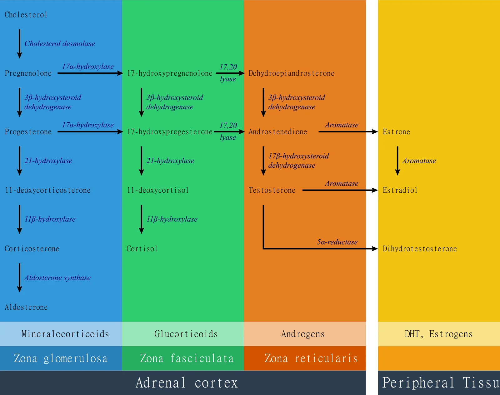
Long description
A colour-coded pathway diagram showing cholesterol converted to mineralocorticoids (aldosterone), glucocorticoids (cortisol) and androgens (androstenedione/testosterone) in the zona glomerulosa, zona fasciculata and zona reticularis of the adrenal cortex, plus DHT, oestrogens and other products in peripheral tissue, with enzymes including 21-hydroxylase, 11β-hydroxylase, 17α-hydroxylase, aldosterone synthase, aromatase, 17,20-lyase and others labelled along the arrows.

- Cortisol deficiency. The block in cortisol synthesis removes negative feedback on the pituitary, so ACTH rises continuously. ACTH drives the adrenal cortex to hypertrophy and to produce everything it can upstream of the block. [1]
- Androgen excess ("overflow"). 17-OHP (and other precursors) accumulate and are diverted into androgen biosynthesis, mainly via CYP17A1 to androstenedione and then testosterone, and via the "backdoor" pathway to dihydrotestosterone. The degree of virilisation in a female infant correlates with the severity and duration of this androgen exposure. [1][2]
- Mineralocorticoid deficiency ("underfill"). In salt-wasting disease there is not enough 21-hydroxylase activity to make aldosterone either. The newborn therefore loses sodium, retains potassium, and dehydrates. Cortisol deficiency worsens the picture by reducing vascular responsiveness to catecholamines and glomerular filtration. [5]
- Why only girls look different at birth. Testosterone and dihydrotestosterone are the same hormones a male fetus makes from his own testes. Excess androgen therefore virilises a 46,XX fetus (enlarging the clitoris and fusing the labioscrotal folds) but has no visible effect on a 46,XY fetus, whose external genitalia are already virilised. The uterus, fallopian tubes and upper vagina develop normally because androgens do not oppose Mullerian development. [1][4]
A diagrammatic way to picture it: the adrenal makes too much ACTH, too little cortisol, too little aldosterone in severe cases, and too many androgens that land on a female external genitalia. [1]
The hallmarks at birth are virilisation, with normal internal female anatomy on ultrasound:
- Clitoromegaly (sometimes large enough to be mistaken for a penis).
- Partial or complete fusion of the labioscrotal folds ("scrotalisation"), sometimes with a single perineal opening (urogenital sinus).
- Normally formed labia minora and no palpable gonads in the "scrotum" (a clue against a 46,XY DSD, where testes would be felt).
The severity of virilisation is graded from near-normal to penile-appearing clitoris with a single perineal orifice, using the Prader scale in practice. The degree is not reliably predicted by genotype alone. [1][2]
- Poor feeding, vomiting, lethargy in the first 3-10 days.
- Dehydration and weight loss.
- Hyperkalaemia first, then hyponatraemia (potassium rises before sodium falls, often around day 7-10).
- Hypovolaemic shock, sometimes mislabelled as sepsis.
Males with salt-wasting disease have no genital ambiguity, so they rely entirely on screening; this is why classic CAH is more often diagnosed late in boys unless newborn screening exists. [1][5]
No salt wasting at birth. Girls are virilised (often less dramatically) and, if untreated, go on to have rapid childhood growth, tall stature then short final height, premature pubarche, and later menstrual problems. Boys have precocious puberty from adrenal androgens. [1]
Presentation depends on age and is driven by androgen excess, not cortisol deficiency:
- Childhood: premature pubarche (pubic hair before age 8), rapid growth, tall stature with advanced bone age.
- Adolescence: acne, hirsutism (often starts after age 10 and worsens with age), oily skin, frontal hair thinning in older women.
- Adults: irregular or infrequent periods, infertility, sometimes androgenic (male-pattern) balding; the academic risk of hirsutism rises with age.
- Many women are asymptomatic and are found only by kindred testing. [4]
- Blood pressure and pulse sitting and standing, plus skin for hyperpigmentation, striae, acne and hirsutism.
- External genitalia in an infant with an adequate light and magnification; look specifically for gonads in the labioscrotal folds.
- Anthropometry in children (height velocity, weight, bone age).
- Gynaecological history and examination in adolescent girls: menstrual pattern, anatomy, vaginal introitus, possible stenosis after prior surgery. [1][2]
Red flagRed flags in a virilised infant
Any infant with ambiguous or unusually virilised genitalia needs measurement of 17-OHP and electrolytes within the first 48-72 hours, because salt wasting can be fatal by the end of the first week. Do not wait for newborn screening results if the clinical picture fits.
Virilisation in a 46,XX infant has a short differential. Androgen excess in a woman has a longer one.
| Condition | Key separating features |
|---|---|
| CAH (21OHD) | Virilised 46,XX infant; raised 17-OHP (basal or stimulated); salt wasting in SW form. [1][4] |
| 11-beta-hydroxylase deficiency | Virilised 46,XX infant (like 21OHD); hypertension and hypokalaemia from deoxycorticosterone excess rather than salt loss. [7] |
| 3-beta-hydroxysteroid dehydrogenase deficiency | Severe salt wasting; milder virilisation; abnormal (delta-5) steroid profile. [7] |
| 17-alpha-hydroxylase deficiency | No virilisation at birth in a 46,XX infant; presents later with hypertension and primary amenorrhoea. [7] |
| P450 oxidoreductase deficiency | Variable; can include skeletal and craniofacial abnormalities (Antley-Bixler phenotype). [7] |
| Aromatase deficiency | Virilised mother and child; very rare. [4] |
| Androgen-secreting tumour (ovary/adrenal) | Rapidly progressive virilisation in an adult; very high androgens; suppressed gonadotropins. [2] |
| Polycystic ovary syndrome (PCOS) | Common cause of hirsutism/acne in women; 17-OHP normal or only mildly raised; no salt wasting. [4] |
| Idiopathic hirsutism | Normal androgens; normal periods; diagnosis of exclusion. [4] |
| Exogenous androgen | History gives it. [2] |
The two questions that matter most at the bedside are: "Is the baby salt wasting?" (separates 21OHD/3BHSD from 11OHD) and "Is the 17-OHP genuinely high?" (separates CAH from PCOS and idiopathic hirsutism).
- Serum 17-OHP (early morning, before 08:00, ideally in the follicular phase in a cycling woman) by LC-MS/MS where possible. Immunoassay is less specific and cross-reacts with other steroids. [1][4]
- Electrolytes, glucose, plasma renin activity (or concentration) and blood pressure in any infant or in any adult with possible salt loss. [3][5]
- Cosyntropin (ACTH) stimulation test to confirm: baseline then 60-minute 17-OHP (and cortisol), using 250 micrograms in older children and adults; 15 micrograms/kg in infants under 1 year. [6] (from dynamic testing review) [7]
- Karyotype for any infant with ambiguous genitalia.
A basal or stimulated 17-OHP above 30 nmol/L (about 1000 ng/dL) essentially diagnoses 21OHD, and classic 21OHD values are usually 10-100 times higher. [3]
Values below that cut-off can be difficult. In a woman with CAH symptoms:
- Morning (07:30-08:00) 17-OHP under 200 ng/dL generally makes non-classic CAH unlikely. [4]
- Morning 17-OHP above 200 ng/dL should prompt full ACTH-stimulation testing. [4]
- After cosyntropin, 17-OHP in non-classic CAH is typically 1,500-10,000 ng/dL, whereas classic disease is above 20,000 ng/dL. [4]
- An elevated result should be interpreted with a full adrenal steroid profile to exclude the rarer enzyme defects. [1]
A normal 17-OHP in a woman with hyperandrogenism does not exclude other causes; the other investigations (androstenedione, testosterone, DHEAS, gonadotrophins) are used to stage androgen excess, as described below.
Reserve genotyping for equivocal stimulation tests, patients already on glucocorticoids, or genetic counselling. Genotype predicts mineralocorticoid deficiency better than the extent of genital virilisation. [1]
| Test | Value that supports 21OHD | Interpretation |
|---|---|---|
| Early-morning basal 17-OHP (symptomatic) | Greater than 30 nmol/L (1000 ng/dL) | Diagnostic of 21OHD with very rare exceptions. [3] |
| Early-morning basal 17-OHP (screening, NCCAH) | Greater than 200 ng/dL | Captures about 90% of non-classic cases; arrange ACTH-stimulation testing. [4] |
| 17-OHP 60 min after cosyntropin | Under 1000 ng/dL: unlikely | Normal (no CAH). [4] |
| Stimulated 17-OHP in NCCAH | 1500-10000 ng/dL | Typical range for non-classic disease. [4] |
| Stimulated 17-OHP in classic disease | Above 20000 ng/dL | Typical of classic CAH. [4] |
| Stimulated 17-OHP (screening threshold) | Above 10 ng/mL (30 nmol/L) | Indicates CAH; above 100 ng/mL indicates classic, 10-100 ng/mL non-classic. [6] [7] |
| Cortisol after cosyntropin | Under 14 to 18 microgram/dL (under 400-500 nmol/L) | Suboptimal cortisol reserve; used in non-classic disease to decide stress dosing. [1] |
In women on treatment, 17-OHP is the most volatile marker: it is 100-1000 times higher than androgens and swings with every dose. Suppressing it into the normal range almost always means over-treatment. Adrenal androgens (androstenedione, testosterone, and the 11-oxygenated androgens) and the clinical picture (menstruation, skin, blood pressure) matter more for guiding therapy. [2]
Women attempting to conceive have one precise biochemical target: progesterone should be under 2 nmol/L (under 0.6 ng/mL) in the follicular phase. [2]
- Immediate treatment when salt wasting is present: intravenous saline and glucose plus intravenous hydrocortisone.
- Maintenance from the first days of life:
- Hydrocortisone (the preferred glucocorticoid in growing children): 10-15 mg/m2/day divided into three doses. [6]
- Fludrocortisone: 150 micrograms/m2/day (about 50 micrograms/day) in infancy, titrated to renin, blood pressure and electrolytes. [5]
- Sodium chloride supplement in infancy (commonly up to 10-12 mmol/kg/day at first), usually not needed after weaning at 6-8 months. [5]
- Doses are adjusted at each infant visit; the requirement for mineralocorticoid is higher in the first year because of a relative aldosterone resistance. [5]
- Hydrocortisone 10-15 mg/m2/day in divided doses (never long-acting potent glucocorticoids chronically in growing children, because of growth suppression). [1][6]
- Fludrocortisone in salt-wasters, titrated by plasma renin, serum potassium and blood pressure; salt supplements in early childhood.
- Monitoring every 3 months under 18 months, then every 4 months, with growth velocity, blood pressure, bone age (annual under age 2) and, in boys, testicular ultrasound. [1]
- Bone age is an integrated (lagging) marker of antecedent control. [3]
- Stress dosing: increase glucocorticoid (but not mineralocorticoid) dosage during febrile illness above 38.5 C, dehydration, major surgery with general anaesthesia and major trauma. [1]
Treat only if there is inappropriately early or rapidly progressive pubarche, accelerated bone age, overt virilisation, or later patient-important hyperandrogenism/infertility. Doses are lower; hydrocortisone 10-15 mg/m2/day divided is the usual. [4][1]
- Hydrocortisone remains first-line in many women: typically 3 doses of 5-10 mg with meals, which rarely causes Cushingoid features. [2]
- If control on hydrocortisone alone is inadequate, add the smallest effective long-acting glucocorticoid: prednisolone 0.5-2 mg (often 1 mg) at night; use dexamethasone 0.25-0.375 mg/day only in selected circumstances (for example, to shrink testicular adrenal rest tumours or when progesterone must be suppressed before conception). [2]
- Avoid prednisone where conversion to active drug is inconsistent, and avoid over-suppressing the adrenal entirely. [2]
- Fludrocortisone: typical adult dose 0.05-0.2 mg once daily in salt-wasters; titrate to keep renin low, potassium normal, and blood pressure normal without orthostasis or salt craving. Women with non-classic disease do not need mineralocorticoid. [2][5]
- Do not aim for a normal 17-OHP: a morning 17-OHP that is still raised but androgens controlled is acceptable and avoids over-treatment. [2]
For women with CAH who are not trying to conceive, androgen excess can be managed without intensifying glucocorticoids:
- Combined oestrogen-progestogen oral contraception raises sex-hormone-binding globulin and regularises menses when progesterone is not chronically high. [2]
- Spironolactone is effective and inexpensive for hirsutism and acne; in classic CAH its mineralocorticoid antagonism usually means a fludrocortisone dose increase to protect volume. [2]
- Mechanical methods (laser, electrolysis, waxing, eflornithine) give additional benefit. [2]
- Surgery is performed only in centres with an experienced multidisciplinary team. [1]
- The trend is to delay surgery until the child is old enough to participate, while informing families of all options, including delay. [1]
- Early vaginoplasty (urogenital mobilisation) and neurovascular-sparing clitoroplasty may be offered for severe virilisation. [1]
- Long-term issues include vaginal stenosis, fistula, stricture and recurrent urinary tract infections. [2]
- Newborn girls with CAH should not be equated with other disorders of sex development when decisions about sex of rearing are considered; the Endocrine Society maintains functional anatomy and fertility as the goal. [1]
- Women with 21OHD can have normal fertility when androgen and progesterone are controlled. For conception, aim for progesterone under 2 nmol/L (under 0.6 ng/mL) in the follicular phase; this usually needs a bedtime dose of a long-acting glucocorticoid. [2]
- Non-classic women who are infertile or have had miscarriage should be treated with a glucocorticoid that does not cross the placenta (hydrocortisone or prednisolone, not dexamethasone). [1][4]
- During pregnancy: continue pre-pregnancy hydrocortisone (or prednisolone) and fludrocortisone doses, increasing the glucocorticoid in the second/third trimester if symptoms of insufficiency occur, give stress doses during labour and delivery, and avoid dexamethasone throughout. [1]
- Arrange an experienced obstetric specialist for the birthing plan, and a glucocorticoid stress dose in labour. [1]
- Miscarriage rates fall markedly after diagnosis and treatment in non-classic disease. [4]
- Bilateral adrenalectomy to restore fertility is discouraged; it trades one form of adrenal insufficiency for a more severe one, and adrenal rest tumours can still appear. [2]
- Every patient should wear/carry medical identification and be equipped with a glucocorticoid injection kit and taught parenteral self-administration (or lay administration by a parent/guardian). [1]
- Increase glucocorticoid (not mineralocorticoid) dosing for febrile illness above 38.5 C, dehydration or gastroenteritis, major surgery and major trauma. [1]
- Everyday mental stress, minor illness and routine exercise do not need extra steroids. [1]
- Adrenal crisis: intravenous hydrocortisone, saline and glucose.
- Annual examination: blood pressure, body mass index, Cushingoid features, plus biochemical monitoring for glucocorticoid and mineralocorticoid adequacy. [1]
- Bone mineral density screening if there has been a long period of higher-than-average glucocorticoid dosing or any non-traumatic fracture. [1]
- No routine adrenal imaging; reserve it for poor control, a lapse in treatment, or suspected mass. [1]
- Gynaecological assessment at transition to adult care for functional anatomy and menstruation, and mental-health support where needed (body image, sexuality, surgical scarring). [1][2]
- No routine screening for cardiac/metabolic disease beyond the general population standard, although obesity and blood pressure should be addressed. [1]
A gap in care at adolescence and young adulthood is common, and many patients stop medication for years without suffering an immediate crisis (a paradox of 21OHD compared with Addison disease). Planning the transition several years before it happens, with joint paediatric-adult clinics, improves retention. [2]
- Salt-losing crisis and death in untreated neonates (mortality estimates range from 0% to 4% in screened populations but higher where screening is absent). [1]
- Virilisation and its surgical consequences (vaginal stenosis, fistula, stricture, frequent urinary tract infections). [2]
- Short stature from androgen-mediated premature epiphyseal fusion and from glucocorticoid exposure. [2][3]
- Metabolic morbidities (hypertension, dyslipidaemia, insulin resistance, obesity, non-physiologic "non-dipping" blood pressure) in adolescents and adults. [2][5]
- Reduced bone mineral density with higher-dose chronic glucocorticoid therapy; osteoporosis is uncommon. [2]
- Infertility (anovulation/hyperandrogenaemia, raised follicular-phase progesterone creating a functional luteal phase, vaginal stenosis) in uncontrolled disease. [2]
- Adrenal rest tumours (in or near the ovaries, especially after bilateral adrenalectomy) and adrenal tumours (myelolipoma). [2]
- Mental-health and psychosexual problems relating to body image, ambiguous genitalia and prior surgery. [1]
With diagnosis at birth (screening plus electrolyte monitoring), adequate replacement and a coordinated transition to adult care, most women with 21OHD live a normal life. Predictors of later problems are: a long childhood history of poor control, chronic dexamethasone or prednisone over-treatment (causing Cushingoid features and bone loss), prolonged amenorrhoea or persistent hyperandrogenism, and discontinuity of care around adolescence. [1][2]
- Newborn screening for 21OHD is recommended in every programme: first-tier 17-OHP assay (stratified by gestational age) plus a second-tier LC-MS/MS profile, because this improves the positive predictive value and shortens time to diagnosis. [1]
- Screened incidence is about 1:14,000 to 1:18,000; symptomatic infants benefit most from early diagnosis (shorter hospitalisation, less hyponatraemia). [1]
- Genetic counselling for affected individuals, partners planning pregnancy, and adults diagnosed with non-classic CAH, because a woman with CAH can have a child with classic CAH if her partner carries a severe allele. [1][4]
- Sick-day education and a written action plan prevent adrenal crises. [1]
Covered in the Management section above. The key principles are: no dexamethasone at any point (it crosses the placenta and affects the fetus), continue hydrocortisone/prednisolone plus fludrocortisone, increase the glucocorticoid when control worsens, and stress-dose for labour. [1]
Classic CAH is treated throughout childhood with hydrocortisone (never chronic long-acting glucocorticoids) plus fludrocortisone in salt-wasters; monitoring includes growth, bone age and, in boys, testicular ultrasound. Stress dosing applies. [1][6]
Gynaecological and mental-health review at transition; the gynaecological history specifically checks for vaginal stenosis or abnormal bleeding. [1]
Continue glucocorticoid and mineralocorticoid with annual review; pay particular attention to bone density and blood pressure; do not over-suppress. [1]
Reduced fertility, TARTs and adrenal rest tumours are rarer in women but adrenal rest tissue can appear, particularly after bilateral adrenalectomy. [2]
- Where newborn screening is unavailable, examine every virilised female infant's electrolytes in the first week of life, and look specifically for salt wasting in any baby girl with ambiguous genitalia. [1][5]
- Hydrocortisone and fludrocortisone (with salt supplementation in infancy) are the low-cost mainstay; salt supplements can be home-prepared but volumes must be taught carefully. [5]
- Use the simplest possible regimen and teach sick-day rules in the family's language.
- Refer to the paediatric endocrinology/urology team that can manage genital surgery and fertility; where local surgery is unavailable, referral is safer than delayed or repeated operations.
A girl or woman in whom CAH is suspected
- PresentationVirilised 46,XX newborn, salt-losing infant, premature pubarche, hirsutism/acne/oligomenorrhoea in a woman
- First investigationsEarly-morning serum 17-OHP (LC-MS/MS), electrolytes, glucose, plasma renin, blood pressure ± karyotype in an infant
- Question: 17-OHP greater than 30 nmol/L (1000 ng/dL)?
Yes
- CAH confirmed clinicallySalt-wasting treatment as an emergency if electrolytes are abnormal
- Cosyntropin test and adrenal profile if results are borderline or the patient is already on steroids250 micrograms adult dose; 15 micrograms/kg under 1 year
No, but symptoms persist
- Question: Early-morning 17-OHP above 200 ng/dL?
Yes
- ACTH (cosyntropin) stimulation: stimulated 17-OHP usually 1500-10000 ng/dL in NCCAH, above 20000 in classicCollect cortisol too, to prove reserve
No
- CAH unlikely: investigate PCOS, androgen-secreting tumour, idiopathic hirsutism, non-endocrine causes
- Classify and treatClassic (salt-wasting or simple-virilising) vs non-classic; hydrocortisone ± fludrocortisone; stress-dosing plan; gynaecology and fertility plan
- Plan follow-upGrowth/bone age in children; blood pressure, renin, androgens; transition to adult care; fertility counselling
- CAH is most often 21-hydroxylase deficiency (about 95% of cases), inherited in an autosomal recessive pattern; the CYP21A2 genotype predicts severity far better than the birth genital appearance.
- In 46,XX infants, 21OHD causes virilisation (clitoromegaly, labioscrotal fusion) but leaves the uterus, tubes and ovaries female; about three-quarters of severe cases also lose salt and can present with a fatal crisis in the first week.
- Non-classic CAH is common (about 1:200 in the white population) and presents later with progressive androgen excess, not cortisol deficiency.
- Diagnosis rests on raised 17-OHP; a basal level above 30 nmol/L (1000 ng/dL) is diagnostic, 1500-10000 ng/dL after stimulation indicates non-classic, and above 20000 ng/dL classic disease.
- Hydrocortisone is the preferred glucocorticoid in growing children and in most adults; long-acting glucocorticoids should not be used chronically in children, and dexamethasone should be reserved for specific situations. [1]
- Salt-wasting infants need fludrocortisone (about 150 micrograms/m2/day initially) with sodium supplements, titrated by renin, blood pressure and electrolytes.
- In adults, aim for clinical control and (in women) controlled androgens with an acceptable 17-OHP; suppress progesterone below 2 nmol/L (0.6 ng/mL) when conception is desired.
- Pregnancy: avoid dexamethasone entirely; continue hydrocortisone/prednisolone plus fludrocortisone, increase the glucocorticoid if control worsens, and stress-dose for labour.
- Surgery is increasingly delayed so the patient can take part in the decision; long-term problems are stenosis, fistula and infection.
- Newborn screening (17-OHP plus second-tier LC-MS/MS) is the single most effective preventive measure.
Answer each one in your head first, then open the answer.
A newborn girl is found to have clitoromegaly and partial fusion of the labioscrotal folds. Pelvic ultrasound shows a normal uterus and ovaries. Which investigations should be ordered within the first 48 hours and why?
Show answerHide answer
Serum 17-hydroxyprogesterone (ideally by LC-MS/MS, with an early-morning or age-appropriate sample), electrolytes, glucose and plasma renin, plus blood pressure. The first two rule in or out congenital adrenal hyperplasia; the latter look for salt wasting, which is the life-threatening part of classic 21-hydroxylase deficiency and can present by day 3-10. A karyotype confirms 46,XX if the phenotype is confusing. [5]
How do you distinguish salt-wasting from simple-virilising 21-hydroxylase deficiency in a female infant, and why does the distinction matter?
Show answerHide answer
Both are virilised. In salt-wasting disease, aldosterone is deficient and the infant develops hyperkalaemia, then hyponatraemia, dehydration and shock; in simple-virilising disease there is enough aldosterone to avoid this. The distinction dictates whether fludrocortisone and salt supplements are needed from birth and how closely electrolytes must be monitored.
A 28-year-old woman presents with hirsutism and irregular periods. Morning 17-OHP is 320 ng/dL. What is the next step and how is the diagnosis confirmed?
Show answerHide answer
Cosyntropin (ACTH) stimulation testing: baseline then 60-minute 17-OHP and cortisol. A stimulated 17-OHP of 1500-10000 ng/dL supports non-classic CAH; above 20000 ng/dL suggests classic disease. Also check androstenedione, testosterone, DHEAS and gonadotrophins, and exclude other causes such as PCOS and an androgen-secreting tumour.
Why is hydrocortisone preferred over dexamethasone for chronic treatment in children with CAH, and what is the usual dose?
Show answerHide answer
A woman with classic CAH wishes to conceive. What single biochemical target matters most and what dose regimen usually achieves it?
Show answerHide answer
Progesterone should be suppressed below 2 nmol/L (0.6 ng/mL) in the follicular phase, because raised adrenal-derived progesterone creates a functional luteal phase that impairs both menstruation and fertility. This usually needs an evening long-acting glucocorticoid such as 0.5-2 mg prednisolone (infrequently dexamethasone, 0.25 mg). [2]
How should CAH be managed throughout pregnancy?
Show answerHide answer
Continue pre-pregnancy hydrocortisone (or prednisolone) and fludrocortisone, increasing the glucocorticoid dose if control deteriorates (especially in the second and third trimesters), give stress doses during labour and delivery, avoid dexamethasone, and involve an experienced obstetrician. [1]
Related MedGrasp topics to read around this one: Disorders of sex development; Hirsutism and androgen excess; Primary amenorrhoea; Secondary amenorrhoea; Polycystic ovary syndrome; Infertility; Cortisol and adrenal insufficiency; Hyperandrogenism approach; Paediatric precocious puberty; Turner syndrome; adrenal tumours.
- Speiser PW, Arlt W, Auchus RJ, et al. Congenital adrenal hyperplasia due to steroid 21-hydroxylase deficiency: an Endocrine Society clinical practice guideline. J Clin Endocrinol Metab 2018;103:4043-4088.. Accessed .
- Auchus RJ, Arlt W. Approach to the Patient: The Adult With Congenital Adrenal Hyperplasia. J Clin Endocrinol Metab 2013;98:2645-2655.. Accessed .
- Sarafoglou K, Merke DP, Reisch N, et al. Interpretation of steroid biomarkers in 21-hydroxylase deficiency and their use in disease management. J Clin Endocrinol Metab 2023;108:2154-2175.. Accessed .
- Trapp CM, Oberfield SE. Recommendations for treatment of nonclassic congenital adrenal hyperplasia (NCCAH): an update. Steroids 2012;77:342-346.. Accessed .
- Padidela R, Hindmarsh PC. Mineralocorticoid deficiency and treatment in congenital adrenal hyperplasia. Int J Pediatr Endocrinol 2010;2010:656925.. Accessed .
- Ng SM, Stepien KM, Krishan A. Glucocorticoid replacement regimens for treating congenital adrenal hyperplasia. Cochrane Database Syst Rev 2020;3:CD012517.. Accessed .
- Auchus RJ. The uncommon forms of congenital adrenal hyperplasia. Curr Opin Endocrinol Diabetes Obes 2022;29:263-270.. Accessed .
Figure credits
Educational content for medical students and doctors. It is not a substitute for clinical judgement or local guidelines.