Anaemia: approach and iron deficiency
How anaemia is defined and sorted by red cell size, how iron is absorbed and stored, why deficiency develops, and how to treat it.
- Anaemia means a haemoglobin concentration below the normal range for that person; the WHO cut-offs are haemoglobin under 130 g/L in men, under 120 g/L in non-pregnant women and under 110 g/L in pregnant women.[2]
- The mean cell volume (MCV) sorts most anaemia into three groups: microcytic (under 82 fL), normocytic (82 to 98 fL) and macrocytic (over 98 fL). Iron deficiency is the most common cause overall.[3]
- A reticulocyte count then separates a marrow that is responding (bleeding or haemolysis) from one that is not producing enough cells (deficiency, chronic disease, marrow failure).[3]
- The laboratory signature of iron deficiency is a low ferritin with high total iron binding capacity and low serum iron; thalassaemia trait gives microcytosis with normal iron stores.[1][3]
- Oral iron (one tablet a day of ferrous sulphate, fumarate or gluconate) is first line; a haemoglobin rise of at least 10 g/L within 2 weeks is expected, and a rise under 10 g/L after 2 weeks predicts failure.[1]
- In men and postmenopausal women, new iron deficiency anaemia usually means look for gastrointestinal blood loss, with gastroscopy and colonoscopy first.[1]
By the end of this page you should be able to:
- Define anaemia using the WHO thresholds and recognise when a haemoglobin is low for this person.
- Classify an anaemia by MCV and build a short differential for each size.
- Use the reticulocyte count to decide whether the marrow is underproducing or losing and destroying cells.
- Explain iron absorption, transport and storage, and how hepcidin controls them. [1]
- Recognise the laboratory pattern of iron deficiency and tell it from anaemia of chronic disease and thalassaemia trait.
- List the causes of iron deficiency, including the reasons to investigate the gut in adults.
- Prescribe oral iron, plan an alternate-day regimen, know when to use intravenous iron, set transfusion thresholds, and monitor response.
- Describe Plummer-Vinson syndrome and adapt anaemia care to a low-resource setting.
Anaemia is a lower than normal haemoglobin concentration. The WHO cut-offs for adults at sea level are haemoglobin under 130 g/L in men 15 years or older, under 120 g/L in non-pregnant women 15 years or older, and under 110 g/L in pregnant women.[2] For individual patients the BSG recommends using the lower limit of normal for the laboratory that performed the test, aligned with the WHO lower limits.[1] Severity is conventionally graded as mild, moderate and severe using WHO cut-offs; a haemoglobin under 80 g/L is severe in adults, while in pregnancy the corresponding cut-off is under 70 g/L.[2] Normal values shift with altitude and smoking, so in highland or smoking populations haemoglobin is adjusted before applying these cut-offs.[2]
Severity, not just the label, matters: a patient can be anaemic without symptoms, and the degree of anaemia predicts the need to investigate. NICE in the UK advises fast-track referral for suspected lower gastrointestinal cancer when iron deficiency anaemia presents with haemoglobin under 110 g/L in men or under 100 g/L in non-menstruating women, although cancer certainly occurs at higher levels.[1]
Classification by MCV is the first step. A normal MCV is 82 to 98 fL; values below 82 fL are microcytic and above 98 fL are macrocytic.[3] The table below gives the causes you should associate with each size. Anaemia of chronic disease is the common mimic: it is usually normocytic early and becomes microcytic late, when iron restriction becomes prominent.[3]
| MCV group | Typical causes |
|---|---|
| Microcytic (under 82 fL) | Iron deficiency anaemia; thalassaemia trait; anaemia of chronic disease (late); sideroblastic anaemia; lead exposure |
| Normocytic (82-98 fL) | Anaemia of chronic disease; renal insufficiency; haemolysis or acute blood loss (early); marrow failure; mixed microcytic plus macrocytic deficiency; early iron, B12 or folate deficiency |
| Macrocytic (over 98 fL) | Vitamin B12 or folate deficiency (megaloblastic); liver disease and alcohol; drugs such as hydroxyurea or methotrexate; myelodysplasia; hypothyroidism; reticulocytosis |
Because MCV is an average it hides mixed populations. A normal MCV with a raised RDW can mean simultaneous microcytic and macrocytic processes, most classically combined iron deficiency with vitamin B12 deficiency in someone with coeliac disease or autoimmune gastritis.[3]
Anaemia is common at every age. In the developed world iron deficiency anaemia affects 2 to 5% of adult men and postmenopausal women.[3] Worldwide about 30% of the population, roughly 1300 million people, has anaemia, and the prevalence is much higher in developing countries than in developed settings, with nutritional, parasitic and infectious causes dominating.[3] Women of reproductive age and preschool children are most often affected. About half of all anaemia worldwide is iron deficiency anaemia. In the UK iron deficiency anaemia accounts for more than 57,000 emergency admissions a year.[1] Age, sex, haemoglobin and MCV are all independent predictors of gastrointestinal cancer risk in iron deficiency, which is why the search for a cause is most intense in older adults, men and postmenopausal women.[1]
Absolute iron deficiency means body iron stores are inadequate to meet demand. Functional (restrictive) iron deficiency means iron is present in stores but not released for red cell production, usually because inflammation or raised hepcidin is blocking mobilisation.[1] The anaemia of chronic disease is the commonest example, and chronic kidney disease and chronic heart failure are important settings where functional deficiency sits alongside true deficiency. Serum ferritin cannot reliably separate the two on its own when inflammation is present, which is why transferrin saturation, reticulocyte haemoglobin and response to a trial of oral iron are used.[1]
Iron deficiency is never a diagnosis in itself; it is the result of one or more of four mechanisms: inadequate intake, impaired absorption, increased demand, and blood loss. The BSG table of causes lists gastrointestinal tumours, inflammatory disease and vascular malformations (angiodysplasia), hookworm and other parasites, menstrual and urinary losses, malabsorption from coeliac disease, Crohn's disease and previous gastric or small bowel surgery, atrophic gastritis and Helicobacter pylori infection, drugs such as NSAIDs and proton pump inhibitors, and high hepcidin states including endurance exercise.[1]
Specific risk factors to ask about include chronic overt blood loss (nosebleeds, heavy periods), blood donation, inadequate dietary intake, long-term NSAID or proton pump inhibitor use, previous gastrointestinal surgery or bariatric surgery, and a diet rich in absorption inhibitors (tea, coffee, calcium, phytates and oxalates taken with iron).[1] In pregnant and lactating women, in growing children and adolescents, demand can outstrip even an adequate diet. In undernourished populations, and in settings with hookworm, inadequate intake and parasitic blood loss combine.
Iron deficiency is often multifactorial, and two pathologies are found in a minority of cases, especially in older adults. A family history of true iron-refractory iron deficiency anaemia is rare but points to a genetic disorder of the hepcidin pathway.[1]
Iron stores can be depleted while haemoglobin is still normal, a stage called non-anaemic iron deficiency. The ferritin is low but the full blood count is not. Because underlying gastrointestinal cancer is rare in this situation, routine endoscopic investigation is not justified in premenopausal women, but the threshold is low in men, postmenopausal women, and anyone with symptoms or a family history of gastrointestinal disease.[1]
The BSG estimates that about a third of men and postmenopausal women presenting with iron deficiency anaemia have an underlying pathological abnormality, most commonly in the gut.[1] For this reason unexplained iron deficiency anaemia in at-risk adults is an indication for expedited secondary care referral in the UK. For men and postmenopausal women with a new diagnosis, gastroscopy and colonoscopy are generally the first-line gastrointestinal investigations, with CT colonography a reasonable alternative when colonoscopy is unsuitable.[1] Every case should at least have urinalysis and screening for coeliac disease; coeliac disease is found in 3 to 5% of unexplained iron deficiency anaemia.[1]
Younger men with confirmed iron deficiency anaemia also require the same algorithm as older patients, because the yield of pathology is considerably higher than in women of the same age.[1] Premenopausal women rarely need endoscopy unless there are red flags: age over 50, hysterectomy or non-menstruating status, symptoms, a strong family history, recurrent or disproportionate anaemia, or suspicion of malabsorption.[1] Older adults often have multifactorial deficiency, and the risks and benefits of invasive investigation must be weighed individually when frailty or comorbidity is present.[1]
When bidirectional endoscopy is negative but iron deficiency persists or recurs, or the response to iron is inadequate, the next step is examination of the small bowel, usually capsule endoscopy, together with renal imaging.[1] A routine blood count is checked periodically after successful treatment, perhaps every 6 months at first, because recurrence in a minority of cases may reveal previously silent disease.[1]
Understanding iron metabolism explains almost every sign and treatment choice. There are only about 3 to 4 g of iron in an adult, and most of it is inside haemoglobin in circulating red cells. The daily loss of 1 to 2 mg (skin and gut shedding, menstruation in women) has to be replaced by absorption from the duodenum. Absorption is therefore tightly controlled.
Dietary iron exists as haem iron (from myoglobin and haemoglobin in meat) and non-haem iron (everything else). Haem iron is absorbed more reliably. Non-haem iron reaches the small intestine mostly as ferric (Fe3+) iron, and gastric acid converts it to ferrous (Fe2+) iron, which is absorbed in the duodenum and upper jejunum. The stomach therefore contributes to optimum iron absorption by reducing ferric to ferrous iron, and drugs or surgery that raise gastric pH or bypass the duodenum reduce absorption.[3][1] Atrophic gastritis, Helicobacter pylori infection, proton pump inhibitors, gastrectomy and small bowel resection all deprive absorption.[1]
Iron absorption rises when stores are low and when erythropoietic demand is high, and falls when stores are replete. Hepcidin, made by the liver, is the dominant brake: it blocks iron export from enterocytes and macrophages through ferroportin. When hepcidin is high, little iron enters the blood; when it is low, iron pours in. Inflammation raises hepcidin, which is why infection and chronic inflammatory disease produce functional iron deficiency. Hepcidin follows a diurnal rhythm and rises after an oral iron dose, so a second dose taken within 24 hours is absorbed less well.[1] In one study, a 60 mg dose of elemental iron raised hepcidin for 24 hours and cut subsequent absorption by 35 to 45%.[1] Endurance exercise is another hepcidin-raising state.[1]
Iron absorption is also heavily affected by what is swallowed with it. Food can reduce bioavailability by up to 75%, and tea, coffee, calcium, phytates, oxalates and flavonoids bind iron in the gut. The effect of tea fades within about 60 minutes. Vitamin C, long recommended as an enhancer, has not improved haemoglobin response or iron loading in a large randomised trial, so co-administration is not recommended.[1]
Once absorbed, iron binds to transferrin, the plasma transport protein, and travels to the marrow and other tissues. Transferrin concentration is usually expressed as total iron binding capacity (TIBC). Transferrin saturation (serum iron divided by TIBC, expressed as a percentage) normally runs around 20 to 45%; a saturation under 20% indicates iron restriction. In storage, iron is held inside ferritin, found mainly in macrophages and hepatocytes. Only a small fraction (called serum ferritin) circulates, but serum ferritin concentration reliably reflects total body iron stores. Each gram per litre of haemoglobin contains about 3 mg of iron, so replenishing the haemoglobin deficit plus the missing stores requires a few grams of iron in heavy deficiency. [1]
The central relationship for diagnosis is this: serum ferritin is low in absolute iron deficiency and normal or raised in functional deficiency and chronic disease; transferrin is high in absolute deficiency and low or normal in chronic disease; serum iron is low in both. Serum ferritin is an acute-phase reactant, so it can look normal despite absent stores when inflammation is present.[1][3]
In the early stage, stores fall and ferritin drops, but haemoglobin and MCV are still normal (non-anaemic iron deficiency). Once stores are exhausted, transferrin saturation falls and erythropoiesis becomes iron-restricted. Red cells are produced small and pale, so MCV and MCH (mean cell haemoglobin) fall and MCH is probably the earlier and more machine-independent of the two. RDW rises because red cell size becomes variable. Eventually haemoglobin falls and symptoms appear. With ongoing blood loss, the marrow's output falls behind the loss, and the anaemia becomes symptomatic.[1]
Microcytosis without anaemia is characteristic of thalassaemia trait but also of iron deficiency with polycythaemia, such as in some high-altitude or smoking populations.[3]
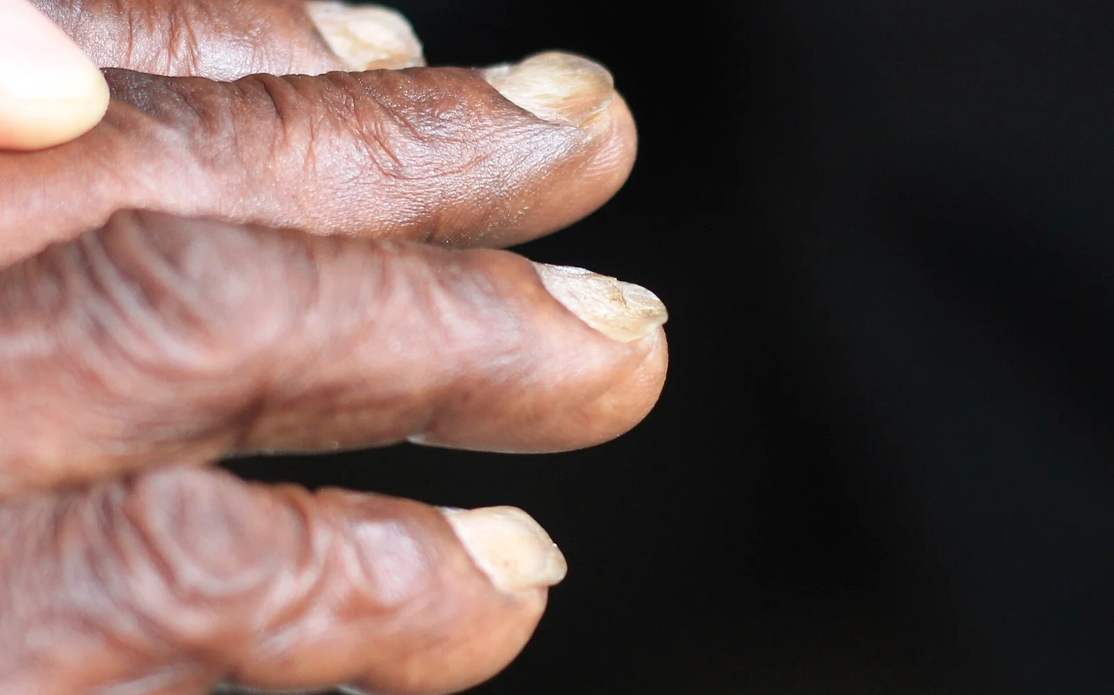
Figure 1Koilonychia iron deficiency anemia
Long description
Koilonychia in iron deficiency anaemia: thin, flattened fingernails that lose their normal curve.

Mild anaemia often produces no symptoms. Moderate and severe anaemia produces the symptoms common to anaemia of any cause: fatigue, breathlessness on exertion, dizziness, palpitations and reduced exercise tolerance. The signs of anaemia include pallor of the conjunctivae, face, nail beds and palmar creases; the absence of pallor does not exclude anaemia.[3] In older adults, anaemia is associated with worsening physical performance, cognitive function and frailty.[1]
Iron deficiency has its own features, which appear on top of the anaemia picture: angular stomatitis, glossitis, koilonychia (spoon-shaped nails), restless legs syndrome, pagophagia (a craving for ice), and blue sclerae.[1] These are the clues that a microcytic anaemia is iron-related rather than thalassaemia. Pica (eating non-food items) is also well described.
- Symptoms suggesting gastrointestinal cancer: weight loss, altered bowel habit, dysphagia, overt bleeding, or a change in stool calibre.[1]
- Haemolysis, with jaundice, dark urine or splenomegaly, points away from iron deficiency.
- Neurological symptoms raise vitamin B12 deficiency.
- Features of heart failure or angina, which lower the acceptable haemoglobin and push towards transfusion.
Long-standing iron deficiency can produce Plummer-Vinson syndrome (Paterson-Brown-Kelly syndrome in the UK): an extremely rare disorder of postcricoid dysphagia, iron deficiency anaemia and upper oesophageal webs, seen most often in middle-aged women. It is rare where nutritional status has improved. It is associated with iron-dependent epithelial atrophy and mucosal injury in the upper oesophagus and regression is often seen with iron repletion; webs may need endoscopic dilatation. Patients carry an increased risk of hypopharyngeal and upper oesophageal cancer, so long-term surveillance matters.[4]
The main question is whether an anaemia is iron deficiency, anaemia of chronic disease, thalassaemia trait, or another process. The table below uses the parameters that separate them best. Ferritin separates absolute iron deficiency from chronic disease when it is clearly low, but it is an acute-phase reactant and therefore loses specificity whenever infection or inflammation is present.[1][3]
| Parameter | Iron deficiency anaemia | Anaemia of chronic disease | Thalassaemia trait |
|---|---|---|---|
| MCV | Low, usually falling | Normal early, low late | Low, often out of proportion to the anaemia |
| MCH | Low early | Low late | Low |
| Red cell count | Normal or low | Normal or low | Normal, often raised (spurious polycythaemia) |
| RDW | Raised (over 15) | Normal | Normal |
| Serum iron | Low | Low | Normal |
| Transferrin (TIBC) | High | Low or normal | Normal |
| Transferrin saturation | Low | Low | Normal or raised |
| Ferritin | Low (under 15 at absent stores) | Normal or high | Normal |
| Serum ferritin response to inflammation | Can look normal despite empty stores | Raised | Not affected |
| Serum transferrin receptor | Raised | Normal | Raised |
Two further points from the same source material: heterozygous thalassaemia (trait) is the commonest cause of microcytosis after iron deficiency and has normal RDW, which is why measuring RDW plus iron studies usually separates the two without bone marrow.[3] Microcytosis with normal ferritin points towards hereditary microcytosis rather than iron deficiency, and at that point haemoglobin electrophoresis or genetic testing is the next step.[3] A low MCV alone is not enough to diagnose iron deficiency, and empirical iron can disguise a haemoglobinopathy and delay correct management.
Mixed states are common, especially combined iron deficiency and anaemia of chronic disease (with blood loss or inflammation on top of chronic disease), where ferritin can be low or normal and transferrin low.[3]
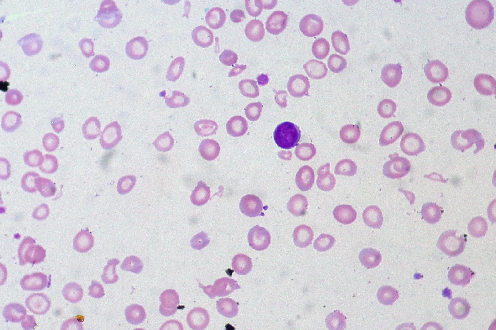
Figure 2Iron-deficiency Anemia, Peripheral Blood Smear (4422704616)
Long description
Peripheral blood smear in iron deficiency anaemia showing small pale (hypochromic microcytic) red cells and pencil-shaped cells.

- Full blood count with MCV, MCH, RDW and red cell count. MCV and MCH should be read together because they usually fall or rise in parallel.[3]
- Reticulocyte count, used both as an index (values roughly interpreted by the RPI) and as an absolute count. A high reticulocyte count in anaemia means haemolysis or bleeding; a low one means underproduction.
- Iron studies: serum iron, total iron binding capacity or transferrin, transferrin saturation, and ferritin.
- Serum ferritin: the single most useful marker of iron deficiency; a value under 15 µg/L means absent iron stores and under 30 µg/L generally means low body iron stores. A cut-off of 45 µg/L gives a specificity of about 0.92 and is the level at which investigation for iron deficiency should be seriously considered, especially when inflammation is present. A ferritin over 150 µg/L effectively excludes absolute iron deficiency even with inflammation.[1]
- Serum transferrin receptor and reticulocyte haemoglobin, where available, refine the picture in chronic kidney disease and inflammatory bowel disease.[1]
- Vitamin B12 and folate when any macrocytic element exists, because mixed deficiency is common.[3]
- Creatinine and liver function, and glucose when indicated.
A good response to iron therapy, defined as a haemoglobin rise of at least 10 g/L within 2 weeks, is highly suggestive of absolute iron deficiency even when iron studies are equivocal. A therapeutic trial of oral iron over a few weeks therefore doubles as a diagnostic test. Under-10 g/L rise after 2 weeks strongly predicts subsequent failure.[1]
The approach that separates production problems from loss or destruction is built on the reticulocyte count. Anaemia stimulates erythropoietin, so when the marrow can respond it should pour out young cells (regenerative anaemia): the RPI is above 3 and the cause is bleeding or haemolysis. When it cannot, RPI stays at or under 3 despite low haemoglobin (hypo-regenerative anaemia): deficiency, chronic disease, renal failure or marrow disease. Chronic haemorrhage is a twist worth remembering: prolonged bleeding depletes iron and converts a regenerative anaemia into a hypo-regenerative microcytic one.[3]
Approach to anaemia: production versus loss or destruction
- Anaemia confirmedHaemoglobin below the WHO cut-off; review RBC indices
- Question: Is the marrow responding? (reticulocyte production index)
Regenerative: RPI above 3
- Blood loss or haemolysisAcute or chronic bleeding, or red cell destruction. Look for the source and the trigger.
- If bleeding is prolongedChronic haemorrhage slowly becomes hypo-regenerative and microcytic as iron stores empty.
Hypo-regenerative: RPI under 3
- Question: Which cell size?
Microcytic (under 82 fL)
- Iron deficiency, thalassaemia trait, anaemia of chronic disease, sideroblastic anaemia
Normocytic (82-98 fL)
- Chronic disease, renal failure, marrow failure, mixed deficiency, early iron deficiency
Macrocytic (over 98 fL)
- Vitamin B12 or folate deficiency, liver disease, alcohol, drugs, myelodysplasia
The only diagnostic criteria for iron deficiency anaemia are: anaemia plus evidence of absent iron stores (ferritin under 15 µg/L) or a ferritin between 15 and 45 µg/L with supporting evidence (low transferrin saturation, high TIBC, high red cell transferrin receptor, raised percentage of hypochromic cells, or a haemoglobin rise of at least 10 g/L within 2 weeks of iron).[1] Staging is by severity of anaemia (under 80 g/L severe in adults) and by whether stores remain. Iron deficiency evolves through three stages: depleted stores with normal haemoglobin (non-anaemic), iron-restricted erythropoiesis with a falling MCV, and frank anaemia.
Every patient with confirmed iron deficiency anaemia needs a renal tract check, coeliac screening, and, where indicated, endoscopic evaluation. In men and postmenopausal women, gastroscopy and colonoscopy are generally first line; CT colonography is reasonable if colonoscopy is contraindicated. Where both procedures are performed at the same session, this is efficient and dual pathology is not rare. An underlying cancer is found in a small but important minority of cases.[1]
Iron replacement therapy aims to relieve symptoms, normalise haemoglobin and quality of life, replete marrow iron stores, and improve physiological function. Treatment and investigation proceed in parallel; oral iron is not deferred while awaiting investigation unless colonoscopy is within the next two weeks, because colonoscopy preparation requires pausing iron.[1]
The initial recommendation is one tablet daily of ferrous sulphate, ferrous fumarate or ferrous gluconate. Traditional salts are inexpensive, effective and safe, and they remain standard. Modified-release preparations release iron beyond the main absorption sites, do not enhance absorption or reduce side effects, and are not recommended. Ferric maltol is an alternative licensed preparation for iron deficiency of any cause.[1]
Suggested regimen details:
- One ferrous sulphate 200 mg tablet provides 65 mg elemental iron; one fumarate tablet provides 69 mg. The target daily dose is 50 to 100 mg elemental iron. [1]
- Take on a fasting stomach, first thing in the morning or between meals, because food cuts bioavailability by up to 75%. Avoid tea, coffee and calcium-rich foods around the dose; the tea effect fades within about an hour.
- Do not co-administer vitamin C: a large randomised trial showed no benefit for haemoglobin response, iron loading or tolerance.
- Expect a haemoglobin rise of at least 10 g/L within 2 weeks in responders, and a helpful rise within days.
- Monitor in the first 4 weeks. A rise of 20 g/L or into the normal range by 28 days is acceptable on lower or alternate-day regimens. A rise under 10 g/L at 2 weeks on daily dosing is strongly predictive of failure.[1]
Alternate-day regimens make physiological sense because hepcidin stays raised for 24 hours after a dose. Alternate-day dosing increases fractional and total iron absorption in iron-depleted women, and 200 mg elemental iron on alternate days gave almost twice the absorption of 100 mg on consecutive days. A randomised trial in iron deficiency anaemia found daily 60 mg twice daily raised haemoglobin faster than 120 mg on alternate days, but the two groups reached similar haemoglobin once the same total dose had been given, with less nausea on the alternate-day regimen. Once-daily or alternate-day lower dose iron is also better tolerated in older adults. Intermittent (less than daily) iron is at least as effective as daily dosing for raising haemoglobin in young women and pregnancy, with fewer gastrointestinal side effects.[1]
Gastrointestinal side effects (nausea, constipation, dyspepsia) are commoner with oral iron than with placebo and with parenteral iron, but discontinuation rates in trials are relatively low; modified-release preparations do not solve the problem. If standard salts are not tolerated, options are a reduced dose of one tablet every other day, ferric maltol, or parenteral iron.[1]
Treatment continues for about 3 months after haemoglobin normalises, to replete marrow stores; 2 months may suffice in someone who was almost iron replete, and longer is needed when blood loss, malabsorption or inflammation continues. Afterwards a blood count is monitored, perhaps every 3 months for a year then every 6 months. Ferritin need not be checked routinely once haemoglobin is normal.[1]
Intravenous iron replenishes stores faster but produces a similar haemoglobin rise in most patients, so the oral route is preferred on cost and convenience. Reserve intravenous iron for when oral iron is contraindicated, ineffective or not tolerated, and consider it early when oral therapy is unlikely to work or when rapid correction is needed: significant ongoing bleeding, malabsorption (gastrointestinal disease or post-bariatric surgery), combined iron deficiency and anaemia of inflammation, severe dysphagia, non-compliance, intolerance, or failure of a response at 2 to 4 weeks.[1]
Common preparations include ferric carboxymaltose, ferric derisomaltose (iron isomaltoside), iron sucrose and (low molecular weight) iron dextran, given in one or two infusions for the total-dose preparations and multiple infusions for sucrose. The Ganzoni formula (roughly 500 mg per every 10 g/L haemoglobin deficit plus stores) is the conventional dosing tool. Response is checked at 2 to 4 weeks, then as above. Intravenous iron is contraindicated in ongoing bacteraemia, deferred when infection is active, and requires precautions where hypersensitivity occurs; hypophosphataemia is commoner with ferric carboxymaltose.[1]
Blood transfusion is rarely needed. Most patients with slowly developing anaemia adapt, and intravenous iron produces a clinically meaningful rise within a week. Transfuse only those with severe symptomatic or circulatory compromise. Use packed red cells in accordance with local guidelines with a restrictive target haemoglobin of 70 to 90 g/L (80 to 100 g/L with unstable coronary artery disease). Each unit contains about 200 mg elemental iron, so a transfusion does not fix a large store deficit, and oral or intravenous iron must follow.[1]
Treatment of non-anaemic iron deficiency improves fatigue, mental and physical quality of life in symptomatic premenopausal women, and safe iron replacement can be offered where there are symptoms. In iron deficiency without anaemia, routine endoscopic investigation is not justified without additional pointers.[1]
- Pregnancy: mild deficiency is common; encourage oral iron and follow guidelines. If investigation is needed before delivery, follow the joint obstetric and gastroenterology guidance on which endoscopic and imaging tests are acceptable; colonoscopy is reserved for pressing indications only.[1]
- Young women: investigate only with red flags after coeliac screening. Recognise heavy menstrual loss as the commonest cause, and treat iron with attention to tolerance, adherence and re-checking haemoglobin after delivery or treatment of menorrhagia.
- Young men: investigate as for older patients, because pathology is common.[1]
- Older adults: deficiency is usually multifactorial (poor diet, reduced absorption, antiplatelet or anticoagulant blood loss, atrophic gastritis); imaging both the upper and lower gastrointestinal tract is more often justified.[1]
- Chronic kidney disease and chronic heart failure: iron deficiency coexists with functional deficiency; use specialist guidelines, because the ferritin and transferrin saturation thresholds that define iron deficiency in these conditions are different from those in the general population, and intravenous iron improves functional capacity where indicated.[1]
- Inflammatory bowel disease: one third of active disease is iron deficient; European guidelines cap oral iron at 100 mg elemental iron daily and recommend intravenous iron for moderate to severe anaemia (haemoglobin under 100 g/L) or intolerance.[1]
Iron deficiency that goes untreated produces anaemia with its own consequences: impaired concentration, reduced exercise tolerance, poorer cognition in older adults, heart failure in severe cases, and adverse maternal and fetal outcomes in pregnancy. Specific iron deficiency complications include Plummer-Vinson syndrome (above), restless legs and pica. Investigation itself carries risks if it is delayed (missed cancer) or done unnecessarily. Functional iron deficiency in chronic disease worsens prognosis in heart failure and chronic kidney disease.[1]
The WHO cut-offs for defining anaemia and its severity remain the standard frame of reference, and are especially useful where laboratories are scarce. Haemoglobin under 80 g/L is severe in adults and under 70 g/L severe in pregnancy; below these levels urgent action is needed.[2] Point-of-care haemoglobinometers allow testing in the field, and the result must be adjusted for altitude and smoking before cut-offs are applied.[2]
In resource-limited settings, the common causes shift: hookworm and other parasites, malaria where endemic, undernutrition, and (in women) frequent pregnancies and blood loss all dominate alongside the same diseases as elsewhere. Where only a blood count and basic iron studies are available, rely on ferritin (the best single test, with the caveat about inflammation), MCV, transferrin saturation where measurable, MCH and response to an oral iron trial. Where endoscopy is unavailable, treat identified deficiency, screen for coeliac disease where serology is accessible, eradicate parasites where hookworm is prevalent, optimise diet, and refer suspected serious pathology when facilities allow. Long-term intermittent iron programmes and fortification of staples are the public health answer, and the WHO framework on anaemia reduction describes the multisector plan.[2]
Practical details useful anywhere:
- Ferrous sulphate 200 mg once daily is cheap, effective taken fasted; where food must accompany it for tolerance, accept the bioavailability loss.
- Mobile or nurse-led anaemia pathways work well where GI endoscopy is distant.
- Pregnant women need particular attention because mild anaemia is common and both mother and fetus benefit from correction.
Pregnancy, young women, young men, older adults, chronic kidney and heart disease and inflammatory bowel disease are covered in Special situations. Children and adolescents are not covered in detail here: the same iron deficiency principles apply, but MCV reference ranges differ and inherited red cell disorders must be excluded.
- Thalassaemia and other haemoglobinopathies
- Anaemia of chronic disease
- Megaloblastic anaemia: vitamin B12 and folate deficiency
- Coeliac disease
- Colorectal cancer
- Chronic kidney disease
- Chronic heart failure
- Inflammatory bowel disease
- Transfusion and blood products
- Anaemia is haemoglobin below the age- and sex-appropriate normal range; WHO cut-offs are men under 130 g/L, non-pregnant women under 120 g/L and pregnant women under 110 g/L.
- One tablet daily of ferrous sulphate, fumarate or gluconate is the standard first treatment for iron deficiency anaemia; alternate-day dosing may be better tolerated with similar ultimate absorption.
- Sort anaemia by MCV: microcytic (under 82 fL), normocytic (82-98 fL), macrocytic (over 98 fL). Iron deficiency is the commonest cause worldwide and usual cause of microcytic anaemia.
- Then use the reticulocyte count: an RPI above 3 means responding marrow (bleeding or haemolysis); under 3 means underproduction (deficiency, chronic disease, renal or marrow disease).
- Iron deficiency's signature is low ferritin, high TIBC and low serum iron; anaemia of chronic disease is low or normal iron, low or normal transferrin, normal or high ferritin; thalassaemia trait has low MCV with normal iron studies and normal RDW.
- A ferritin under 15 µg/L means absent iron stores; 15 to 30 means depleted stores, and over 150 µg/L excludes absolute deficiency even with inflammation; 45 µg/L is the practical level at which to investigate.
- Oral iron should raise haemoglobin by at least 10 g/L within 2 weeks; a smaller rise predicts failure and should push you to parenteral iron.
- Treat for about 3 months after the haemoglobin normalises to replete stores.
- Transfuse only for severe symptoms or haemodynamic compromise, with a restrictive target haemoglobin of 70 to 90 g/L (80 to 100 g/L with unstable coronary artery disease).
- Investigate men and postmenopausal women with new iron deficiency anaemia with gastroscopy and colonoscopy first, after screening for coeliac disease.
- Plummer-Vinson syndrome is the rare dysphagia-iron deficiency-upper oesophageal web triad, with a small increase in cancer risk at those sites. [4]
- In low-resource settings use the WHO thresholds, a point-of-care haemoglobin value adjusted for altitude and smoking, and treat deficiency while looking for parasites, undernutrition and accessible gastrointestinal disease.
Answer each one in your head first, then open the answer.
A 62-year-old man has haemoglobin 98 g/L, MCV 76 fL, ferritin 8 µg/L and transferrin saturation 6%. He has no abdominal symptoms. What is the diagnosis, and what should happen next?
Show answerHide answer
Iron deficiency anaemia with empty stores. Next steps are a full history for blood loss and risk factors, urinalysis and coeliac screening, then gastroscopy plus colonoscopy as the first-line gastrointestinal evaluation, with treatment started in parallel.
How does a 70-year-old woman with iron deficiency anaemia, an MCV of 104 fL and no blood loss on history become iron deficient? Is that even possible?
Show answerHide answer
It is possible but should make you reconsider the diagnosis: an isolated iron deficiency anaemia is always microcytic, so an MCV of 104 fL means the anaemia is mainly macrocytic, for example vitamin B12 or folate deficiency. A low ferritin could coexist in a combined deficiency, classically in coeliac disease or autoimmune gastritis, but the blood count should be re-examined before labelling it simple iron deficiency.
A patient with rheumatoid arthritis has haemoglobin 102 g/L, MCV 84 fL, ferritin 85 µg/L and transferrin saturation 12%. Is this iron deficiency anaemia?
Show answerHide answer
No. These are classic for anaemia of chronic disease with possible functional iron restriction: ferritin is normal or raised and serum iron and transferrin saturation are low. Functional iron deficiency may still contribute, and a haemoglobin rise of at least 10 g/L after 2 weeks of oral iron, or reticulocyte haemoglobin, would help decide whether additional iron is useful. Check for active inflammation and coexisting true deficiency.
Why does alternate-day oral iron work at least as well as daily dosing in iron deficiency anaemia?
Show answerHide answer
A dose of iron raises hepcidin for about 24 hours, blocking absorption from a dose taken the next day. In iron-depleted women, 200 mg elemental iron on alternate days gave almost twice the overall absorption of 100 mg on consecutive days. In trials the daily group reached haemoglobin faster, but both reached similar haemoglobin once the same total dose was given, with less nausea on the alternate-day schedule.
A pregnant woman at 24 weeks has haemoglobin 100 g/L with microcytic hypochromic cells. She eats a poor iron diet. How do you manage her?
Show answerHide answer
Treat with oral ferrous sulfate, fumarate or gluconate daily, check tolerance, and re-check haemoglobin in 2 to 4 weeks expecting a rise of at least 10 g/L in 2 weeks. Look for and treat hookworm or parasitic infection where relevant. Reserve endoscopy for red flags; gastroscopy is considered safe and MR enterography can be used after the first trimester, while colonoscopy is reserved for pressing indications. [1]
A 35-year-old woman has a 3-month history of solids sticking in her throat, microcytic anaemia with ferritin 5 µg/L and upper oesophageal webs on barium swallow. What is the syndrome, and what two risks must you address?
Show answerHide answer
Plummer-Vinson syndrome: postcricoid dysphagia, iron deficiency anaemia and upper oesophageal webs, usually in middle-aged women. Treat the iron deficiency (iron repletion often relieves dysphagia) and dilate the web if it persists; arrange long-term surveillance because of the increased risk of hypopharyngeal and proximal oesophageal cancer.
- Snook J, Bhala N, Beales ILP, et al. British Society of Gastroenterology guidelines for the management of iron deficiency anaemia in adults. Gut 2021;70(11):2030-2051. doi:10.1136/gutjnl-2021-325210. Accessed .
- World Health Organization. Guideline on haemoglobin cutoffs to define anaemia in individuals and populations. Geneva: WHO; 2024. ISBN 978-92-4-008854-2. Accessed .
- Moreno Chulilla JA, Romero Colás MS, Gutiérrez Martín M. Classification of anemia for gastroenterologists. World J Gastroenterol 2009;15(37):4627-4637. Accessed .
- Kassama Z, Goosenberg E. Plummer-Vinson syndrome. In: StatPearls [Internet]. Treasure Island (FL): StatPearls Publishing; 2026. Accessed .
Figure credits
Educational content for medical students and doctors. It is not a substitute for clinical judgement or local guidelines.