Autoimmune haemolytic anaemia
Autoimmune haemolytic anaemia: warm IgG versus cold IgM, DAT patterns, blood film, treatment, Evans syndrome, low-resource care.
- Autoimmune haemolytic anaemia (AIHA) is destruction of the body's own red blood cells by autoantibodies; it is a relatively uncommon cause of anaemia with an annual incidence of 1-2 cases per 100,000 persons.[1]
- Warm AIHA (about two-thirds of cases) is driven by IgG that reacts at body temperature and destroys red cells mainly in the spleen (extravascular haemolysis); cold AIHA (15-20% of cases) is driven by IgM that activates complement and destroys cells in the circulation.[1] [2]
- Both are recognised by a positive direct antiglobulin test (DAT, "Coombs" test), but the pattern differs: warm usually IgG (with or without C3d); cold IgM leaves only C3d on the cells.[1]
- First-line treatment of warm AIHA is prednisone 1.0-1.5 mg/kg/day; rituximab is preferred for steroid-refractory warm disease and as first-line for cold agglutinin disease.[2][1]
- Splenectomy still has a place in warm disease but is used less often since rituximab; avoid cold exposure in cold antibody disease, and be cautious with transfusions.[2]
By the end of this page you should be able to:
- Explain the difference between warm (IgG, extravascular) and cold (IgM, complement-mediated) AIHA, including the associated conditions.
- Interpret the DAT pattern and blood film in each type.
- Investigate a patient with suspected AIHA and recognise Evans syndrome.
- Choose and dose first- and second-line treatment for warm and cold disease, and manage transfusions safely.
- Adapt care when tests, rituximab or complement inhibitors are not available.
DefinitionAutoimmune haemolytic anaemia (AIHA)
AIHA is anaemia caused by autoantibodies that bind to an individual's own red blood cells (RBCs) and accelerate their destruction. The antibody is usually present on essentially all RBCs, so the direct antiglobulin test (DAT) is the key serological test. AIHA may be primary (idiopathic) or secondary to another disease.[1]
AIHA is classified mainly by the thermal behaviour and class of the autoantibody:
- Warm AIHA: antibody most reactive at 37 degrees C; usually IgG.
- Cold antibody AIHA: antibody reactive in the cold (below 30 degrees C); usually IgM. This includes cold agglutinin syndrome (CAS), usually secondary to infection, and cold agglutinin disease (CAD), a chronic clonal B-cell disorder.
- Mixed AIHA: both a warm IgG and a cold IgM antibody coexist.
- Paroxysmal cold haemoglobinuria (PCH): a cold-reactive IgG that fixes complement and causes intravascular haemolysis.
- Drug-induced immune haemolytic anaemia and direct antiglobulin test-negative AIHA are also part of the spectrum.[1]
| Feature | Warm AIHA | Cold agglutinin disease / syndrome | Paroxysmal cold haemoglobinuria |
|---|---|---|---|
| Share of cases | About two-thirds[1] | 15-20%[1] | Rare, mainly children[1] |
| Antibody | IgG (rarely IgA/IgM) | IgM (polyclonal in CAS, monoclonal in CAD) | IgG, biphasic cold-reactive |
| Site of destruction | Extravascular (spleen, liver) | Intravascular and some extravascular | Intravascular |
| Blood film | Spherocytes common | Agglutination on the slide; spherocytes can occur | Red-cell phagocytosis by neutrophils may be seen |
| DAT | IgG, or IgG + C3d (C3d only in a minority) | C3d only | C3d only |
| Associated conditions | Lupus, CLL, lymphoma, drugs (e.g. methyldopa) | Mycoplasma (CAS), EBV (CAS); chronic monoclonal disease (CAD) | Viral/bacterial infections in children |
| Cold symptoms | No | Acrocyanosis, Raynaud, haemoglobinuria in cold | Haemoglobinuria after cold |
| First-line treatment | Corticosteroids | Rituximab, keep warm, complement inhibition | Supportive; self-limited |
The yearly occurrence of AIHA is about 1-2 cases per 100,000 persons.[1] Warm AIHA is the most common type (about two-thirds of cases), cold antibody disease is the second (15-20%), and mixed forms are third.[1] Most series report about two-thirds of adult cases as primary (idiopathic) and the rest as secondary.[1]
Overall outcome varies with the type. Mortality is low (around 4%) in children with isolated AIHA, but rises to around 10% when AIHA is combined with immune thrombocytopenia (Evans syndrome).[2] Warm IgM disease and mixed or atypical forms tend to run more severe courses.[2]
About half of warm AIHA has no identifiable cause; the rest are secondary to an underlying disease or drug.[1] Secondary warm disease is associated with autoimmune conditions (notably systemic lupus erythematosus), chronic lymphocytic leukaemia and non-Hodgkin lymphoma, other haematological malignancies and, less often, solid tumours.[1]
Cold agglutinin syndrome arises after infections, classically Mycoplasma pneumoniae pneumonia and Epstein-Barr virus infectious mononucleosis, and resolves as the infection clears.[1][3] Cold agglutinin disease (primary CAD) is a chronic clonal B-cell lymphoproliferative disorder producing a monoclonal IgM against the I/i red-cell antigen system.[1]
Drug-induced immune haemolytic anaemia is rare. Methyldopa is the prototype "true autoantibody" mechanism: it can induce an IgG autoantibody that mimics warm AIHA and persists for months after the drug is stopped.[1] Other drugs (penicillins, cephalosporins) more often act as haptens and cause acute intravascular haemolysis.[1]
Other risk factors include female sex (warm disease) and, for CAD, older age. A positive DAT alone is not enough: in one study 51% of DAT-positive patients did not have haemolysis, so confirm haemolysis before diagnosing AIHA.[1]
The clinical picture follows from which antibody is involved:
- IgG (warm AIHA) has a high affinity for the Fc receptors of splenic macrophages. Red cells are partially eaten ("biting"), losing membrane and becoming spherocytes, which are trapped in the spleen's red pulp. Haemolysis is therefore usually extravascular and the spleen is often enlarged.[1]
- IgM cold antibody activates the classical complement pathway efficiently. In cold agglutinin disease the intravascular haemolysis is complement-mediated, although some clearance of C3b-coated cells by hepatic Kupffer cells also occurs.[1]
- PCH uses an IgG "biphasic haemolysin" (the Donath-Landsteiner antibody): it binds red cells in the cold and fixes complement as the blood warms to 37 degrees C, causing intravascular haemolysis.[2][3]
Mixed AIHA has both components, so haemolysis is both intravascular and extravascular.[1]
A minority of patients have reduced marrow compensation (reticulocytopenia), reported in about 20% of adults and 39% of children; these patients can become profoundly anaemic very quickly.[2]
Symptoms depend on how fast haemolysis occurs and how well the marrow compensates. Common features are fatigue, dyspnoea, palpitations, pallor and jaundice (unconjugated hyperbilirubinaemia). Dark urine may signal intravascular haemolysis. In cold antibody disease, cold exposure provokes acrocyanosis, Raynaud phenomenon and sometimes gangrene; the urine may darken in the cold.[1]
Examination may show pallor, mild jaundice and splenomegaly (warm disease). Lymphadenopathy or hepatosplenomegaly suggests a secondary (especially lymphoproliferative) cause.
Severity ranges from mild compensated anaemia to life-threatening haemolysis. Rapid, severe courses are more common in mixed and atypical (including warm IgM) disease.[2]
AIHA must be separated from other causes of haemolysis (hereditary spherocytosis, G6PD deficiency, sickle disease, thrombotic microangiopathy, paroxysmal nocturnal haemoglobinuria) and from anaemia without haemolysis. The DAT helps: most other haemolytic anaemias are DAT-negative.
| Condition | Clue that separates it from AIHA |
|---|---|
| Hereditary spherocytosis | Family history, splenomegaly from childhood, DAT negative, osmotic fragility abnormal |
| G6PD deficiency | Oxidant trigger, bite cells on film, normal DAT, male sex more often |
| Paroxysmal nocturnal haemoglobinuria | Flow cytometry for CD55/CD59 deficiency, nocturnal haemoglobinuria |
| Thrombotic microangiopathy (TTP/HUS) | Schistocytes, low platelets, normal DAT, multi-organ ischaemia |
| Mechanical prosthetic valve haemolysis | Schistocytes, known valve, normal DAT |
| Infection-related haemolysis (e.g. malaria) | Travel, smear, DAT negative (unless mixed) |
Figure 1Coombs test schematic
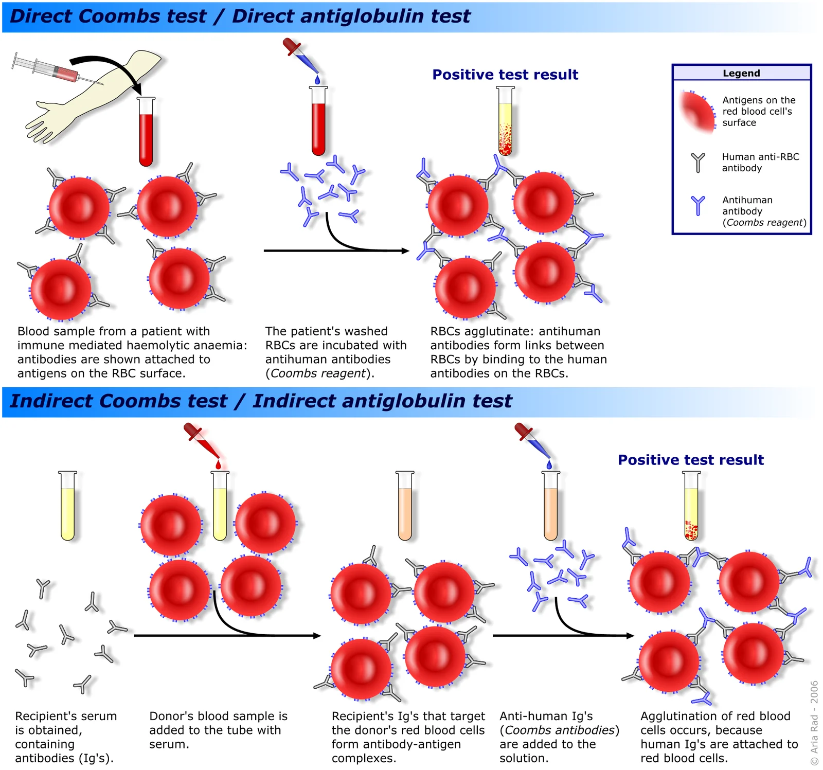
Long description
Schematic of the direct antiglobulin (Coombs) test, which detects antibodies already bound to the patient's red cells, and the indirect test, which detects antibodies in the patient's serum; agglutination after adding anti-human globulin is a positive result.

Confirm haemolysis first: falling haemoglobin, high LDH, high unconjugated bilirubin, low haptoglobin, and reticulocytosis (though 20% of adults and 39% of children have reticulocytopenia).[2]
Direct antiglobulin test (DAT) is the central test. Use a polyspecific reagent first (anti-IgG plus anti-C3d), then monospecific reagents:
- Warm AIHA: IgG alone (about 67%), IgG plus C3d (about 20%), or C3d only (about 13%).[1]
- Cold antibody AIHA: C3d only.[1]
- Mixed AIHA: IgG plus C3d, with a cold IgM component.[1]
- PCH: C3d only; the diagnosis is established with the Donath-Landsteiner test (biphasic haemolysis of P-antigen-positive cells).[1][2]
- DAT-negative AIHA: 3-10% of cases, diagnosed by exclusion; treat as warm AIHA.[3]
The antibody may appear in the serum and eluate; in cold disease the eluate is negative because IgM is free in plasma and only C3d remains on the cells.
Blood film. Warm disease often shows spherocytes (membrane loss from macrophage "biting"). Cold disease shows red-cell agglutination on the slide, sometimes only in the cooler parts of the smear; spherocytes can also appear. In PCH, erythrophagocytosis by neutrophils may be seen. Confirm by repeating the film after warming the sample to 37 degrees C when cold agglutination is suspected.
Cold agglutinin titre (tested at 4 degrees C) is 64 or higher in cold antibody disease, with a thermal amplitude up to 30 degrees C.[1]
Other tests. A detailed drug history; testing for lupus, CLL/lymphoma when secondary warm disease is suspected; infection screening for CAS (Mycoplasma/EBV). Cross-matching studies (warm autoadsorption or allogeneic adsorption) detect alloantibodies, which occur in 12-40% of AIHA patients.[2]
Therapy setting. Mixed AIHA accounts for 7-8% of cases and is marked by severe haemolysis (haemoglobin under 6 g/dL at onset is common in mixed and atypical forms) with a chronic relapsing course.[2] [1]
Approach to suspected AIHA
- Confirm haemolysislow haemoglobin, high LDH, bilirubin and reticulocytes, low haptoglobin
- Exclude other causesinherited haemolysis, TTP, PNH, mechanical valves
- DAT (polyspecific)anti-IgG plus anti-C3d
- Question: DAT result?
Positive
- Monospecific DAT: anti-IgG and anti-C3d
- Question: Pattern
IgG (or IgG + C3d)
- Warm AIHAscreen for lupus, CLL, lymphoma, drugs
C3d only
- Question: Antibody type
IgM cold agglutinin
- CAS or CAD
Biphasic IgG (Donath-Landsteiner)
- PCH
IgG + C3d plus cold IgM
- Mixed AIHA
Negative
- DAT-negative AIHAtreat as warm AIHA after excluding other causes
There is no single score; the diagnosis rests on:
- Evidence of haemolysis (biochemical and/or reticulocytosis).
- A positive DAT (or the rare DAT-negative form).
- Exclusion of other causes of haemolysis.
- Identification of the antibody type (temperature, class, thermal amplitude) and of any secondary cause.
Severity is judged on the haemoglobin, rate of fall, reticulocyte response and symptoms. Patients with haemoglobin below 6 g/dL, reticulocytopenia or intravascular haemolysis are emergencies.[2]
Corticosteroids are first-line.[2] Give prednisone (or prednisolone) 1.0-1.5 mg/kg/day (about 1 mg/kg/day in most adult regimens) for 1-3 weeks until the haemoglobin exceeds 10 g/dL.[2] Expected response is 70-85%; long-term remission after stopping the drug occurs in only about one in three, half need maintenance doses, 20-30% need second-line therapy, and fewer than 20% are cured by steroids alone.[2]
Corticosteroid taper is slow: 10-15 mg reduction per week to 20-30 mg/day, then 5 mg every 1-2 weeks to 15 mg, then 2.5 mg every 2 weeks, with at least 3-4 months at 10 mg/day or less before stopping.[2] Patients on prolonged steroids need bisphosphonates, vitamin D, calcium and folic acid.[2]
In severe haemolysis or Evans syndrome, use intravenous methylprednisolone 100-200 mg/day for 10-14 days, or 250-1000 mg/day for 1-3 days.[2]
Rituximab (375 mg/m^2 weekly for 4 weeks) is the preferred second-line drug for warm AIHA and is increasingly used before splenectomy; overall response is about 83-87% (complete response 54-60%), with disease-free survival of 72% at one year and 56% at two years.[2] Retreatment can be effective.[2] In Evans syndrome, rituximab gives an overall response of about 83% (66% complete), up to 94% in newer series.[2] Side effects include infusion reactions and a small risk (about 7%) of infection, including hepatitis B reactivation; give antiviral prophylaxis when appropriate.[2]
Splenectomy remains a second-line option for warm AIHA: a partial or complete remission is obtained in about two-thirds of patients (38-82%), with a presumed cure rate of up to 20%.[2] Its success cannot be predicted from steroid response or scan findings. Drawbacks are surgical complications (laparoscopic 0.5-1.6% versus conventional surgery about 6%) and overwhelming post-splenectomy sepsis (risk 3.3-5%, mortality up to 50%) from encapsulated organisms.[2] Vaccinate against pneumococcus, meningococcus and Haemophilus influenzae type b at least two weeks before surgery.[2][4] Rituximab is now often preferred before splenectomy, especially in severe disease or young children.[2]
Other second-line options (azathioprine 100-150 mg/day, cyclophosphamide 100 mg/day, cyclosporine, mycophenolate mofetil) are used in refractory disease;[2] intravenous immunoglobulin has limited, short-lived efficacy (response about 40%, only 15% reach haemoglobin 10 g/dL or higher; better in children at 54%).[2] Plasma exchange is a temporising measure in severe warm AIHA but is category III/last-ditch. Haematopoietic stem-cell transplantation is reserved for refractory disease.
General care. Avoid cold exposure; keep the patient warm (room temperature 37-40 degrees C, warm clothing, warm intravenous fluids and blood warmed before transfusion). Low-titre, asymptomatic CAD often needs only cold protection and occasional transfusion.[2] [1]
CAD requiring therapy. Rituximab is now recommended as first-line, alone or with fludarabine or bendamustine; anti-complement therapy with sutimlimab blocks the classical pathway; eculizumab blocks terminal complement; daratumumab is an option for relapsed disease.[1][2]
Sutimlimab (ENJAYMO) for adults with CAD: 6,500 mg intravenously if body weight is 39 to less than 75 kg, or 7,500 mg if 75 kg or more; weekly for the first two weeks, then every two weeks. Vaccinate against encapsulated bacteria at least two weeks before starting.[4] It reduces transfusion dependence and rapid intravascular haemolysis.
Cold agglutinin syndrome is usually self-limited once the infection (Mycoplasma, EBV) resolves; supportive care and warmth suffice. In severe cases sutimlimab has been used.[1]
PCH. Usually self-limited in children after viral infection. Severe cases may need transfusion; steroids may be tried but effectiveness is hard to judge because the haemolysis is transient. Eculizumab was ineffective in one steroid-refractory case with myeloma. Plasma exchange and rituximab have been used in severe disease.[2]
Stop the suspected drug. Hapten (drug-dependent) haemolysis is acute and intravascular; methyldopa-type haemolysis is extravascular, can persist for months after stopping the drug, and is treated like warm AIHA if severe.[1]
Evans syndrome is the simultaneous or sequential combination of AIHA with immune thrombocytopenia (and sometimes autoimmune neutropenia).[3] It behaves more severely than isolated AIHA, with mortality around 10% in children.[2] Treat as warm AIHA (corticosteroids, then rituximab), with added attention to bleeding and the need for earlier second-line therapy; response to rituximab is good (around 83%, 66% complete).[2]
Transfusion practice. Never withhold RBC transfusion from a critically ill AIHA patient because of incompatibility: warm autoantibodies are often pan-reactive and transfused blood survives similarly to the patient's own cells.[2] Prefer ABO- and RhD-matched units, and give them when alloantibodies are reasonably excluded (alloantibodies occur in 12-40% of AIHA).[2] In less urgent cases, extended phenotyping and cross-matching are advisable; complex adsorption studies may be needed.[2] Transfuse slowly when possible (not more than 1 mL/kg/h) and avoid overtransfusion, particularly in older patients. Use leukodepleted cells. A rise in haemolysis after transfusion should prompt a search for an unsuspected alloantibody, not automatic blame on AIHA.[2] [1]
In CAD specifically, keep the patient and the infusion limb warm, use an in-line blood warmer, avoid cold intravenous fluids and high-plasma products.[2]
- Overwhelming sepsis from encapsulated organisms after splenectomy (risk 3.3-5%, mortality up to 50%).[2]
- Thromboembolism and pulmonary hypertension after splenectomy.[2]
- Severe haemolysis with haemoglobinuria, pigment nephropathy and renal strain.
- Cold injury: acrocyanosis, Raynaud and (rarely) gangrene in cold antibody disease.[1]
- Treatment complications: hepatitis B reactivation and rare progressive multifocal leukoencephalopathy with rituximab; long-term steroid toxicity.[2]
- Warm AIHA responds to first-line corticosteroids in 70-85% of patients, but only about one-third remain in long-term remission after stopping the drug; fewer than 20% are cured by steroids alone.[2]
- Rituximab maintains disease-free survival of about 72% at one year and 56% at two years.[2]
- Splenectomy gives remission in about two-thirds, with a presumed cure rate up to 20%.[2]
- CAD is chronic; rituximab gives an overall response of about 58% (complete response about 4.5%).[2]
- PCH is usually self-limited, especially in children.
- Overall mortality is low in children (around 4%) but rises to around 10% with Evans syndrome.[2] Mixed/atypical forms and severe intravascular haemolysis carry the worst prognosis.[2]
Children. Many childhood cases are post-infectious and transient; CAS from Mycoplasma/EBV is the classic example. Reticulocytopenia is commoner in children (39%) and worsens urgency. Mortality in isolated childhood AIHA is about 4%, rising to about 10% with Evans syndrome; splenectomy and rituximab have particular roles in severe disease.[2]
Pregnancy. Management should be multidisciplinary (haematology, obstetrics, transfusion medicine). Corticosteroids and IVIG are generally preferred first-line; rituximab and splenectomy are usually reserved for refractory disease. Transfusion and fetal monitoring are tailored to severity.
Older adults and frail patients. Comorbidity (especially cardiac and pulmonary disease) lowers the threshold to transfuse and favours avoiding prolonged high-dose steroids; early rituximab may be safer than splenectomy. Complement inhibitors (sutimlimab) are an important option in CAD.
Access to tests and targeted drugs is uneven. Where resources are limited: diagnose with basic haemolysis markers (LDH, bilirubin, haptoglobin, reticulocyte count) and the DAT; rely on a careful drug and infection history; keep treatment simple with corticosteroids as the mainstay; protect patients with cold antibody disease from cold (a cheap, effective intervention); transfuse only when necessary and follow strict cross-match and infusion-rate safety; and build referral links to centres able to deliver rituximab, splenectomy or complement inhibitors.[5]
- AIHA is a relatively uncommon (1-2 per 100 000 per year) anaemia from autoantibodies against RBCs; roughly two-thirds is warm IgG disease.[1]
- Warm (IgG, extravascular) versus cold (IgM, complement) mechanisms explain the clinical findings, blood film and treatment.
- A positive DAT is essential, but interpretation depends on whether it is IgG, C3d or both; PCH needs the Donath-Landsteiner test.[1][2]
- Warm disease: first-line prednisone 1.0-1.5 mg/kg/day for 1-3 weeks; slow taper; severe cases IV methylprednisolone.[2]
- Second-line for warm disease: rituximab 375 mg/m^2 weekly for 4 weeks; splenectomy in selected patients after vaccination.[2]
- Cold disease: avoid cold; rituximab first-line; sutimlimab dosed by weight for CAD.[1][4]
- Transfuse safely: ABO/Rh matched, exclude alloantibodies (12-40%), slow infusion (not more than 1 mL/kg/h), avoid overtransfusion.[2]
- Evans syndrome is AIHA plus ITP, more severe, with higher mortality.[2][3]
- Low-resource care centres on simple diagnostics, steroids, cold protection and referral networks.[5]
Answer each one in your head first, then open the answer.
A 55-year-old woman has fatigue and jaundice. Haemoglobin is 78 g/L, LDH raised, haptoglobin low, and the DAT is positive for IgG only. What type of AIHA is most likely, and what is the usual first treatment?
Show answerHide answer
Warm AIHA, driven by IgG causing extravascular haemolysis in the spleen. First-line treatment is oral prednisone (or prednisolone) 1.0-1.5 mg/kg/day for 1-3 weeks until haemoglobin exceeds 10 g/dL, with a slow taper. Look for secondary causes such as lupus, CLL and lymphoma. [2]
A 25-year-old man develops severe anaemia after Mycoplasma pneumonia. His DAT is positive for C3d only and his blood film shows red-cell agglutination. What type of AIHA is this, and how does management differ from warm disease?
Show answerHide answer
Cold agglutinin syndrome (secondary to infection). It is IgM-mediated, complement-driven and self-limited. Keep the patient warm, avoid cold exposure and transfuse carefully with a blood warmer; steroids are not first-line, unlike warm AIHA. Rituximab is considered if severe haemolysis persists.
Which DAT patterns are expected in warm AIHA, cold agglutinin disease, mixed AIHA and paroxysmal cold haemoglobinuria?
Show answerHide answer
Warm AIHA: IgG alone (about 67%), IgG plus C3d (20%) or C3d only (13%). Cold antibody AIHA: C3d only. Mixed AIHA: IgG plus C3d. PCH: C3d only, diagnosed with the Donath-Landsteiner test.
A patient with warm AIHA needs red cells urgently but no compatible unit can be found. What should you do?
Show answerHide answer
Do not withhold transfusion in a critical situation. Give ABO- and RhD-matched red cells (warm autoantibodies are pan-reactive and transfused cells survive similarly), with alloantibodies reasonably excluded; transfuse slowly (not more than 1 mL/kg/h) and avoid overtransfusion. Seek leucodepleted units.
A woman presents with AIHA and a platelet count of 15 x 10^9/L. What syndrome does this suggest, and which treatment is particularly effective?
Show answerHide answer
Evans syndrome (AIHA plus immune thrombocytopenia). It is more severe, with mortality around 10% in children. Corticosteroids are first-line; rituximab is particularly effective (overall response about 83%, complete response about 66%). [2]
What dose and schedule of sutimlimab is used for cold agglutinin disease in adults?
Show answerHide answer
6,500 mg intravenously if body weight is 39 to less than 75 kg, or 7,500 mg if 75 kg or more: weekly for the first two weeks, then every two weeks. Vaccinate against encapsulated bacteria at least two weeks beforehand.
Name one key intervention for AIHA care in low-resource settings that costs little and is highly effective.
Show answerHide answer
Cold protection for cold agglutinin disease (keeping the patient warm and transfusing warmed blood). Simple, cheap measures and clear referral pathways make the biggest difference.
What is the most feared complication of splenectomy in AIHA, and what preventative step must precede it?
Show answerHide answer
Overwhelming sepsis from encapsulated organisms (risk 3.3-5%, mortality up to 50%). Vaccinate against pneumococcus, meningococcus and Haemophilus influenzae type b at least two weeks before surgery, and consider antibiotic prophylaxis.
Warm AIHA and its associations: systemic lupus erythematosus; chronic lymphocytic leukaemia; non-Hodgkin lymphoma; drug-induced immune haemolytic anaemia. Related blood disorders: disseminated intravascular coagulation; thrombotic thrombocytopenic purpura and haemolytic uraemic syndrome; sickle cell disease; haemophilia and bleeding; von Willebrand disease. Emergency evaluation of anaemia and jaundice appears in the approach-to-anaemia and liver-related topics. No topic links are embedded here; navigate via the haematology hub.
- Loriamini M, Cserti-Gazdewich C, Branch DR. Autoimmune Hemolytic Anemias: Classifications, Pathophysiology, Diagnoses and Management. Int J Mol Sci. 2024;25(8):4296. PMCID:PMC11049952. Accessed .
- Zanella A, Barcellini W. Treatment of autoimmune hemolytic anemias. Haematologica. 2014;99(10):1547-1554. PMCID:PMC4181250. Accessed .
- Berentsen S, et al. The choice of new treatments in autoimmune hemolytic anemia: how to pick from the basket?. Front Immunol. 2023. PMCID:PMC10165002. Accessed .
- Recordati Rare Diseases Inc. ENJAYMO (sutimlimab-jome) prescribing information. US Food and Drug Administration, 2022. Accessed .
- Various authors. Management of autoimmune haemolytic anaemia in low-to-middle income countries: current challenges and the way forward. PMC. 2024. PMCID:PMC11004394. Accessed .
Figure credits
Educational content for medical students and doctors. It is not a substitute for clinical judgement or local guidelines.