Haemolytic anaemias: spherocytosis, G6PD deficiency and PNH
How red cells are destroyed, how to recognise haemolysis, and the causes, tests and treatments for spherocytosis, G6PD deficiency and PNH.
- A haemolytic anaemia is any anaemia caused by red blood cells (RBCs) being destroyed faster than the bone marrow can replace them. The marrow compensates, so reticulocytes rise.
- The first move is not the diagnosis but the proof of haemolysis: raised reticulocytes, raised lactate dehydrogenase (LDH), raised unconjugated bilirubin, low or absent haptoglobin, and a characteristic blood film, with the direct antiglobulin test (DAT) to separate immune from non-immune causes.
- Haemolysis is either intravascular (cells rupture in the circulation, free haemoglobin, shock-wave low haptoglobin and haemoglobinuria) or extravascular (macrophages in the spleen and liver remove damaged cells, bilirubin and splenomegaly dominate).
- The three named conditions are hereditary (membrane defects, G6PD deficiency) or acquired complement-mediated (PNH). All three share markers of haemolysis; they are separated by the blood film, special tests and history.
- Spherocytosis damages the membrane (ankyrin and spectrin); G6PD deficiency is an X-linked enzyme defect unmasked by oxidant stress (drugs, infection, fava beans); PNH is an acquired defect in the PIGA gene that leaves cells without CD55 and CD59, so complement attacks them. [1]
- Treatment is cause-specific: folate and splenectomy for spherocytosis; trigger avoidance and supportive care for G6PD deficiency; complement inhibition (eculizumab or ravulizumab) for PNH, plus thrombosis prevention where needed. [6]
By the end of this page you should be able to:
- Define haemolysis and state the laboratory pattern that proves it.
- Distinguish intravascular from extravascular haemolysis and give the classic examples of each.
- Explain the pathophysiology of hereditary spherocytosis, G6PD deficiency and PNH, and relate each to its blood film.
- Plan the investigation of a haemolytic anaemia, including DAT, osmotic fragility and eosin-5-maleimide (EMA) binding, G6PD assay timing, and CD55/CD59 flow cytometry.
- State the management of each condition, including the indication for splenectomy, trigger avoidance in G6PD deficiency, and the dosing and safety monitoring of eculizumab and ravulizumab.
- Recognise the red flags that suggest haemolysis and the low-resource alternatives when specialised tests are unavailable.
DefinitionHaemolytic anaemia
An anaemia in which the circulating lifespan of red blood cells is shortened and the bone marrow cannot fully compensate, so anaemia develops. It is defined in the laboratory by evidence of accelerated red cell destruction together with a compensatory rise in erythropoiesis, not by anaemia alone.
Haemolysis is not a single disease. It is a final common pathway produced by damage that occurs either inside the red cell (intrinsic defects, almost always inherited in this topic) or outside it (extrinsic, acquired). Intrinsic causes include abnormalities of the membrane (hereditary spherocytosis), the enzymes (G6PD deficiency), and the haemoglobin (sickle cell disease, thalassaemia, not covered here). Extrinsic causes include immune attack (autoimmune or drug-induced haemolysis), mechanical shearing (microangiopathy, prosthetic valves), infection (malaria) and oxidant stress.
A normal red cell survives about 120 days in the circulation.[1] When the lifespan falls below the marrow's capacity to replace cells, anaemia results. Haemoglobin released from destroyed cells is processed in two ways: in the spleen and liver macrophages (extravascular) or directly into plasma (intravascular), and this determines the clinical and laboratory picture.
- Hereditary spherocytosis affects up to one person in 2,000 in many populations, and about 1 in 2,000 to 1 in 3,000 in Caucasian populations.[1][2] It is the most common inherited red cell membrane disorder.
- G6PD deficiency affects more than 400 million people worldwide, making it the most common human enzyme deficiency. In some populations, up to one person in ten is affected; prevalence exceeds 20% in parts of western and southern Africa and is above 10% in many parts of Asia.[1] Prevalence in the Americas is under 5%.[1]
- PNH is an ultra-rare disease. Clinically significant PNH is quoted at roughly 0.5 to 1.5 cases per million, while the prevalence of any clone size (including tiny clones found in people with bone marrow failure) is far higher, about 15.9 per million in Europe.[3]
- G6PD deficiency and sickle-cell trait overlap geographically with endemic malaria. PNH occurs at any age, but usually presents in adults in their 30s; men and women are equally affected.[3]
Figure 1Hereditary Spherocytosis smear 2010-03-17
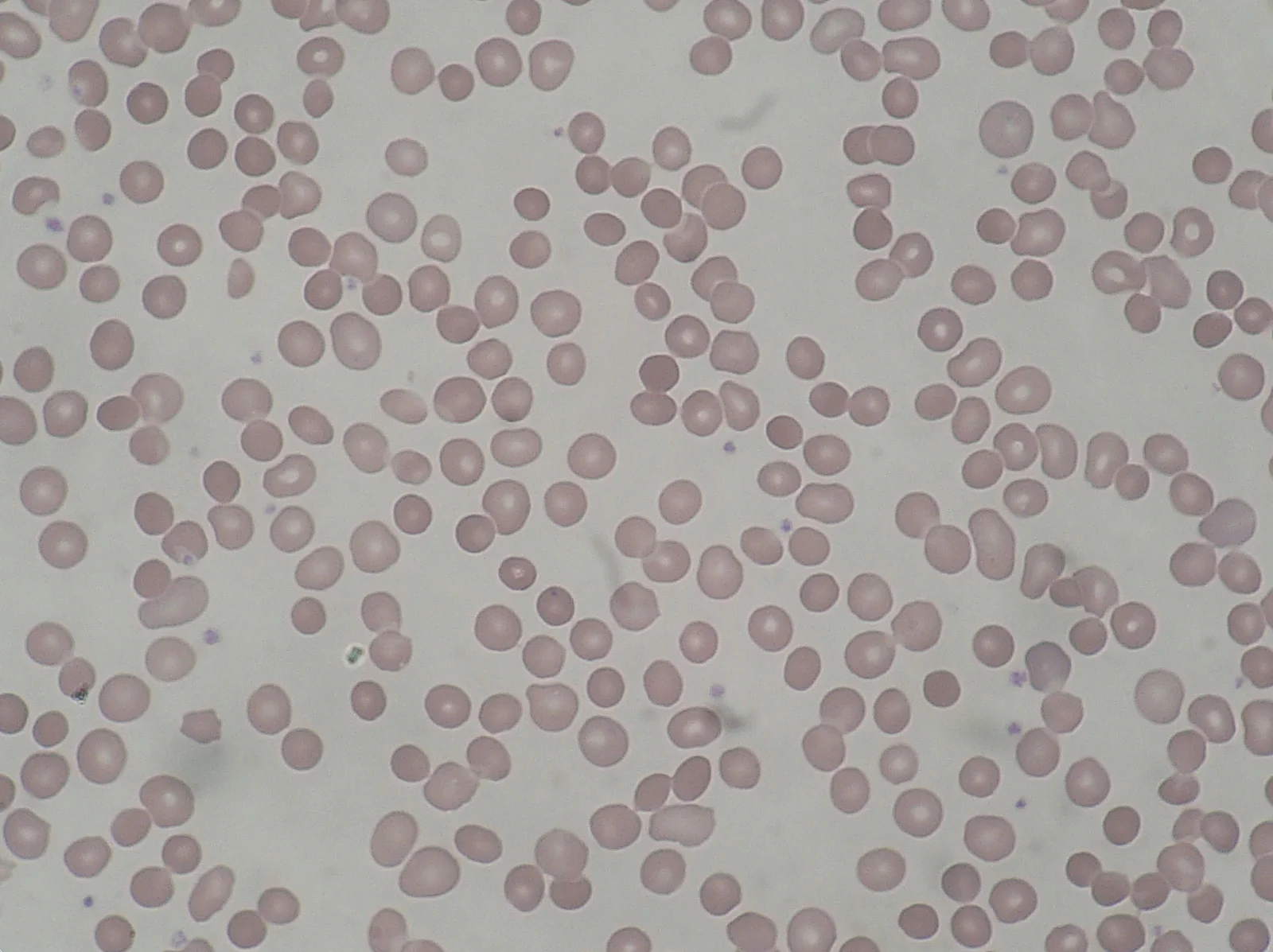
Long description
Blood smear in hereditary spherocytosis: many small, dense, round red cells without central pallor (spherocytes).

- Inheritance is autosomal dominant in about 75% of cases, most often involving mutations in ANK1, SLC4A1 (band 3) and SPTB; de novo mutations are common. The remaining cases are autosomal recessive, often biallelic defects in SPTA1 or EPB42.[2]
- The result is loss of membrane surface area. The cell becomes a sphere (a spherocyte) with a decreased surface-to-volume ratio, and is trapped in the splenic red pulp.
- X-linked recessive. The gene sits on the X chromosome, so a male with one mutant allele is deficient, and a female carrier has two populations of cells. Because PNH and X-linked PIGA mutations are acquired, not inherited, the inheritance pattern differs entirely.
- G6PD sits in the pentose phosphate pathway and is the main generator of NADPH in the red cell. NADPH maintains glutathione in its reduced form, which protects the cell against oxidative damage. Red cells have no mitochondria and no alternate source of NADPH, so they are entirely dependent on G6PD.[1]
- Triggering oxidants overwhelm the reduced glutathione reserve and cause haemoglobin denaturation and Heinz body formation. Triggers include fava beans, infection (the commonest trigger, especially pneumonia, hepatitis A and B, and cytomegalovirus), and drugs: sulphonamides, nitrofurantoin, dapsone, primaquine, aspirin, and many others (including acetaminophen, chloramphenicol, vitamin C, fluoroquinolones).[1] Over 400 G6PD variants are recognised; the G6PD A-variant is less sensitive to fava bean-induced stress than the Mediterranean variant.[1]
- PNH is an acquired clonal disorder. Mutations arise in the PIGA gene on the X chromosome (position p22.2). Because PIGA mutations are acquired after X-inactivation, men and women are equally affected.[3]
- PIGA encodes the first enzyme in glycosylphosphatidylinositol (GPI) anchor synthesis. Without a functioning GPI anchor, the red cell surface lacks CD55 and CD59, both of which are regulators of complement. CD55 restrains complement activation at C3b; CD59 blocks terminal complement activation and membrane attack complex formation. Their absence allows uncontrolled complement activation and haemolysis.[3]
- PNH arises on a background of bone marrow failure, so every patient with aplastic anaemia or hypoplastic myelodysplastic syndrome should be screened.[3]
- The red cell is damaged faster than the marrow can replace it.
- In extravascular haemolysis, damaged or rigid red cells are recognised by macrophages in the splenic red pulp and to a lesser extent the liver, where they are phagocytosed. Haemoglobin is broken down to unconjugated bilirubin, iron is recycled, and the spleen enlarges. The blood film shows spherocytes; reticulocytes rise.
- In intravascular haemolysis, the cell membrane ruptures inside the circulation, releasing free haemoglobin into the plasma. Free haemoglobin is filtered by the kidneys (haemoglobinuria) and scavenged by haptoglobin (which becomes very low or undetectable). LDH is released from the red cell cytoplasm and rises markedly. Bilirubin rises later. [1]
- In G6PD deficiency the attack is oxidative: denatured haemoglobin precipitates as Heinz bodies, splenic macrophages remove the Heinz bodies ("bite cells"), and the cell may rupture in the circulation, so both intravascular and extravascular haemolysis occur. [1]
- In PNH the attack is complement-mediated inside the circulation: CD55 and CD59 are absent, the membrane attack complex punches holes in the cell, and free haemoglobin is released. This is predominantly intravascular haemolysis.
- The marrow responds to shortened red cell survival by increasing erythropoiesis; the reticulocyte count rises within days of an acute haemolytic insult. [1]
Clinical clues: jaundice (usually with unconjugated hyperbilirubinaemia), dark urine, pallor, tachycardia, splenomegaly (extravascular), fatigue; in PNH, abdominal pain, dysphagia or erectile dysfunction from smooth muscle relaxation failure caused by nitric oxide scavenging by free haemoglobin.
The laboratory proof of haemolysis is a cluster of findings, not a single test:
- Raised reticulocytes (the marrow's compensation).
- Raised LDH (released from destroyed red cells).
- Raised unconjugated (indirect) bilirubin, with normal or near-normal liver enzymes and no obstruction.
- Low or undetectable haptoglobin (it is consumed binding free haemoglobin).
- The blood film gives the diagnosis in many cases: spherocytes, bite cells, schistocytes, sickle cells.
- Direct antiglobulin test (DAT, direct Coombs test) separates immune haemolysis (positive DAT) from the intrinsic and mechanical causes (negative DAT).
Approach to suspected haemolysis
- Anaemia with jaundice or dark urineCheck reticulocytes, LDH, unconjugated bilirubin, haptoglobin, blood film
- Prove haemolysisReticulocytes raised; LDH raised; unconjugated bilirubin raised; haptoglobin low
- Question: Direct antiglobulin test (DAT)?
Positive
- Immune haemolysisWarm or cold autoimmune haemolysis, or drug-induced; look for trigger and underlying cause
Negative
- Question: Blood film?
Spherocytes
- Hereditary spherocytosis (or membrane defect)Osmotic fragility, EMA binding, genetic panel
Bite cells or blister cells
- G6PD deficiencyG6PD enzyme assay after recovery; oxidant history
Schistocytes
- Microangiopathic haemolysisTTP, HUS, DIC, malignant hypertension; mechanical valve if prosthetic
Normal/minimal change
- Consider PNHCD55/CD59 flow cytometry with FLAER; test also for aplastic anaemia or MDS
- Management by cause, then treat complications
| Marker | What it indicates |
|---|---|
| Reticulocytes | Raised in every haemolytic anaemia as the marrow compensates. |
| LDH | Raised; particularly high in intravascular haemolysis, especially PNH. |
| Unconjugated bilirubin | Raised from macrophage breakdown of haemoglobin; liver enzymes remain normal. |
| Haptoglobin | Low or undetectable in intravascular haemolysis; may be normal in pure extravascular haemolysis. |
| Blood film | Spherocytes, bite cells, schistocytes, target cells and sickle cells point to specific diagnoses. |
| Direct antiglobulin test | Positive in immune haemolysis; negative in intrinsic membrane, enzyme, haemoglobin and mechanical causes. |
| Feature | Intravascular | Extravascular |
|---|---|---|
| Site of destruction | Inside the circulation | Macrophages in spleen and liver |
| Main mechanism | Membrane rupture (complement, mechanical shearing) | Phagocytosis of damaged or rigid cells |
| LDH | Markedly raised | Raised, usually less extreme |
| Haptoglobin | Very low or absent | Low or normal |
| Plasma haemoglobin / haemoglobinuria | Present | Absent |
| Bilirubin (unconjugated) | Raised, often later | Raised |
| Blood film | Schistocytes, bite cells possible | Spherocytes |
| Spleen | Normal size (unless underlying disorder) | Enlarged |
| Classic examples | PNH, mechanical haemolysis, severe microangiopathy, G6PD (partly) | Hereditary spherocytosis, warm autoimmune haemolysis, sickle cell disease, thalassaemia |
- Age at presentation is variable. Neonatal jaundice is common; the patient may present later with asymptomatic splenomegaly, anaemia, pigmented gallstones or an acute haemolytic crisis during infection.[1]
- Splenomegaly is usual. Jaundice is mild and chronic, often deepening with infection.
- The blood film shows spherocytes, but spherocytes are not specific to this disorder: they also appear in warm autoimmune haemolysis and in other red cell injury. An elevated mean corpuscular haemoglobin concentration (MCHC), above 36.5 g/dL, is a strong pointer.[1][2]
- Gallstones at a young age are a hallmark clue.
- Most people are asymptomatic. Haemolysis is triggered by oxidative stress: drugs (sulphonamides, nitrofurantoin, dapsone, primaquine and most sulfa drugs), infection and fava beans.[1]
- Episodic acute intravascular haemolysis with jaundice, dark urine and abdominal pain; sometimes severe anaemia requiring transfusion, with haemoglobin falling under 8 g/dL in the dramatic episodes.[1]
- Neonatal hyperbilirubinemia can be severe.
- The blood film (during a crisis) shows Heinz bodies on supravital stain and bite cells (degmacytes) on the routine film; blister cells may appear.[1]
- Splenomegaly can occur during an acute attack.
- Classic chronic intravascular haemolysis with marked LDH elevation; haemoglobinuria, which may be episodic and visible; anaemia and reticulocytosis; low haptoglobin.[3] [1]
- Haemoglobin 20 g/L or less below normal is common; the severity varies.
- Symptoms from smooth muscle dystonia (nitric oxide scavenged by free haemoglobin): abdominal pain, oesophageal spasm with dysphagia, and erectile dysfunction.[3]
- Fatigue, breathlessness and pallor dominate.
- Venous and arterial thrombosis, often at unusual sites (hepatic, portal, splenic, splanchnic, cerebral), is a major cause of morbidity and was the leading cause of death before complement inhibitors.[3] 70 to 80% of thromboses are venous and 20 to 30% arterial, with deep venous thrombosis, pulmonary embolism and intra-abdominal events particularly represented.[3]
- Bone marrow failure often co-exists; pancytopenia should prompt screening.
- All PNH patients with an acquired Coombs-negative haemolytic anaemia picture should be considered, even with minor haemolysis.
- A fall in haemoglobin with a sharply rising LDH and a vanishing haptoglobin.
- Dark urine in a patient on a new drug or with fever.
- Neurological symptoms, chest pain or haematuria in possible microangiopathy or thrombosis.
- Any thrombosis at an unusual site without another explanation.
The full haemolytic anaemia differential is wide; the blood film is the most useful sorting tool. Mechanisms can be grouped as intrinsic (membrane, enzyme, haemoglobin defects) and extrinsic (immune, mechanical, infectious, and oxidant causes).[1]
| Condition | Category | Mechanism | Location | Diagnostic test |
|---|---|---|---|---|
| Microangiopathic haemolysis | Extrinsic | Mechanical shearing | Intravascular | Blood film, coagulation screen |
| Warm autoimmune haemolysis | Extrinsic | IgG-mediated opsonisation | Extravascular (spleen) | Positive DAT |
| Hereditary spherocytosis | Intrinsic | Membrane defect (ankyrin/spectrin) | Extravascular (spleen) | Osmotic fragility, EMA binding |
| Thalassaemia | Intrinsic | Globin gene mutation | Extravascular (spleen) | Haemoglobin analysis |
| Sickle cell disease | Intrinsic | Haemoglobin S polymerisation | Extravascular | Haemoglobin analysis |
| G6PD deficiency | Intrinsic | Oxidative stress denatures haemoglobin | Both (intra and extra) | G6PD assay after recovery |
| PNH | Intrinsic (acquired) | Complement attack on CD55/CD59-deficient cells | Intravascular | CD55/CD59 flow cytometry |
| Mechanical (prosthetic valve, microangiopathy) | Extrinsic | Mechanical shearing | Intravascular | Blood film, echo/doppler |
| Malaria | Extrinsic/intrinsic | Parasite invasion and splenic clearance | Both | Thick and thin films |
The DAT is negative in all of these intrinsic, mechanical and infectious causes except warm autoimmune haemolysis, which is the common mimic that must be excluded early.
- Full blood count and reticulocyte count.
- Peripheral blood film reviewed by an experienced haematologist.
- LDH, total and direct (conjugated) bilirubin, haptoglobin.
- DAT.
- Folate and vitamin B12 levels (chronic haemolysis depletes folate).
- Hereditary spherocytosis. Osmotic fragility (fresh and incubated) and EMA binding by flow cytometry, which measures fluorescence of a dye intercalating into membrane proteins; fluorescence is reduced when membrane proteins (usually ankyrin and spectrin) are defective.[1][2] Ektacytometry (osmotic gradient deformability) is used in specialist centres, and a genetic panel can confirm the variant. An MCHC above 36.5 g/dL is highly suggestive.[1]
- G6PD deficiency. Direct enzyme assay by spectrophotometric measurement of NADPH formation is the gold standard; a fluorescent spot test is the usual screen. Assay several months after an acute episode, because recently haemolysed cells have better enzyme activity and can produce a falsely normal result.[1] DNA testing for known mutations is useful in female heterozygotes or borderline results.
- PNH. High-sensitivity flow cytometry on peripheral blood, assessing CD55 and CD59 on red cells (plus FLAER on granulocytes and monocytes, which is the most sensitive approach, with CD24, CD66b and CD16 on neutrophils and CD14 on monocytes). Because haemolysis selectively removes GPI-anchor-deficient red cells, the white cell clone is more informative in haemolytic PNH, and a bone marrow examination is needed only with concurrent cytopenias.[3]
- Other causes. Thick and thin films (and rapid testing) for malaria; echocardiography and Doppler for a prosthetic valve; ADAMTS13 and coagulation profile for microangiopathy; hepatitis serology and autoantibody panel where infection or autoimmunity is suspected.
Two points matter in practice. First, no single test diagnoses hereditary spherocytosis with certainty: the EMA test and osmotic fragility are both used because sensitivity varies with genotype. Second, in acute G6PD haemolysis the enzyme assay is misleading for weeks afterwards, so the diagnosis is often clinical and confirmed later.
PNH is classified by haemolytic intensity, bone marrow findings and clone size:[3]
- Classic (haemolytic) PNH: florid intravascular haemolysis (markedly abnormal LDH, sometimes visible haemoglobinuria), a cellular marrow with erythroid hyperplasia, and a large clone of PNH cells.
- PNH in the setting of bone marrow failure: mild haemolysis, a hypoplastic or dysplastic marrow, and usually a small clone (under 10%).
- Subclinical PNH: no clinical or biochemical haemolysis; a tiny clone (under 1%) in a person with bone marrow failure. [3]
A raised LDH above 1.5 times the upper limit of normal counts as high disease activity, and unexplained thrombosis or cytopenias with a negative DAT should prompt testing even when haemolysis is absent.[3]
Hereditary spherocytosis severity (by haemoglobin) is useful for deciding on splenectomy: asymptomatic (male haemoglobin above 14 g/dL, female above 12), mild (above 11 g/dL), moderate (8 to 11 g/dL) and severe (under 8 g/dL).[2] [4]
G6PD variants are grouped by the World Health Organization into classes by residual enzyme activity and clinical effect: the most severe variants cause chronic haemolysis, the Mediterranean variant causes intermittent severe acute haemolysis, the African A- variant causes milder intermittent haemolysis, and the mildest groups cause no clinical problem.[1]
- Treat the patient, not the number: oxygen, hydration and transfusion when the haemoglobin falls under 8 g/dL or symptoms of inadequate oxygen delivery appear.[1]
- Stop the offending drug or remove the oxidant trigger.
- Investigate and treat infection.
- Folate supplementation during the recovery phase, because chronic haemolysis consumes folate.
- Folate supplementation is standard in chronic haemolysis to support the increased erythropoietic drive.
- Splenectomy removes the principal site of destruction and raises the haemoglobin. It is reserved for moderate to severe disease, transfusion dependence, disabling symptoms or complications, and is weighed carefully against the lifelong risk of overwhelming post-splenectomy infection and thrombosis. Subtotal or partial splenectomy is increasingly favoured in children and is associated with better retention of splenic immune function and a lower thrombotic risk.[4]
- Cholecystectomy when gallstones are present, often at the same operation.
- Vaccination against pneumococcus, Haemophilus influenzae type b and meningococcus, plus antibiotic prophylaxis after splenectomy, follows national asplenia guidance.
- Genetic counselling and family testing; male and female lines are both affected in autosomal dominant disease, so a parent with mild disease may be identified only after the child presents.
- Remove the trigger: stop the drug, treat the infection, avoid fava beans.
- Supportive care: fluids, rest; transfuse for severe or symptomatic anaemia, particularly haemoglobin under 8 g/dL with evidence of organ hypoxia.[1]
- Long-term management is trigger avoidance. Affected individuals and carriers need a written list of drugs to avoid (sulphonamides, nitrofurantoin, dapsone, primaquine and related oxidants) and should be warned about fava beans and mothballs containing naphthalene.[1]
- Newborns with neonatal jaundice need phototherapy or exchange transfusion as clinically indicated; early recognition matters.
- Screening is not uniform internationally, despite the World Health Organization recommendation in populations with prevalence above 5%.
- Indications for complement inhibition include PNH-related thrombosis (a medical emergency), symptomatic intravascular haemolysis with anaemia and raised LDH above 1.5 times the upper limit of normal, PNH complications (renal impairment, pulmonary hypertension), and pregnancy with a clone above 20% (eculizumab).[3]
- Eculizumab (Soliris), intravenous: 600 mg weekly for the first 4 weeks, then 900 mg at week 5, then 900 mg every 2 weeks. It reduces haemolysis and transfusion requirement.[5]
- Ravulizumab (Ultomiris), intravenous, weight-based, with longer-acting maintenance every 4 or 8 weeks depending on body weight (for example, 2,400 mg loading then 3,000 mg every 8 weeks for 40 to 59 kg; 2,700 mg then 3,300 mg every 8 weeks for 60 to 99 kg; 3,000 mg then 3,600 mg every 8 weeks for 100 kg or more, with smaller children dosed by weight).[6]
- Both agents block terminal complement (C5), not the underlying clone. Monitoring during treatment uses LDH, haemoglobin, reticulocyte count, renal and liver function and clone size.
- Vaccination against meningococcus (serogroups A, C, W, Y and B) at least 2 weeks before the first dose is mandatory with both agents; both carry a boxed warning for meningococcal infection and are available only through a restricted programme.[5][6] Urgent treatment with antibiotic prophylaxis is given when vaccination is incomplete.
- Thrombosis is treated with anticoagulation, and complement inhibitor therapy should not alter chronic anticoagulation management on its own. Central lines and thrombosis at unusual sites require urgent haematology input.
- Bone marrow failure is treated on its own terms (immunosuppression or transplant), even when a PNH clone is present. Medication changes or stopping a complement inhibitor require close monitoring because haemolysis returns.
- PNH clone size is monitored regularly, more often in the first year after diagnosis and when the clone is changing or immunosuppressive therapy is given; follow the local haematology protocol.[3]
- Autoimmune haemolysis. Treatment differs for warm and cold antibody disease and follows local haematology guidance, with corticosteroids, rituximab or splenectomy in selected cases.
- Microangiopathic haemolysis. Treat the cause: plasma exchange for thrombotic thrombocytopenic purpura, supportive care and control of hypertension for haemolytic uraemic syndrome, withdrawal of causative drug (e.g. quinine, ciclosporin).
- Mechanical haemolysis from a prosthetic valve: optimise valve position and flow, treat iron deficiency (which worsens the anaemia), and consider surgical revision if haemolysis is severe or progressive.
- Malaria: antimalarial therapy treats both the infection and the haemolysis; transfuse for severe anaemia.
- Oxidant drugs (dapsone, sulphonamides, primaquine, nitrofurantoin): stop the drug and screen for G6PD deficiency before re-exposure.
- Gallstones at a young age in hereditary spherocytosis.
- Iron overload from chronic extravascular haemolysis and transfusion.
- Thrombosis in PNH, including hepatic and portal vein thrombosis and pulmonary embolism; thrombosis accounts for 22 to 67% of deaths in the pre-complement era and 23 to 30% cumulative incidence over 8 to 10 years.[3]
- Infection and sepsis after splenectomy, and meningococcal infection during complement inhibitor therapy.
- Renal impairment from chronic haemoglobinuria in PNH.
- Post-splenectomy vascular events, including pulmonary hypertension, a documented long-term risk after splenectomy in hereditary spherocytosis.
- Hereditary spherocytosis is compatible with a normal lifespan; splenectomy cures the haemolysis but trades it for infection and thrombosis risk.
- G6PD deficiency carries an excellent prognosis once triggers are avoided; the dangerous period is the acute haemolytic crisis.
- PNH had a median survival of 10 to 22 years before eculizumab; with complement inhibition, life expectancy in countries with treatment availability has nearly normalised.[3]
- No population screening for hereditary spherocytosis; an MCHC above 36.5 g/dL on a routine blood count should prompt testing.[1]
- G6PD deficiency screening is not in the routine neonatal panel in most countries; the World Health Organization recommends it where prevalence exceeds 5%. Africa and the Middle East, where prevalence is highest, have the lowest screening rates.[1]
- Newborn screening for sickle cell disease covers the commonest haemoglobinopathy in many countries but not all.
- PNH should be screened for in every patient with aplastic anaemia or hypoplastic myelodysplastic syndrome, and in all DAT-negative haemolytic anaemias and unexplained thrombosis at unusual sites.[3]
- Avoiding oxidants prevents most G6PD crises.
- Asplenic patients need vaccination and antibiotic cover, and families should carry a patient alert card.
- Neonates. Hereditary spherocytosis and G6PD deficiency both present with severe unconjugated hyperbilirubinaemia; early phototherapy and exchange transfusion follow local jaundice guidelines.
- Pregnancy. PNH clone above 20% is a complement inhibitor indication (eculizumab); pregnancy itself worsens haemolysis.
- Older adults. Mechanical haemolysis from a prosthetic valve is more common with age, and cardiovascular disease may limit transfusion tolerance.
- Children. Partial splenectomy is preferred over total splenectomy when possible, to reduce infection risk while improving haemolysis.
- Where laboratory capacity is limited, the proof of haemolysis is still available: anaemia with a raised reticulocyte count, raised LDH and low haptoglobin together establish it. Unconjugated bilirubin and urine urobilinogen are widely measurable.
- The blood film remains the cheapest diagnostic tool and is available wherever a microscope and Romanowsky stain exist.
- Osmotic fragility testing with a simple hypotonic saline series and an EMA test need flow cytometry, which may not exist: in that case the combination of spherocytes, a family history and a raised MCHC supports the diagnosis of spherocytosis, and treatment proceeds on clinical grounds.
- G6PD screening by the fluorescent spot test is cheap and field-usable; where MCHC enzyme assay is unavailable, a clinical diagnosis after an oxidant exposure plus cessation of the drug is lifesaving.
- PNH diagnosis needs high-sensitivity flow cytometry and is often unavailable outside referral centres; treatment with complement inhibitors is also unavailable or unaffordable in many settings. In those circumstances, meticulous support (transfusion, anticoagulation for thrombosis, and iron chelation) plus referral to a national haematology centre is the realistic plan.
- Splenectomy for severe hereditary spherocytosis is a definitive and relatively low-technology operation, but vaccination, antibiotic prophylaxis and long-term infection awareness are essential.
- Blood transfusion access, rather than specialist testing, is often the binding constraint in severe haemolytic crises.
- Haemolytic anaemia is anaemia from premature red cell destruction; it is proven by raised reticulocytes, raised LDH, raised unconjugated bilirubin and low haptoglobin, with a characteristic blood film.
- Intravascular haemolysis (PNH, mechanical shearing, microangiopathy) produces the sharpest rises in LDH, low haptoglobin and haemoglobinuria.
- Extravascular haemolysis (hereditary spherocytosis, warm autoimmune, thalassaemia, sickle cell disease) dominates the bilirubin and splenomegaly.
- Hereditary spherocytosis is a membrane defect (ankyrin, spectrin, band 3, protein 4.2), producing osmotically fragile spherocytes destroyed in the spleen, diagnosed by osmotic fragility and EMA binding, and treated with folate and, for severe disease, splenectomy.
- G6PD deficiency is an X-linked enzyme defect of the pentose phosphate pathway, leaving red cells unable to handle oxidant stress; Heinz bodies and bite cells appear in crises, and trigger avoidance is the whole of long-term management.
- PNH is an acquired PIGA mutation leaving cells without CD55 and CD59, so complement attacks them in the circulation; thrombosis is common and complement inhibition (eculizumab, ravulizumab) transforms prognosis. [3]
- A negative DAT is required to call an intrinsic, mechanical or infectious haemolysis, and excludes warm autoimmune haemolysis.
- Always check folate and B12 in chronic haemolysis.
- The flowchart, tables and the low-resource section above are the practical core of this topic.
Answer each one in your head first, then open the answer.
A 24-year-old woman has pallor, jaundice and dark urine. Her haemoglobin is 7.2 g/dL, reticulocytes 12%, LDH 1,800 U/L, unconjugated bilirubin 4.1 mg/dL and haptoglobin undetectable. The DAT is negative. What diagnosis is most likely and which single confirmatory test best supports it?
Show answerHide answer
PNH. The picture is acute intravascular haemolysis with a negative DAT. Support it with high-sensitivity flow cytometry showing CD55/CD59 deficiency (FLAER on granulocytes/monocytes) and a high LDH above 1.5 times normal.
Why must a G6PD enzyme assay be delayed after a haemolytic crisis?
Show answerHide answer
The cells that remain after the crisis are the ones with relatively better enzyme activity, so testing during or shortly after the episode can give a falsely normal result. Reassay weeks to months later, once a new cohort of red cells has entered the circulation.
A 3-year-old child presents with neonatal jaundice treated with phototherapy, anaemia, splenomegaly and pigment gallstones. The MCHC is 38 g/dL and the blood film shows spherocytes. What is the diagnosis and what is the role of folate?
Show answerHide answer
Hereditary spherocytosis. Folate supplementation supports the increased red cell production in chronic haemolysis and prevents megaloblastic worsening.
Which two complement regulators are missing on the surface of PNH red cells, and what does each normally do?
Show answerHide answer
CD55 and CD59. CD55 restrains complement activation at C3b; CD59 blocks terminal complement activation and membrane attack complex formation. Their absence lets complement punch holes in the cell.
A patient with PNH-related thrombosis is started on a complement inhibitor. State the eculizumab induction and maintenance schedule and the mandatory safety steps before the first dose.
Show answerHide answer
Eculizumab 600 mg intravenously weekly for 4 weeks, 900 mg at week 5, then 900 mg every 2 weeks. Vaccination against meningococcus serogroups A, C, W, Y and B at least 2 weeks before the first dose; antibiotic prophylaxis if urgent treatment cannot wait; enrolment in the restricted programme; meningococcal safety counselling and a patient safety card. [5]
Why are spherocytes found in both hereditary spherocytosis and warm autoimmune haemolysis, and how is the distinction made?
Show answerHide answer
Both diseases remove membrane surface and reduce the surface-to-volume ratio. Hereditary spherocytosis gives a positive osmotic fragility and reduced EMA binding with a negative DAT; warm autoimmune haemolysis gives a strongly positive DAT and is not associated with a raised MCHC above 36.5 g/dL.
- Aplastic anaemia and bone marrow failure syndromes (when PNH co-exists)
- Thalassaemia and sickle cell disease (intrinsic haemoglobin defects)
- Autoimmune haemolytic anaemia and the direct antiglobulin test
- Malaria (intravascular and extravascular haemolysis)
- Iron-deficiency anaemia (chronic mechanical haemolysis)
- Splenectomy, asplenia and infection prophylaxis
- Bussing K, Brehl N, Inman A, Davidson D. Educational Case: Glucose-6-phosphate dehydrogenase (G6PD) deficiency anemia. Academic Pathology 2026;13(3):100282. doi:10.1016/j.acpath.2026.100282. Accessed .
- Zaninoni A, Fermo E, Vercellati C, et al. Decoding the Ektacytometric Landscape of Hereditary Spherocytosis: Insights from 204 Cases. Diagnostics (Basel) 2026;16(14):2257. doi:10.3390/diagnostics16142257. Accessed .
- Griffin M, Kelly R, Bradley C, et al. Integrating paroxysmal nocturnal hemoglobinuria (PNH) into mainstream hematological curriculum training. HemaSphere 2026;10(10):e70484. doi:10.1002/hem3.70484. Accessed .
- Turpaev K, Bovt E, Shakhidzhanov S, et al. An overview of hereditary spherocytosis and the curative effects of splenectomy. Frontiers in Physiology 2025;16:1497588. doi:10.3389/fphys.2025.1497588. Accessed .
- Alexion Pharmaceuticals Inc. (prescribing information). SOLIRIS (eculizumab) injection, solution, concentrate. DailyMed U.S. National Library of Medicine, label updated 2026-07-08. setid ebcd67fa-b4d1-4a22-b33d-ee8bf6b9c722. Accessed .
- Alexion Pharmaceuticals Inc. (prescribing information). ULTOMIRIS (ravulizumab-cwvz) injection, solution, concentrate. DailyMed U.S. National Library of Medicine, label updated 2025-10-09. setid a9a590d9-0217-43c7-908d-e62a71279791. Accessed .
Figure credits
Educational content for medical students and doctors. It is not a substitute for clinical judgement or local guidelines.