Normal haemostasis and the coagulation cascade
How blood stops bleeding: vessel spasm, platelet plug, coagulation cascade, natural anticoagulants, fibrinolysis, and the tests used.
- Haemostasis is the body's way of stopping bleeding from a damaged vessel, using vasoconstriction, a temporary platelet plug and then a solid fibrin clot.
- Platelets stick to exposed subendothelium through von Willebrand factor and the GPIb receptor, then activate and clump through fibrinogen bound to GPIIb/IIIa.
- Plasma coagulation builds fibrin in a cascade (intrinsic, extrinsic and common pathways) and is nowadays described more accurately by a cell based model built around thrombin.
- Natural anticoagulants (antithrombin, protein C and protein S, tissue factor pathway inhibitor) and fibrinolysis (plasmin) keep the clot local and temporary.
- Common tests map onto specific steps: platelet count and PFA-100 on primary haemostasis; PT on the extrinsic pathway; aPTT on the intrinsic pathway; thrombin time and fibrinogen on the common end; D-dimer on fibrinolysis.
By the end of this page you should be able to:
- Describe the four stages of haemostasis after a vessel injury, in order.
- Explain platelet adhesion, activation and aggregation, naming the receptors and ligands involved.
- List the clotting factors of the intrinsic, extrinsic and common pathways and state which are vitamin K dependent.
- Contrast the classical cascade with the cell based model of coagulation.
- Describe the natural anticoagulants and the fibrinolytic pathway, including D-dimer formation.
- Choose the right screening and confirmatory test for a bleeding problem and interpret a prolonged PT or aPTT.
- Outline a safe minimum work up for a prolonged clotting time in a low resource setting.
Haemostasis (from the Greek for stopping bleeding) is the set of processes that keep the circulation intact after a vessel is injured. It is often split into primary haemostasis (the vessel wall and platelets) and secondary haemostasis (the plasma coagulation cascade), followed by resolution of the clot. This page covers the normal physiology of each step and the laboratory tests that reflect them. Companion topics on von Willebrand disease and haemophilia build on this one.
Figure 1Blood clot formation
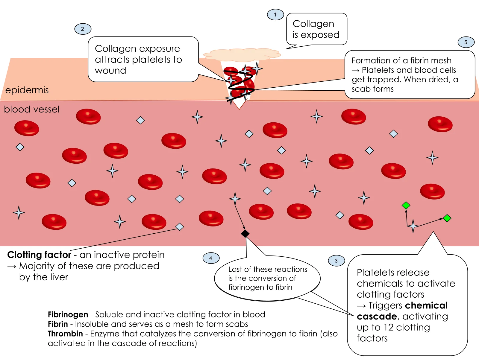
Long description
Blood clot formation in simple steps: a wound exposes collagen, platelets stick and are activated, they release chemicals that trigger the clotting cascade, and fibrinogen is converted to fibrin, whose mesh traps platelets and blood cells to form a clot.

When a vessel is cut or its endothelium damaged, the vessel narrows (vasoconstriction) and the normally hidden subendothelial matrix is exposed to the blood. Endothelins and thromboxane help this constriction. Vasoconstriction alone cannot seal even a small wound; it only buys seconds while platelets arrive. At the same time the endothelial cells release von Willebrand factor (vWF), which tethers platelets and also carries coagulation factor VIII. [1]
The first contact between a circulating platelet and the injured wall is between platelet GPIb (part of the GPIb/IX/V complex) and vWF that has stuck to exposed collagen; this interaction is strengthened by high shear stress, which is why it matters particularly in arterioles. The platelet then grips the collagen firmly through GPVI and integrin alpha2 beta1. Without this sequence, as in Bernard-Soulier syndrome where the GPIb/IX/V complex is deficient, a platelet plug cannot start even when the platelet count is normal. [1]
Adhesion plus local agonists (ADP, thromboxane A2, serotonin and thrombin itself) change the platelet from a resting disc to a spiky, spreading cell. Intracellular calcium rises, granules move to the surface and release their contents: ADP, serotonin, platelet factor 4, beta-thromboglobulin, thrombospondin, fibronectin, vWF and platelet derived growth factor. These substances recruit and activate more platelets, amplify the signal (ADP through P2Y1 and P2Y12 receptors), enhance vasoconstriction (thromboxane A2), stabilise the developing mass (thrombospondin) and promote later vessel repair. [1]
The critical event is activation of the platelet GPIIb/IIIa receptor (integrin alpha IIb beta 3). In its resting state this receptor cannot bind fibrinogen; signalling from inside the platelet (inside-out signalling, driven by calcium and the agonists above) changes its shape so that it binds fibrinogen with high affinity. Fibrinogen is bivalent, so one molecule bridges two platelets by their GPIIb/IIIa receptors and the mass grows. vWF can substitute for fibrinogen, especially under high shear. Early on the plug is fragile; once thrombin is generated, fibrin reinforces it into a durable clot. [1][2]
Exam trapPrimary haemostasis can fail with a normal platelet count
Glanzmann thrombasthenia (defective or absent GPIIb/IIIa) and Bernard-Soulier syndrome (defective GPIb/IX/V) both produce mucocutaneous bleeding with a normal platelet count. A low platelet count is not the only, or even the commonest, cause of failure of primary haemostasis. [1]
Figure 2Coagulation Cascade and Major Classes of Anticoagulants
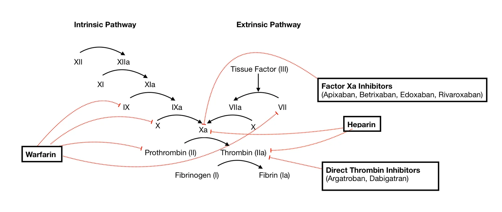
Long description
The coagulation cascade with its intrinsic pathway (XII, XI, IX), extrinsic pathway (tissue factor and VII) and common pathway (X, prothrombin, thrombin, fibrinogen to fibrin), and where warfarin, heparin, factor Xa inhibitors and direct thrombin inhibitors act.

Plasma coagulation is a chain of zymogen activations. Enzymes (mostly serine proteases) activate the next enzyme in the chain until thrombin converts fibrinogen to fibrin. The cascade is traditionally divided into three pathways. In the body, tissue factor is the trigger; the contact activation pathway contributes little to normal haemostasis (people with severe factor XII deficiency do not bleed abnormally). [1][2]
| Factor | Name | Pathway | Vitamin K dependent |
|---|---|---|---|
| I | Fibrinogen | Common | No |
| II | Prothrombin | Common | Yes |
| III | Tissue factor (tissue thromboplastin) | Extrinsic | No |
| IV | Calcium ions | All steps | No |
| V | Proaccelerin (labile factor) | Extrinsic and intrinsic | No |
| VI | (unused, historical name for Va) | - | - |
| VII | Proconvertin (stable factor) | Extrinsic | Yes |
| VIII | Antihemophilic factor A (vWF carrier) | Intrinsic | No |
| IX | Antihemophilic factor B (Christmas factor) | Intrinsic | Yes |
| X | Stuart-Prower factor | Extrinsic and intrinsic (common) | Yes |
| XI | Antihemophilic factor C | Intrinsic | No |
| XII | Hageman factor (contact activation) | Intrinsic (initiates in vitro) | No |
| XIII | Fibrin stabilising factor | Common (cross links fibrin) | No |
Notes on the table: factors II, VII, IX and X are synthesised in the liver in inactive forms and need vitamin K for gamma-carboxylation; protein C and protein S, the natural anticoagulants discussed below, are also vitamin K dependent. Factor VIII is not vitamin K dependent but its plasma half life depends on von Willebrand factor. [2]
Steps in brief:
- Extrinsic pathway. Tissue factor, exposed on subendothelial cells outside the vessel, binds factor VII and activates it (VIIa); the TF-VIIa complex directly activates factor X. This arm completes within seconds. [1][2]
- Intrinsic pathway. Contact with negatively charged surfaces (collagen, or glass in the laboratory) activates XII, then XI, then IX in complex with its cofactor VIIIa (activated by thrombin), which activates factor X. This arm completes in minutes. [1][2]
- Common pathway. Factor Xa with its cofactor Va forms the prothrombinase complex, which converts prothrombin (II) to thrombin (IIa). Thrombin converts fibrinogen (I) to fibrin, and activates factor XIII to XIIIa, which cross links the fibrin into a stable mesh. [1][2]
The classical cascade explains the laboratory tests well but not how clotting actually happens in a living person. The cell based model puts tissue factor and thrombin at the centre and is built in three phases. Factor XII and prekallikrein contribute little to haemostasis in vivo and are left out. [1]
- Initiation. On a tissue factor bearing cell, the TF-VIIa complex forms and activates factors X and IX. This produces a small amount of thrombin. Platelets passing by are themselves activated by tissue factor, von Willebrand factor and this early thrombin. [1]
- Amplification. The activated platelets provide the surface. Thrombin binds GPIb and activates factors V, VIII and XI on that surface, building the cofactor complexes needed for the next phase. [1]
- Propagation. On the activated platelet surface these complexes generate a large burst of thrombin, which converts fibrinogen to fibrin and activates factor XIII. [1]
In both models, thrombin is the central enzyme: it creates fibrin, activates cofactors and platelets, and switches itself off through feedback when bound to thrombomodulin.
Thrombin is the hub of secondary haemostasis. It converts fibrinogen to fibrin, activates factors V, VIII and XIII, activates platelets via protease activated receptors, and, once bound to endothelial thrombomodulin, activates protein C. This positive feedback (thrombin makes more thrombin) is why the cascade amplifies so quickly, while the switch to protein C activation is what keeps it local. Thrombin's actions happen on phospholipid surfaces: tissue factor bearing cells and activated platelets provide the negatively charged membranes (with calcium as factor IV) on which the tenase and prothrombinase complexes assemble. Without these surfaces there is no useful amount of thrombin; this is why haemophilia (lack of factor VIII or IX) is a bleeding problem while factor XII deficiency is not. [1][2]
Factors II, VII, IX and X (and protein C and protein S) are made in the liver as inactive precursors. Vitamin K is the cofactor for gamma-glutamyl carboxylase, which adds carboxyl groups to glutamate residues on these proteins so that they can bind calcium and attach to phospholipid membranes. Without vitamin K (dietary lack, fat malabsorption, or warfarin) the proteins are released in inactive forms and hepatic gamma-carboxylation fails; the result is a bleeding tendency. Because factor VII has the shortest half life, the PT/international normalised ratio (INR) rises first, followed later by the aPTT. [2]
A clot must stay where it is needed. Four endothelial and plasma systems limit it:
- Antithrombin (formerly antithrombin III). A circulating inhibitor that inactivates thrombin and factor Xa (also IXa, XIa, XIIa). Its action is accelerated by heparan sulphate on endothelial cells or by heparin. [1][2]
- Protein C and protein S. Thrombin bound to endothelial thrombomodulin changes its substrate specificity and activates protein C; activated protein C, with protein S as cofactor, inactivates factors Va and VIIIa. Both are vitamin K dependent. [1][2]
- Tissue factor pathway inhibitor (TFPI). TFPI binds and inhibits the TF-VIIa complex (the extrinsic tenase), shutting down the initiating signal. [1][2]
- Prostacyclin (PGI2) and nitric oxide from intact endothelium keep circulating platelets from activating where there is no injury. [1]
Because protein C and protein S depend on vitamin K, giving vitamin K antagonists (warfarin) transiently produces a procoagulant state by dropping protein C before the procoagulant factors have fully fallen. This is the basis for overlapping heparin when starting warfarin. [1]
Factor XII, prekallikrein and high molecular weight kininogen start the contact activation (intrinsic) pathway in a glass tube, but they are not essential for normal haemostasis: people with severe factor XII deficiency do not bleed. That is why the PT and aPTT screen different things: PT bypasses the contact step, while aPTT depends on it. Factor XII results should be interpreted in the context of the clinical picture, not as a bleeding risk. [1][2]
A formed fibrin mesh needs factor XIIIa to create covalent cross links between fibrin monomers. Without factor XIII, a clot forms but is unstable and can break down quickly, leading to delayed bleeding after wounds or surgery and poor wound healing. Factor XIII is also a transglutaminase and works best once thrombin has deaminated it. Platelet contraction (clot retraction, driven by actin and myosin) then brings the wound edges together and squeezes out serum, while the outer surface is shed as repair proceeds. [1][2]
Fibrinolysis removes the clot once the vessel has healed. Plasminogen, trapped in the fibrin mesh as it forms, is converted to plasmin by tissue plasminogen activator (tPA) released from endothelium. Plasmin digests fibrin into fibrin degradation products, one of which is D-dimer. The process is controlled by alpha2-antiplasmin (which blocks plasmin) and by plasminogen activator inhibitor-1 (which blocks tPA). In the laboratory, D-dimer is used to help exclude venous thromboembolism, for example pulmonary embolism. [1][2]
The tests below map onto specific steps of haemostasis. Ranges and reference intervals vary by laboratory and reagent, so the local laboratory's interval always takes precedence. Numbers given for mixing studies are the indices used, not diagnostic thresholds for a patient.
| Test | What it reflects | Typical abnormality |
|---|---|---|
| Platelet count | Number of circulating platelets | Thrombocytopenia: bleeding risk rises |
| Bleeding time / PFA-100 | Platelet adhesion and aggregation under shear | Prolonged in platelet function disorders and von Willebrand disease |
| PT (prothrombin time) / INR | Extrinsic and common pathways, vitamin K dependent factors | Prolonged in factor VII deficiency, vitamin K deficiency, warfarin, liver disease |
| aPTT (activated partial thromboplastin time) | Intrinsic and common pathways | Prolonged in factors VIII, IX, XI, XII deficiency, von Willebrand disease, heparin, lupus anticoagulant |
| Thrombin time (TT) | Conversion of fibrinogen to fibrin, direct thrombin inhibitors | Prolonged by low fibrinogen, dysfibrinogenaemia, direct thrombin inhibitors, heparin |
| Fibrinogen level | Amount of substrate | Low in consumption, severe liver disease, dilution |
| D-dimer | Fibrinolysis / thrombus turnover | Raised when clots are being broken down |
| Mixing studies | Deficiency versus inhibitor (1:1 patient and normal plasma) | Correction favours deficiency; non correction or discordant indices favour inhibitor |
- A normal PT with a prolonged aPTT usually means a problem in the intrinsic pathway: factor VIII, IX or XI, von Willebrand disease (through low factor VIII), heparin contamination or an inhibitor.
- A prolonged PT with a normal aPTT usually means a problem in the extrinsic/common pathway towards factor VII or vitamin K.
- Both prolonged suggests the common pathway or more than one factor (II, V, X), liver synthetic failure, vitamin K deficiency in late form, or a broad inhibitor.
Mixing studies are the next step: patient plasma is mixed with normal pooled plasma in a 1:1 ratio and the clotting time is repeated, immediately and after 1 to 2 hours at 37 degrees. If it corrects (Rosner inhibitor calculation algorithm under 12 to 15 percent, or a Chang percent correction of at least 70 percent), a factor deficiency is likely. If it does not correct, or indices disagree, an inhibitor or residual anticoagulant is the more likely cause. Delayed prolongation after incubation suggests a time dependent inhibitor, such as an autoantibody to factor VIII. [3]
Enhanced ratios are used selectively, not as a routine:
- A 4:1 mix (80 percent patient, 20 percent normal plasma) unmasks weak inhibitors or residual direct oral anticoagulants that are masked at 1:1. [3]
- A 1:4 mix (20 percent patient, 80 percent normal plasma) adds factor replenishment and helps confirm a suspected mild deficiency.
Even a modest reduction in factor activity (around 50 to 60 percent of normal) can push an aPTT past the upper reference limit, depending on the reagent's sensitivity. A prolonged aPTT therefore does not always mean a severe deficiency. Factors VIII, IX and XI are checked for an isolated aPTT; factor XII and other contact factors can be measured when there is no bleeding history, but their clinical significance is limited. When both PT and aPTT are prolonged, assessment extends to common pathway factors II, V and X. [3]
Anticoagulant effects should be excluded early. Residual heparin or direct oral anticoagulants (even at subtherapeutic levels) can produce a reproducible borderline prolongation that mimics an inhibitor. Thrombin time, anti-Xa activity and validated neutralisation are used before factor investigations. Pre-analytical problems (underfilled tubes, high haematocrit, delayed centrifugation, prolonged storage) can produce the same picture and must be excluded first. [3]
Approach to a prolonged PT or aPTT
- Prolonged PT or aPTTConfirmed on repeat testing in a good sample
- Check pre-analytical factorsTube fill, storage, haematocrit, reagent and repeat assay
- Exclude anticoagulant effectThrombin time, anti-Xa or DOAC neutralisation where available
- Question: Which test is prolonged?
PT prolonged, aPTT normal
- Extrinsic and common factor problemFactor VII or vitamin K related factors
- Perform PT mixing study
aPTT prolonged, PT normal
- Intrinsic pathway problemFactors VIII, IX, XI, XII, or von Willebrand disease
- Perform aPTT mixing study
Both PT and aPTT prolonged
- Common pathway or multiple factorsFactors II, V, X, or fibrinogen
- Perform PT and aPTT mixing studies
- Question: Mixing study result
Corrects (for example ICA under 12 to 15 percent, and at least 70 percent correction)
- Factor deficiency or vitamin K deficiency likely
Does not correct, or indices discordant
- Inhibitor, lupus anticoagulant or residual anticoagulant likely: escalate
- If still uncertainEnhanced ratios (4:1 to unmask weak inhibitors, 1:4 to confirm mild deficiency) and targeted factor assays with parallelism
Mixing studies correct approximately in factor deficiency and fail to correct, or show discordant indices, in the presence of an inhibitor or anticoagulant. In borderline results a mild factor deficiency, for example factors at 50 to 60 percent of normal, can itself push the aPTT past the upper reference limit, depending on the reagent used. [3]
- Where laboratory capacity is limited, a bleeding history and family history carry most of the diagnosis. A prolonged aPTT with a normal PT and normal platelets already points to a coagulation factor problem such as von Willebrand disease or haemophilia.
- Local reference intervals matter: reagents differ, so use the interval printed for the assay in use and repeat the test on a fresh sample before acting on a single mildly abnormal result.
- Basic screening (platelet count, PT, aPTT, thrombin time) plus clinical assessment can separate platelet disorders from coagulation factor problems; specific factor assays and mixing study indices need a referral laboratory.
- Fibrinogen concentrate, recombinant factors and PFA-100 are often unavailable; manage trauma and surgical bleeding with pressure, tranexamic acid where licensed, blood and plasma products where stocked, and early transfer to a centre with haematology support.
East Africa
In many East African laboratories the PT and aPTT are available, specialist factor assays are not always, and patients with inherited bleeding disorders may be diagnosed only at surgery. A simple panel (blood count, PT, aPTT) plus a careful bleeding history is still worthwhile; tranexamic acid and whole blood or fresh frozen plasma are the workhorses where factor concentrates are not stocked.
Exam trapPT and aPTT are screening tests, not diagnoses
A prolonged aPTT can follow haemolysis of the sample, an underfilled tube, high haematocrit or heparin contamination as readily as a real factor deficiency. Repeat the test on a fresh, correctly filled sample before starting an expensive deficiency work up. A normal PT and aPTT do not rule out a bleeding tendency: platelet function disorders and mild von Willebrand disease can sit inside the reference interval. [3]
- Haemostasis has four stages: vasoconstriction, platelet plug formation, fibrin clot formation and resolution (fibrinolysis).
- Primary haemostasis depends on vWF binding GPIb on platelets at a site of injury, then activation and aggregation through fibrinogen and GPIIb/IIIa.
- Secondary haemostasis runs through intrinsic, extrinsic and common pathways; the extrinsic arm is the real initiator, while the intrinsic (contact) arm contributes little in vivo.
- The cell based model (initiation, amplification, propagation) is the more accurate picture of how thrombin is actually generated.
- Vitamin K dependent factors are II, VII, IX, X; the natural anticoagulants protein C and protein S are also vitamin K dependent.
- Natural anticoagulants are antithrombin, protein C with protein S, and tissue factor pathway inhibitor; prostacyclin and nitric oxide keep resting platelets quiet.
- Fibrinolysis is driven by plasmin (from tPA-activated plasminogen), controlled by alpha2-antiplasmin and plasminogen activator inhibitor-1; D-dimer is a fibrin breakdown product.
- Test selection follows the step affected: PT for the extrinsic pathway, aPTT for the intrinsic, thrombin time for fibrin conversion, fibrinogen for substrate, D-dimer for fibrinolysis, platelet count and PFA-100 for platelets.
- A prolonged PT with a normal aPTT points to factor VII or vitamin K; the reverse points to factors VIII, IX, XI, XII or von Willebrand disease; both point to the common pathway.
- Mixing studies distinguish factor deficiency (corrects) from inhibitors (does not correct, or discordant indices); escalate borderline results rather than calling them normal.
Answer each one in your head first, then open the answer.
A 60-year-old patient has a raised INR but a normal aPTT. What part of the cascade is most likely affected, and name one common cause.
Show answerHide answer
Vitamin K dependent factor VII (extrinsic pathway) is low; common causes include vitamin K deficiency and warfarin. Factor VII has the shortest half life so the PT rises first.
Why does von Willebrand disease often prolong the aPTT as well as cause mucocutaneous bleeding?
Show answerHide answer
von Willebrand factor carries factor VIII; when vWF is deficient, factor VIII falls and the intrinsic (tenase) pathway slows, prolonging the aPTT, while poor platelet adhesion explains the bleeding.
A laboratory notices a mildly prolonged aPTT that corrects on a 1:1 mix with a Rosner index under 15 percent. What is the next sensible consideration?
Show answerHide answer
Correction favours a factor deficiency; in borderline cases a mild deficiency (even about 50 to 60 percent activity) can prolong the aPTT, so check factors VIII, IX and XI and consider a 1:4 mix before concluding the result is trivial. [3]
Place the three phases of the cell based model in order and say where each happens.
Show answerHide answer
Initiation on a tissue factor bearing cell (a little thrombin); amplification on activated platelets (cofactor activation); propagation again on the activated platelet surface (a large thrombin burst).
How does activated protein C switch thrombin from procoagulant to anticoagulant?
Show answerHide answer
Thrombin bound to endothelial thrombomodulin activates protein C, and activated protein C, with protein S, inactivates factors Va and VIIIa, damping further thrombin generation.
von Willebrand disease; haemophilia A and B; thrombocytopenia; deep vein thrombosis; disseminated intravascular coagulation; vitamin K deficiency; liver disease and coagulation; bleeding history and the bleeding assessment tool; tranexamic acid.
- Stefan M, Filipescu D, Predoi C, et al. Normal Haemostasis, Inherited Bleeding Disorders and Surgery: What Does the Anaesthesiologist Need to Know?. Medicina (Kaunas) 2025;61(6):1087. doi:10.3390/medicina61061087. Accessed .
- OpenStax, Rice University. Anatomy and Physiology (2nd ed): Haemostasis (section 18.5). OpenStax Anatomy and Physiology (open textbook). Accessed .
- Androulakis N, Nioti E, Dilintas A, Kalpadakis C. How I Investigate Borderline Prolonged aPTT: An Integrated Laboratory Approach to Mixing Studies and Factor Analysis. International Journal of Laboratory Hematology 2026;48:951-960. doi:10.1111/ijlh.70156. Accessed .
Figure credits
Educational content for medical students and doctors. It is not a substitute for clinical judgement or local guidelines.