TTP and haemolytic uraemic syndrome
A thrombotic microangiopathy: TTP (ADAMTS13 deficiency) and HUS. Presentation, PLASMIC score, ADAMTS13, plasma exchange, caplacizumab, rituximab, eculizumab.
- Thrombotic microangiopathy is a pattern, not one disease: small vessels fill with platelet-rich thrombi, which shreds red cells as they pass, consumes platelets, and ischaemises organs. Thrombotic thrombocytopenic purpura (TTP) and haemolytic uraemic syndrome (HUS) are the two classic causes.
- TTP is caused by severe deficiency of the enzyme ADAMTS13, which normally cuts ultra-large von Willebrand factor. Without it, platelets clump in arterioles. It is either acquired, from an autoantibody, or congenital, from ADAMTS13 gene mutations. The untreated mortality is about 90 per cent. [1]
- HUS is the same microangiopathy with a kidney emphasis. Most cases follow Shiga-toxin-producing E. coli diarrhoea in young children. A minority is complement-driven (atypical HUS), in which E. coli is not the trigger and kidneys fail unless complement is blocked.
- The two are told apart clinically and by ADAMTS13 activity: less than 10 per cent points to TTP. Plasma exchange, not anticoagulation, is the emergency treatment; platelets are usually withheld.
- Low-resource settings cannot offer plasma exchange, caplacizumab or eculizumab easily, so early recognition, safe supportive care, and rapid referral carry most of the benefit.
By the end of this page you should be able to:
- Define thrombotic microangiopathy and explain why it produces thrombocytopenia, microangiopathic haemolytic anaemia, and organ ischaemia.
- Describe the pathophysiology of TTP, including acquired and congenital ADAMTS13 deficiency.
- Recite the pentad and say why most real patients do not present with all five features.
- Apply the PLASMIC score and interpret an ADAMTS13 activity result. [6]
- Outline the emergency and definitive management of iTTP, including doses and safety limits for plasma exchange, corticosteroids, caplacizumab and rituximab.
- Distinguish STEC-HUS from atypical HUS and plan basic STEC-HUS support and aHUS eculizumab therapy.
- Recognise secondary causes of thrombotic microangiopathy and separate TTP/HUS from DIC and ITP.
- Adapt management when plasma exchange, caplacizumab or eculizumab are unavailable.
DefinitionThrombotic microangiopathy
Thrombotic microangiopathy is the formation of platelet-rich thrombi in small blood vessels (arterioles and capillaries). It consumes platelets, shears red cells into fragments (schistocytes) as they squeeze past thrombi, and produces microangiopathic haemolytic anaemia with thrombocytopenia and ischaemic end-organ damage. It is a pathophysiological pattern with several causes, of which TTP and HUS are the clinically important ones.
TTP is a microangiopathic haemolytic anaemia caused by severe deficiency of ADAMTS13, the von Willebrand factor cleaving protease. It splits into two forms:
- Immune-mediated TTP (iTTP, acquired TTP, about 95 per cent of cases). Autoantibodies, usually IgG, bind and clear ADAMTS13 or block its active site.[1][3]
- Congenital TTP (cTTP, Upshaw-Schulman syndrome, hereditary TTP). Bi-allelic mutations in the ADAMTS13 gene leave the enzyme absent or non-functional from birth.[3]
TTP is defined by severely deficient ADAMTS13 activity, less than 10 per cent of normal (less than 10 IU per dL), in the appropriate clinical context of thrombocytopenia and microangiopathic haemolysis, with or without detectable anti-ADAMTS13 antibodies.[3] The hereditary form is confirmed by repeated absence of antibodies after treatment together with positive ADAMTS13 genetic testing.[3]
HUS is the same microangiopathic process in which the kidney dominates. It divides into:
- STEC-HUS (typical HUS, diarrhoea-associated HUS): usually post-diarrhoeal, from Shiga toxin-producing E. coli (STEC), predominantly in children under five. It accounts for 90 to 95 per cent of HUS.[2]
- Atypical HUS (aHUS, complement-mediated HUS): from genetic or acquired dysregulation of the alternative complement pathway; accounts for 5 to 10 per cent of HUS.[2]
- Secondary HUS: from malignancy, autoimmune disease, genetic mutation, or medication, which activates complement or injures endothelium.[2]
A low ADAMTS13 level is not expected in HUS, so every suspected HUS still needs an ADAMTS13 assay to exclude TTP.[2]
TTP has historically been described by a pentad: microangiopathic haemolytic anaemia, thrombocytopenia, neurological dysfunction, renal dysfunction, and fever (the last four often called Raynaud's pentad in the ISSH literature). In current practice only about 40 per cent of patients show all five, because antibiotics and blood support often prevent fever, and renal and neurological findings may be subtle or delayed.[3] Starting treatment because of unexplained thrombocytopenia and microangiopathic haemolysis, rather than waiting for the full pentad, is what has reduced mortality.
TTP is rare. Its presenting features overlap heavily with other thrombotic microangiopathies, so incidence figures depend on whether the case definition requires a low ADAMTS13. Acquired TTP is more common in women and usually presents in adults; congenital TTP presents from the neonatal period to adulthood and worsens with physiological stress, pregnancy or infection.[1] STEC-HUS follows food- and water-borne outbreaks and is chiefly a disease of children under five.[2] Atypical HUS is also rare and can occur at any age. Secondary TTP occurs in the context of HIV infection, systemic lupus erythematosus, antiphospholipid syndrome and acute pancreatitis, among others.[1]
ADAMTS13 is a plasma metalloprotease made in hepatic stellate cells, endothelial cells and megakaryocytes, which cleaves ultra-large von Willebrand factor multimers secreted by endothelium.[3] Von Willebrand factor tethers platelets to injured or activated endothelium; when multimers remain ultra-large, their platelet binding is excessive and spontaneous platelet clumping occurs in the microcirculation under shear stress.
- In iTTP, autoantibodies against ADAMTS13, mostly IgG, abolish enzyme activity; over 80 per cent of these are inhibitory antibodies detectable in vitro. Levels can be undetectable in about a fifth of patients because of antibody-enzyme complex clearance, so antibody negativity does not exclude iTTP.[3]
- In cTTP, mutations, often compound heterozygous and sometimes homozygous, either block secretion of the protein or produce a defective protease. Over 250 disease-causing mutations are known.[3] [1]
The resulting microthrombi are platelet-rich (not fibrin-rich), which is why coagulation tests stay relatively normal and antiplatelet therapy is not the treatment.
STEC-HUS. Shiga toxin binds receptors on renal endothelium, particularly (but not exclusively) in the kidney, activates complement and provokes endothelial injury and microvascular thrombosis; the toxin is endocytosed and inhibits protein synthesis. Children are affected because receptor density is higher in the young. Presentation usually begins with bloody diarrhoea.[2]
aHUS. Loss-of-function mutations in complement regulators (factor H, factor I, membrane cofactor protein/CD46, thrombomodulin) or gain-of-function mutations in effectors (factor B, C3) remove the brakes from the alternative complement pathway. Any trigger, often an infection, then causes uncontrolled endothelial C5b-9 deposition. C5 cleavage generates C5a and the membrane attack complex, which drive platelet activation and thrombosis.
The presentation is dominated by whichever organs the microthrombi hit first. Common features:
- General and haematological: fatigue, pallor, petechiae and bruising, fever in some.
- Neurological: headache, confusion, altered behaviour, seizures, focal deficits, coma. Symptoms often fluctuate or are transient because microemboli clear intermittently.
- Renal: microscopic haematuria, proteinuria, acute kidney injury, sometimes severe.
- Cardiac and abdominal: chest pain, troponin rise, abdominal pain, nausea, diarrhoea.
- Skin: purpura, not the classic palpable purpura of vasculitis.
Only a minority show the complete pentad. [3]
A child under five, 3 to 10 days after the start of diarrhoea, with pallor, irritability, reduced urine output and oedema. Vomiting occurs in about two-thirds, fever in about a third, and abdominal pain in about 30 per cent of cases.[2] Adults, especially the elderly, can also be affected in outbreaks and have more neurological involvement and higher mortality.[2]
Often nonspecific at first: fatigue, pallor, somnolence, or fluid overload from acute kidney injury. Unlike STEC-HUS there may be no diarrhoea, or any diarrhoea may be bloody and misleading. Relapse after recovery is common.[2]
Purpura and petechiae reflect thrombocytopenia, not vasculitis. Neurological examination may be entirely normal between paroxysms. A renal focus (oliguria, oedema) suggests HUS rather than TTP.
The table compares the thrombotic microangiopathies with DIC and ITP, the two conditions most often confused with them.
| Condition | Platelets | Haemolysis / schistocytes | Coagulation (PT/INR, aPTT, fibrinogen) | ADAMTS13 / trigger | Key clue |
|---|---|---|---|---|---|
| TTP (iTTP or cTTP) | Markedly reduced | Microangiopathic haemolytic anaemia present | Usually normal unless concomitant DIC | ADAMTS13 under 10 per cent | Neurology predominant; PLASMIC high [3] |
| STEC-HUS | Reduced | Present | Usually normal | Shiga toxin; ADAMTS13 usually preserved | Post-diarrhoeal child under 5, renal failure |
| aHUS | Reduced | Present | Usually normal | Complement dysregulation; ADAMTS13 preserved | Relapsing kidney disease, family history |
| DIC | Reduced | May be present | Prolonged PT/INR and aPTT; low fibrinogen; raised D-dimer | Consumption; trigger (sepsis, trauma, obstetrics) | Oozing from lines; massive transfusion state |
| ITP | Isolated low | Absent | Normal | Immune platelet destruction | Well adult or child; otherwise normal FBC and film |
Underlying triggers for TTP and HUS must also be considered: malignant hypertension, pregnancy (pre-eclampsia/HELLP, postpartum), drugs (for example calcineurin inhibitors, quinine-containing agents, ticlopidine), malignancy, bone-marrow transplantation and systemic autoimmune disease. Many of these produce a "secondary thrombotic microangiopathy" that mimics idiopathic TTP but is not ADAMTS13-mediated. Secondary immune TTP specifically occurs with HIV infection, systemic lupus erythematosus, antiphospholipid syndrome and acute pancreatitis.[1] Secondary HUS can be due to malignancy, autoimmune disorders, genetic mutations and medication use.[2]
Figure 1Microangiopathic haemolytic anaemia (Schistocytes and spherocytes)
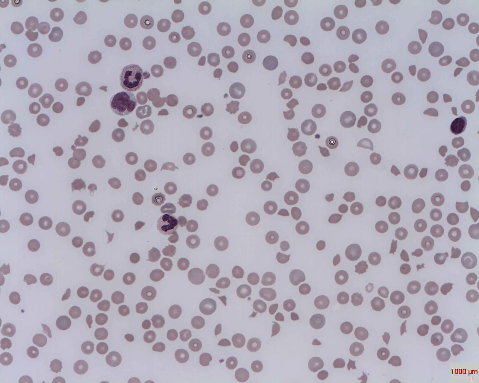
Long description
Blood film in microangiopathic haemolytic anaemia, showing fragmented red cells (schistocytes) and spherocytes among normal cells.

A complete blood count with a carefully reviewed blood film (thrombocytopenia plus schistocytes), lactate dehydrogenase, bilirubin, haptoglobin, reticulocyte count, creatinine and electrolytes, urinalysis, troponin, ECG, coagulation profile (PT/INR, aPTT, fibrinogen, D-dimer), direct antiglobulin (Coombs) test and blood grouping. Coagulation studies are expected to be relatively normal in TTP; abnormal PT/aPTT with high D-dimer suggest DIC.[3]
- Troponin I and ECG: assess silent cardiac ischaemia.[3]
- Brain imaging: if neurological symptoms are present.[3]
- Stool testing: Shiga-toxin PCR and culture whenever diarrhoea is present; if the toxin is already cleared it may be falsely negative despite typical HUS.[2]
- Complement testing and genetics: in suspected aHUS (C3, C4, CH50/AH50, factor H/I/B, anti-factor H antibodies, CD46 expression; genetic panel).
- ADAMTS13 activity and inhibitor assay: must be taken before plasma exchange begins; plasma exchange itself raises ADAMTS13, so the pre-treatment sample is the only reliable one.[3]
Assays report activity as a percentage of normal. Under 10 per cent confirms severe deficiency in a patient who has thrombocytopenia and microangiopathic haemolysis. Severe deficiency is not entirely specific (low activity can occur in severe sepsis and systemic cancer) and, in the hereditary form, a level under 20 per cent after recovery from an acute episode counts as relapse. In large cohorts, under 15 per cent was used as the severe cut-off for some validation work.[1][3][6] [2]
The PLASMIC score is a seven-point score built from bedside variables. It predicts severe ADAMTS13 deficiency (less than or equal to 10 per cent) while the assay is pending, and is designed as a screening tool.[1]
One point is awarded for each of:
- Platelet count less than 30,000 per microlitre. [1]
- Evidence of haemolysis: reticulocyte count greater than 2.5 per cent, or undetectable haptoglobin, or indirect bilirubin greater than 2 mg per dL (roughly greater than 34 micromol per L). [1]
- Mean corpuscular volume less than 90 fL.
- INR less than 1.5.
- Creatinine less than 2.0 mg per dL (roughly less than 177 micromol per L). [1]
- Absence of active cancer.
- Absence of solid-organ or stem-cell transplant in the past.
A higher score means a higher probability of TTP: a score greater than 5 suggests high probability, a score below 5 low probability.[1] In external validation, dichotomising at 6 to 7 versus 0 to 5 produced a positive predictive value of 72 per cent, a negative predictive value of 98 per cent, sensitivity of 90 per cent and specificity of 92 per cent.[6] The score was reported in one cohort with sensitivity of 99 per cent and specificity of 57 per cent. Because of its high negative predictive value, it is most useful for identifying patients who are unlikely to have TTP.[1]
The French clinical score exists as a second scoring approach.[3]
- TTP: thrombocytopenia and microangiopathic haemolysis without another cause, plus ADAMTS13 under 10 per cent. An inhibitor assay (anti-ADAMTS13 IgG or Bethesda units) separates immune-mediated from hereditary disease. Negative inhibitor testing does not exclude iTTP.[3]
- Refractory TTP: persistent platelets under 50,000 per microlitre and LDH greater than 1.5 times the upper limit of normal after five plasma exchanges and appropriate steroid use.[1]
- Clinical response: platelets above 150,000 per microlitre for two consecutive days with normalising LDH and no new organ dysfunction. Clinical remission: a sustained response for more than 30 days after the last plasma exchange.[1]
- STEC-HUS: the triad plus a STEC infection or Shiga toxin (stool PCR/culture), with preserved ADAMTS13.
- aHUS: the triad with complement dysregulation, usually with preserved ADAMTS13 and no STEC.
Suspected thrombotic microangiopathy
- Unexplained thrombocytopenia + schistocytes + LDH riseAnaemia, low haptoglobin, raised bilirubin; troponin; creatinine
- Confirm the patternExclude DIC (coagulation, D-dimer, fibrinogen) and ITP (isolated low platelets)
- Take ADAMTS13, stool Shiga toxin and complement tests before treatment if possible
- Question: Does the case look like TTP (neurology, no diarrhoea, PLASMIC high)?
Yes
- iTTP or cTTP likelyUrgent plasma exchange + corticosteroids; caplacizumab; rituximab for refractory or relapse
Diarrhoea / paediatric contact with STEC
- STEC-HUS likelySupportive care; avoid antibiotics and antimotility agents; dialysis if needed
Relapsing, renal-predominant or familial
- aHUS likelyEculizumab + supportive care; meningococcal vaccination before/with treatment
Clear secondary trigger
- Secondary TMATreat the trigger (malignant hypertension, drugs, pregnancy) and consider complement blockade if TTP excluded
- Monitor plateau: platelets above 150,000 for two days with normalising LDH; watch for relapse (ADAMTS13 under 20 per cent)
TTP is a haematological emergency. Untreated mortality is about 90 per cent; early plasma exchange plus corticosteroids reduce mortality to about 10 to 20 per cent.[1]
Immediate measures
- Admit to high-dependency or intensive care; continuous monitoring of neurological, cardiac and oxygenation status.[3]
- Obtain the ADAMTS13 sample, then start urgent daily plasma exchange; do not delay exchange for the result if clinical suspicion is high.[1][3]
- Avoid prophylactic platelet transfusion unless there is major bleeding or an invasive procedure is unavoidable, because it may precipitate further microthrombi.[1][3]
- Acute blood transfusion is given only for clinical indications.[1]
- Central venous access is placed early; platelet-supported line placement follows local practice.[3]
- Venous thromboembolism prophylaxis: non-pharmacological while platelets are below 50,000 per microlitre, pharmacological (for example low-molecular-weight heparin) once above 50,000 per microlitre, per local protocol.[3]
- Antithrombotic agents are generally avoided unless strongly indicated.[3]
Plasma exchange (plasmapheresis)
Daily plasma exchange removes autoantibodies, ultra-large von Willebrand factor multimers and pro-inflammatory cytokines, while supplying fresh ADAMTS13. The recommended exchange is one plasma volume, usually 40 mL per kg, although 1.0 to 1.5 plasma volumes (about 40 to 60 mL per kg) is widely used, daily until platelets are stable above 150,000 per microlitre for two consecutive days, LDH normalises and organ dysfunction resolves.[1][3] Fresh frozen plasma (or cryo-poor or solvent-detergent plasma) is the replacement fluid. Plasma infusion without exchange supplies only small amounts of ADAMTS13 and is an inadequate substitute, though it can serve as a stopgap.[1][3] In refractory disease twice-daily exchange may help.[1]
Corticosteroids
A strong recommendation supports adding corticosteroids to plasma exchange in a first acute event (and in relapse), over plasma exchange alone. Typical adult dose is prednisone 1 mg per kg per day in patients who are neurologically intact; intravenous methylprednisolone 1000 mg as a single daily dose for three days, or 125 mg two to four times daily, is used when the illness is severe. Corticosteroids continue while exchange continues, then taper on clinical response.[1] They reduce the number of exchange sessions needed and treatment-related complications by suppressing autoantibody production and inflammatory activity.[1]
Caplacizumab
Caplacizumab is a humanised anti-von Willebrand factor (A1 domain) nanobody that blocks platelet adhesion and stops microthrombus formation; it does not reduce autoantibody production, so it must be combined with immunosuppression. Adult indication (12 years and older) is 11 mg intravenously at least 15 minutes before plasma exchange on day one, then 11 mg subcutaneously daily: during exchange after each session, then daily for 30 days beyond the last exchange, extendable by up to 28 days if ADAMTS13 remains suppressed; withhold 7 days before elective surgery. It is avoided with antiplatelet agents or anticoagulants and stopped if clinically significant bleeding occurs (von Willebrand factor concentrate may correct haemostasis). Evidence supports faster platelet recovery and fewer exchanges and exacerbations when added to exchange plus immunosuppression, but guidelines rate it as a suggestion rather than a firm recommendation.[3][4]
Rituximab
Rituximab, an anti-CD20 monoclonal antibody, depletes B cells and the autoantibody source. Guidelines suggest adding it to corticosteroids and exchange for a first event and for relapse (conditional recommendation). It is particularly used when the course is refractory or relapsing, or for prophylaxis in remission with low ADAMTS13 activity. Dosing follows haematology protocols.[3]
Treatment response and duration
Exchanges stop once platelets exceed 150,000 per microlitre for two consecutive days with normalising LDH and no evolving organ damage. Relapse is more likely when ADAMTS13 remains under 20 per cent in remission, which is an indication for rituximab prophylaxis.[1][3]
Acute cTTP is treated with plasma infusion (10 to 15 mL per kg), daily until symptoms resolve and platelets normalise; plasma exchange is used if infusion is inadequate. Maintenance is plasma infusion every 1 to 3 weeks, or, where available, recombinant ADAMTS13, which the ISTH 2025 focused update recommends over FFP for cTTP in remission. Pregnancy in cTTP usually needs prophylactic plasma infusion (strong recommendation) rather than no prophylaxis.[3]
Management is primarily supportive:
- Correct volume depletion aggressively: patients are often fluid depleted and inadequate resuscitation increases the need for renal replacement therapy.[2]
- Half of all patients need dialysis; arrange early nephrology input.[2]
- Blood transfusion as clinically needed; platelets sparingly to avoid thrombotic complications.[2]
- Avoid antibiotics and antimotility agents in STEC-HUS: they are linked to worse outcomes, probably through greater Shiga-toxin exposure. Exceptions: when Shigella dysenteriae or Streptococcus pneumoniae is the cause, antibiotics improve outcomes.[2]
- Electrolyte monitoring, pancreatic enzymes (hyperglycaemia can occur), and neurology/cardiac monitoring; stroke or seizure prophylaxis as needed.[2]
STEC-HUS carries a fair prognosis: overall mortality around 5 per cent, but long-term renal impairment (reduced GFR, hypertension, proteinuria) develops in up to 25 per cent, and deaths occur mainly in adults over 60. [2]
Plasma exchange has not been shown to help STEC-HUS and is not routinely used, except in very severe or atypical presentations.
Early treatment is essential to prevent end-stage kidney disease and death. Eculizumab, a monoclonal antibody against complement C5 that prevents its cleavage and (indirectly) membrane attack complex formation, is the first-line treatment, with plasma exchange as second line, or first line only when eculizumab is unavailable. Outcomes have improved greatly since C5 blockade: ESKD or death in children has fallen from 30 to 50 per cent to about 9 per cent, and in adults from 60 to 6 per cent down to 6 to 15 per cent.[2] [5]
Adult eculizumab regimen: 900 mg intravenously weekly for the first 4 weeks, then 1200 mg for the fifth dose one week later, then 1200 mg every 2 weeks. Paediatric dosing follows body-weight bands (900/600/300 mg induction and maintenance, as per the label). Meningococcal vaccination must be given at least 2 weeks before the first dose, with antibiotic prophylaxis until 2 weeks after vaccination; vaccination alone is unreliable, so monitor for invasive meningococcal infection. Eculizumab is available only through a restricted programme and is given as an intravenous infusion over about 35 minutes in adults.[5]
Relapse is common in aHUS, so treatment is continued with close monitoring; complement inhibitor withdrawal requires specialist input.
Treat the provoking illness. Malignant hypertension is controlled carefully (not too fast), nephrotoxic drugs (such as calcineurin inhibitors) are reduced or switched, pregnancy-related thrombotic microangiopathy is managed with obstetric teams, and autoimmune disease is treated. If TTP is excluded and complement may be driving the microangiopathy, eculizumab or plasma exchange may be considered.
Low C3 suggests complement consumption; ADAMTS13 levels usually remain preserved. None of these tests should delay clearly indicated treatment, because earlier treatment is more effective.[2]
- TTP untreated has a mortality of about 90 per cent; with exchange plus steroids, about 10 to 20 per cent.[1]
- Renal failure: requiring dialysis in up to 50 per cent of HUS patients.[2]
- Neurological: seizures, stroke, visual loss and coma, which account for much of the morbidity and mortality.[2]
- Cardiac: troponin rise, arrhythmia and heart failure; subclinical involvement is common.[3]
- Pancreatitis and hyperglycaemia occur in up to 20 per cent of STEC-HUS.[2]
- Therapy related: plasma-exchange (hypotension, reactions), caplacizumab (bleeding, including life-threatening), plasma (allergy, infection risk, volume overload), central-line complications.
- iTTP: 80 per cent respond to initial treatment; post-treatment mortality 10 to 15 per cent. Common causes of death are myocardial infarction and sudden cardiac or neurological events. Microthrombi recurrence occurs in many; relapse is heralded by ADAMTS13 falling below 20 per cent. Long-term, survivors may experience mood and cognitive disorders, hypertension and renal insufficiency.[1][3]
- STEC-HUS: mortality about 5 per cent, but long-term renal impairment develops in up to 25 per cent, especially after prolonged dialysis. Adults over 60 account for most deaths.[2]
- aHUS: without C5 blockade, 50 per cent of aHUS reached dialysis dependence with 25 per cent mortality; with eculizumab, far fewer progress to ESKD or death.[2]
- Congenital TTP: natural history includes stroke, ESKD, cardiac events and fetal loss if untreated.
- Food safety: reducing STEC infection (correct cooking, hygiene) prevents most HUS. Person-to-person transmission occurs in households and childcare settings.
- Outbreaks: toxin-producing E. coli outbreaks should be reported promptly so that interventions reach contacts (microbiological testing, exclusion from food handling, hand hygiene).
- Vaccination: meningococcal vaccination before eculizumab (and continued prophylaxis) prevents invasive meningococcal disease during complement blockade.
- Remission monitoring in TTP: serial ADAMTS13 in remission, rituximab prophylaxis when activity is low, and vigilance for relapse.
- Children: Most HUS is STEC-HUS; peak under five. Atypical HUS is more dangerous early; complement blockade should not be delayed.
- Pregnancy: TTP can present or relapse in pregnancy and the puerperium. STEC-HUS incidence is described in pregnancy contexts; aHUS may present postpartum. cTTP carries a high fetal loss rate without prophylaxis. Management needs obstetric, haematology and nephrology teams.
- Older adults: worse STEC-HUS prognosis; more neurological manifestations.
- Immunocompromised and transplanted patients: calcineurin-inhibitor related thrombotic microangiopathy, delayed diagnosis, and drug interaction limits.
- A blood film, full blood count, lactate dehydrogenase, creatinine and bilirubin are cheap and diagnose the pattern; the main missing step is often not thinking of thrombotic microangiopathy early.
- ADAMTS13 assays are slow or unavailable. Use clinical suspicion plus the PLASMIC score to decide whom to treat; treat empirically rather than wait. [3] [6]
- Plasma exchange centres are scarce. If transport is possible, transfer urgently for exchange. If not, plasma infusion is an inadequate but useful temporising measure; plasma exchange remains the definitive treatment. [1] [3]
- Caplacizumab and eculizumab are rarely available; plasma exchange and immunosuppression remain the backbone of TTP care. [3]
- Eculizumab's cost and meningococcal vaccination requirements make aHUS in low-resource settings difficult; dialysis availability often determines survival. [2] [5]
- Antibiotics and antimotility agents must be withheld in STEC-HUS; children need careful rehydration and dialysis access. [2]
- Avoid plasma and platelet transfusions unless clearly indicated; send ADAMTS13 samples before plasma is started if there is any chance of testing. [1] [3]
- Thrombotic microangiopathy is one pattern with several causes; its hallmarks are thrombocytopenia, schistocytes, microangiopathic haemolytic anaemia and ischaemic organ damage.
- TTP is caused by ADAMTS13 under 10 per cent: immune-mediated (autoantibody) or congenital (gene mutation). Treat urgently with plasma exchange plus corticosteroids and, where available, caplacizumab and rituximab.
- Do not wait for the full classic pentad before treating; only about 40 per cent of patients show all five features. [3]
- Plasma exchange rather than anticoagulation or antithrombotic drugs treats TTP; it removes antibodies and ultra-large von Willebrand factor and supplies fresh enzyme.
- Platelet transfusions are usually avoided unless bleeding is major.
- STEC-HUS in children follows Shiga-toxin diarrhoea; management is supportive, with careful rehydration, sparing use of platelets, and dialysis when needed; avoid antibiotics and antimotility agents.
- aHUS is complement driven; first-line therapy is eculizumab, with plasma exchange second line, and meningococcal vaccination beforehand.
- The PLASMIC score predicts ADAMTS13 deficiency from bedside variables; a score above 5 makes TTP likely and a score below 5 makes it unlikely.
- Platelets under 50,000 per microlitre with normal coagulation in a fitting clinical picture should trigger exchange without waiting for the ADAMTS13 result.
- Renal nerve damage and long-term complications matter; monitor ADAMTS13 in remission and consider rituximab prophylaxis when it falls below 20 per cent.
- In low-resource settings, recognition and supportive care with urgent referral achieve most of the benefit.
Answer each one in your head first, then open the answer.
A 34-year-old woman has three days of confusion, bruising and oliguria. Platelets are 18,000 per microlitre, LDH is raised and schistocytes are seen. Coagulation is normal. ADAMTS13 activity is under 10 per cent. What is the diagnosis and the first treatment?
Show answerHide answer
Immune-mediated thrombotic thrombocytopenic purpura. Start daily plasma exchange with fresh frozen plasma, add high-dose corticosteroids, and give caplacizumab if available, while avoiding prophylactic platelet transfusion. Draw the ADAMTS13 sample before exchange; if clinical suspicion is high, do not delay exchange for the result. [1][3]
A 4-year-old child develops pallor, oliguria and oedema one week after bloody diarrhoea. Platelets are low and schistocytes are present. How do you manage STEC-HUS, and what must you avoid?
Show answerHide answer
Supportive care: aggressive but judicious rehydration, dialysis if needed, blood as clinically indicated, platelets sparingly. Avoid antibiotics and antimotility agents in this STEC setting because they are linked to worse outcomes. Test stool for Shiga toxin; check ADAMTS13 to exclude TTP. [2]
A 28-year-old man has relapsing microangiopathic haemolysis with renal failure. STEC testing is negative and ADAMTS13 is 45 per cent. What is the likely diagnosis and its specific treatment?
Show answerHide answer
Atypical, complement-mediated haemolytic uraemic syndrome. First-line treatment is eculizumab (900 mg weekly for 4 weeks, then 1200 mg every 2 weeks) after meningococcal vaccination, with plasma exchange if eculizumab is unavailable or clinically indicated. [5]
A patient has thrombocytopenia and haemolysis but normal creatinine, MCV 88 fL, INR 1.1 and no cancer or transplant. Platelets are 22,000 per microlitre. ADAMTS13 is pending. How does the PLASMIC score direct treatment?
Why are platelet transfusions and anticoagulation not the treatment for TTP?
Show answerHide answer
TTP thrombi are platelet-rich but form because of excessively large, uncleaved von Willebrand factor, not because of ordinary coagulation activation. Anticoagulation does not remove the enzyme deficiency or the autoantibody. Platelet transfusions can worsen the picture by adding fuel to microthrombosis and are reserved for major bleeding. [1] [3]
A pregnant woman in her third trimester presents with confusion, low platelets and schistocytes. ADAMTS13 is 4 per cent. Outline the management and a key pregnancy consideration.
Show answerHide answer
Treat as iTTP with daily plasma exchange and corticosteroids, and caplacizumab if appropriate and available. Involve obstetrics; monitor for pre-eclampsia, which can co-exist or mimic. After recovery, monitor ADAMTS13 in remission; rituximab is generally deferred until after delivery. In congenital TTP, pregnancy needs prophylactic plasma infusion. [3]
Acute kidney injury, nephritic syndrome and nephrotic syndrome (renal topics covering intrinsic glomerular disease, which must be separated from the thrombotic microangiopathies). Sepsis and disseminated intravascular coagulation often co-exist and must be excluded by coagulation studies. Iron deficiency anaemia and bone-marrow failure are other causes of cytopenias but lack schistocytes. The platelet disorders index covers immune thrombocytopenic purpura. Thrombophilia and antiphospholipid syndrome overlap with secondary TTP. Complement system disorders and glomerular disease in the renal curriculum provide further background.
- Nuñez Zuno JA, Khaddour K. Thrombotic Thrombocytopenic Purpura: Evaluation and Management. StatPearls [Internet]. Treasure Island (FL): StatPearls Publishing; last updated 2023-09-27. Accessed .
- Bhandari J, Rout P, Sedhai YR. Hemolytic Uremic Syndrome. StatPearls [Internet]. Treasure Island (FL): StatPearls Publishing; last updated 2023-10-19. Accessed .
- Zheng XL, Al-Housni Z, Cataland SR, Coppo P, Geldziler B, Germini F, Iorio A, Keepanasseril A, Masias C, Matsumoto M, McCrae KR, McIntyre J, Mustafa RA, Peyvandi F, Russell L, Tarawneh R, Vesely SK, International Society on Thrombosis and Haemostasis. 2025 focused update of the 2020 ISTH guidelines for management of thrombotic thrombocytopenic purpura. J Thromb Haemost 2025;23(11):3711-3732. Accessed .
- Sanofi-aventis US. CABLIVI (caplacizumab-yhdp) prescribing information. DailyMed (NLM), revised 12/2025. Accessed .
- Alexion Pharmaceuticals. SOLIRIS (eculizumab) prescribing information. DailyMed (NLM), revised 2026. Accessed .
- Li A, Khalighi PR, Wu Q, Garcia DA. External validation of the PLASMIC score: a clinical prediction tool for thrombotic thrombocytopenic purpura diagnosis and treatment. J Thromb Haemost 2018;16:164-169. Accessed .
Figure credits
Educational content for medical students and doctors. It is not a substitute for clinical judgement or local guidelines.