Oropharyngeal and oesophageal candidiasis
Oral thrush and Candida oesophagitis: who is at risk, how they present, how they are diagnosed and treated, with outlines of invasive and vulvovaginal.
- Candidiasis of the mouth and oesophagus is caused by yeasts of the genus Candida, most often Candida albicans. These organisms usually live harmlessly in the mouth and gut and cause disease only when local or systemic host defences fail.
- In the mouth it produces white, creamy plaques that can be wiped away, or red, sore patches in the folds of the mucosa. In the oesophagus it causes pain on swallowing; a course of systemic antifungal therapy is justified before endoscopy in the right clinical setting.
- Diagnosis is usually clinical for the mouth; microscopy of a scraping confirms it. Oesophageal disease is confirmed by endoscopy with biopsy when a therapeutic trial fails.
- Mouth disease is treated with topical azoles or nystatin for mild illness, and oral fluconazole for more extensive disease. Oesophageal disease always needs systemic therapy, usually fluconazole for 14 to 21 days.
- Invasive candidiasis (candidaemia) is treated with an echinocandin first line; vulvovaginal candidiasis is usually treated with a single dose of fluconazole or topical agents.
By the end of this page you should be able to:
- Explain why Candida becomes pathogenic and list the main local and systemic risk factors.
- Recognise the common forms of oropharyngeal candidiasis and distinguish them from other stomatitis.
- Interpret the signs and symptoms of oesophageal candidiasis and decide when endoscopy is needed.
- Choose appropriate antifungal treatment for oropharyngeal and oesophageal disease, including refractory and recurrent cases.
- Outline the management of candidaemia and vulvovaginal candidiasis.
- Adapt management to patients with HIV, dentures, inhaled corticosteroids or limited resources.
DefinitionCandidiasis
Candidiasis is a fungal infection caused by Candida species, most often Candida albicans. Mucosal disease (mouth and oesophagus) is superficial but can extend downwards; invasive candidiasis (candidaemia and deep organ infection) is a separate, life-threatening entity discussed in outline below.
The mouth and oesophagus are usually the first sites affected because swallowed yeast from the oropharynx can settle anywhere from the palate to the stomach. Surface infection is the rule: the fungus grows as a biofilm attached to mucosa and, in the deeper forms, sends filaments into the epithelium. This topic covers mucosal disease in detail and briefly outlines invasive and vulvovaginal disease so that the common drug choices and precautions are in one place.
Oral candidiasis is probably the most common fungal infection of humans. It can occur in healthy people, but it is far more frequent and severe in people with impaired immunity or altered local ecology.
- Candida colonises the mouths of many healthy adults; a systematic review of oral isolates found Candida albicans in about 82% of all isolates from mouth disease, with non-albicans species (notably C. tropicalis, C. glabrata and C. krusei) making up the remainder.[3]
- Inhaled corticosteroid users have a higher risk: a narrative review gives a 5 to 15% prevalence of oral candidiasis among users in the United States and a similar 7 to 14% in Europe.[2]
- Exposure to inhaled corticosteroids almost triples the risk (relative risk 2.89).[2]
- In people living with HIV, Candida is found in the mouth in 62 to 93% of patients, and more than 90% develop oropharyngeal candidiasis at some point in the disease.[2]
- Oropharyngeal candidiasis also affects 23.6% of HIV-infected patients in one reported cohort and, in children, often relapses if first episodes are not treated properly.[3]
The same Candida organism can be harmless or harmful depending on the host. Risk falls into two groups: conditions that depress systemic immunity, and local changes in the mouth that disturb the normal bacteria and mucosal barrier.
Most mucosal disease is caused by Candida albicans, which is dimorphic: it switches from budding yeast to filamentous hyphae when tissue is invaded and is isolated in about 80% of oral lesions. Other species become important in specific settings. C. glabrata shows intrinsic resistance to azoles, the most commonly used topical agents for mucosal infection; C. krusei is also azole-resistant; C. tropicalis and C. parapsilosis occur in mixed colonisation, in denture wearers and in older or immunocompromised patients; C. dubliniensis looks like C. albicans under the microscope but tends to be more virulent.[2][3]
- HIV and other immunosuppression. Advanced HIV, haematological malignancy, chemotherapy, radiotherapy and transplant immunosuppression remove the cell-mediated defence that keeps Candida in check.
- Diabetes mellitus. Hyperglycaemia impairs neutrophil function and glucose-rich saliva and tissue fluids feed the yeast.
- Malnutrition. Both undernutrition and overnutrition are implicated, through impaired immune function, altered mucosal integrity and shifts in oral ecology. Specific deficiencies (iron, zinc, folate, vitamins A, B6, B12 and C) are associated with it.[3]
- Age. Children with severe asthma on high-dose inhaled corticosteroids, and older adults with comorbidity, are at higher risk.
- Malignancy, neutropenia and chronic disease in general.
- Inhaled corticosteroids. Local deposition in the oropharynx suppresses local immunity. The risk rises with dose, duration and poor inhaler technique.
- Systemic antibiotics. Broad-spectrum drugs suppress the competing oral bacterial flora and permit overgrowth.
- Dentures and oral appliances. More than 60% of removable denture wearers harbour Candida on the prosthesis, which acts as a reservoir, especially when hygiene is poor or the denture is worn at night. [2]
- Xerostomia (reduced saliva), oral trauma, cavities, poor oral hygiene, smoking and alcohol.
- Corticosteroid use by any route, and acid-suppressing or other drugs that reduce gastric defences.
A clinical problem that looks like simple thrush often has more than one of these factors. Dentists and doctors should review inhaler technique, drug lists, glycaemic control, denture fit and oral hygiene at the same time as prescribing an antifungal.
Candida colonises the oral cavity of many healthy people, kept in balance by salivary defences (lysozyme, lactoferrin, IgA), competing bacteria on the mucosal surface and intact T-cell immunity. When these fail, a chain of events follows:
- Colonisation. Candida adheres to epithelial cells and to surfaces such as dentures and biofilms, using adhesins and hydrophobic cell-wall proteins.
- Overgrowth and biofilm formation. Loss of bacterial competition (after antibiotics) or loss of local immunity (after inhaled corticosteroids or in HIV) allows numbers to rise. On a surface, the yeasts secrete a polysaccharide extracellular matrix and form a biofilm that protects them and makes them harder to eradicate.
- Epithelial invasion. Under the hyphal form, filaments penetrate the superficial squamous epithelium and trigger a neutrophilic inflammatory response. The inflamed mucosa produces the soreness and erythema; the pseudomembrane is a mixture of fungal elements, desquamated epithelial cells, debris and inflammatory cells.
- Spread. Infection spreads contiguously from the mouth to the oropharynx and oesophagus. The absence of oral thrush never excludes oesophageal disease, because organisms can pass down without visible mouth disease.
- Invasion. In profoundly immunocompromised hosts, Candida can pass through the mucosal barrier into the bloodstream, producing candidaemia and deep organ infection. This is no longer "thrush"; it is invasive candidiasis.
C. glabrata is intrinsically resistant to azoles; biofilm-forming non-albicans species are generally harder to clear with topical agents than planktonic yeast.
Figure 1CandidiasisFromCDCinJPEG03-18-06
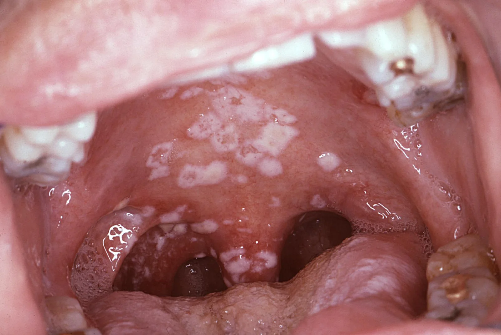
Long description
An intra-oral photograph of a patient with HIV/AIDS who presented with a secondary oral pseudomembranous candidiasis infection.

Oropharyngeal candidiasis is not one rash but several, distinguished by where it sits and whether the white material wipes off.
| Form | Appearance | Typical setting |
|---|---|---|
| Pseudomembranous ("thrush") | White or yellowish cottony plaques on tongue, palate and buccal mucosa that wipe off, leaving red, often sore mucosa | Neonates; immunocompromised hosts; inhaled corticosteroid users |
| Acute erythematous ("antibiotic sore mouth") | Red, sore patches without plaques, often palate and tongue | After broad-spectrum antibiotics |
| Chronic atrophic (denture stomatitis) | Chronic redness under a denture, often with no symptoms | Denture wearers; HIV |
| Angular cheilitis | Cracks, erythema and maceration at the corners of the mouth | Denture wearers; haematinic deficiency |
| Chronic hyperplastic (candidal leukoplakia) | Raised white plaques that cannot be scraped off; sometimes nodular | Middle-aged smokers; potentially serious because of malignant potential |
| Median rhomboid glossitis | Red, atrophic area in the midline of the back of the tongue | Steroid inhaler use; smoking |
Common symptoms are oral burning and soreness, an unpleasant taste (dysgeusia), a sensation of cotton wool in the mouth, dry mouth, and pain on swallowing if the disease extends into the pharynx. In denture wearers the whole under-surface of an upper denture may look red without the patient noticing.
The symptom is odynophagia: pain on swallowing that begins with the first swallow of a meal and is usually described as a burning or sticking feeling behind the sternum. It can be severe enough to stop eating. Dysphagia (difficulty with solids, occasionally liquids), retrosternal pain worsened by hot drinks and weight loss are common. Fever and night sweats appear when the patient is systemically unwell or coinfected.
A crucial negative is that oral thrush may be absent. Because infection spreads from the mouth, its presence raises suspicion but its absence never excludes oesophageal disease. Candida oesophagitis is the commonest infectious oesophagitis in adults; viral causes (herpes simplex and cytomegalovirus) must also be considered in immunocompromised patients, and they can coexist with Candida.
Look at the mouth carefully: the dorsum and sides of the tongue, palate, buccal mucosa, corners of the mouth, and the under-surface of dentures. Palpate gently for lymph nodes. Check the patient's weight, temperature, hydration and, when relevant, HIV status and medications. Ask about inhaler use and technique, antibiotics, dentures and teeth, and whether pain worsens with every swallow rather than with specific foods (which would suggest a mechanical obstruction).
Much of what looks like candidiasis is not, and the lookalikes are worth sorting by site.
- Angular cheilitis can be due to staphylococcus, streptococcus or iron/B-vitamin deficiency rather than Candida.
- Leukoplakia and lichen planus give white patches that do not scrape off; they are not infections.
- Oral hairy leukoplakia (in HIV) is usually on the lateral tongue and has a hairy surface; it is caused by Epstein-Barr virus, not Candida.
- Reflux and pill oesophagitis cause dysphagia and odynophagia without white plaques and without immunosuppression; endoscopy distinguishes them.
- Viral oesophagitis (herpes simplex, cytomegalovirus) produces ulcers rather than plaques.
- Cancer of the mouth or oesophagus can present with painful ulceration, white patches or dysphagia and must be biopsied when the picture is atypical or persistent.
- Geographic tongue, lichen planus and chemotherapy-induced mucositis can mimic thrush in patients on cancer treatment.
A persistent white patch that cannot be scraped off, any unhealing mouth ulcer, or any progressive swallowing problem needs investigation rather than repeated antifungal courses.
Figure 2Mycology algorithm
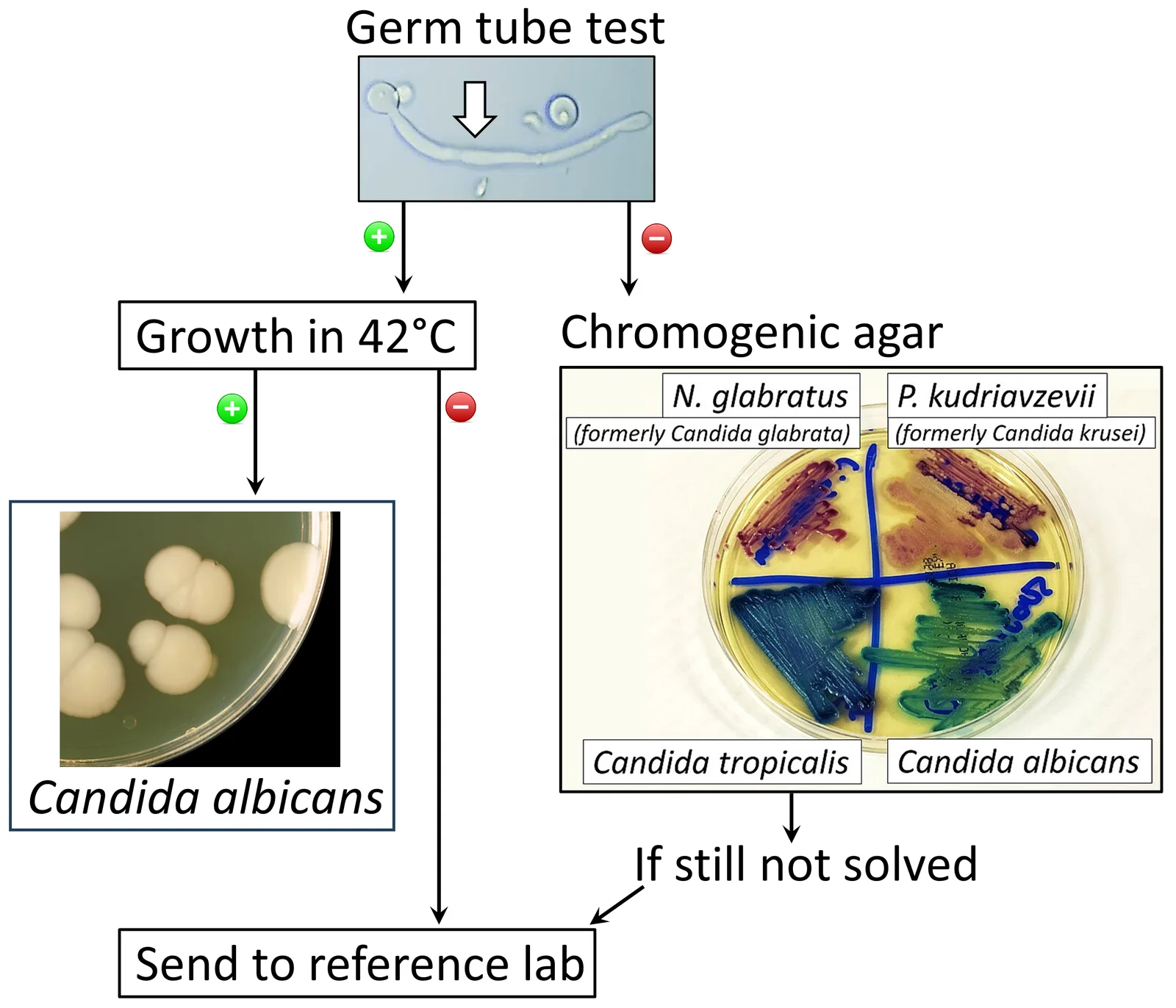
Long description
Diagram of a fungal infection workup algorithm used at a microbiology laboratory, including germ-tube testing of Candida albicans.

Diagnosis of oropharyngeal candidiasis is primarily clinical. A fresh lesion can be confirmed by scraping or smearing material on to a slide and examining it under the microscope; a potassium hydroxide preparation shows budding yeasts and pseudohyphae (filaments). Culture of a mouth rinse, or a swab of a lesion, is useful when infection is refractory, recurrent, resistant or atypical, because it identifies the species and its susceptibility. A biopsy is considered for raised plaques that do not wipe off, ulcers or other features that raise the possibility of malignancy, lichen planus or a non-candidal process.
A diagnostic trial of systemic antifungal therapy is appropriate before endoscopy in a patient with the classic picture of immunosuppression, odynophagia and (usually) oral thrush. Symptoms should start to improve within a few days; if they do not, or if the presentation is atypical, upper endoscopy with biopsy and brushings is needed to confirm Candida and to exclude viral co-infection. Candida produces creamy or yellow-white plaques on an erythematous, oedematous mucosa; biopsy shows budding yeast and pseudohyphae invading viable squamous epithelium, with neutrophilic inflammation and necrotic debris. Special stains such as periodic acid-Schiff and Gomori methenamine silver highlight the fungal wall.
Suspicion rises with odynophagia in an immunocompromised patient, after recent antibiotics, corticosteroids or chemotherapy, and in anyone with advanced HIV. The absence of thrush does not exclude oesophageal disease, and co-infection with two or more organisms is common rather than rare.
In a sick patient with suspected invasive candidiasis, blood cultures (which are slow and insensitive) and careful examination for a source are needed. That is beyond mucosal disease, although candidaemia can follow a breach in the mucosal barrier.
The approach is to match intensity to severity and to treat the cause at the same time.
- Mild disease. Clotrimazole troches, 10 mg five times daily, or a miconazole mucoadhesive buccal 50 mg tablet applied to the mucosal surface once daily for 7 to 14 days. Alternatives are nystatin suspension (100 000 units per mL) 4 to 6 mL four times daily, or 1 to 2 nystatin pastilles (200 000 units each) four times daily for 7 to 14 days.[1]
- Moderate to severe disease. Oral fluconazole 100 to 200 mg daily for 7 to 14 days.[1]
- Fluconazole-refractory disease. Itraconazole solution 200 mg once daily, or posaconazole suspension 400 mg twice daily for 3 days then 400 mg daily for up to 28 days. Alternatives are voriconazole 200 mg twice daily or amphotericin B deoxycholate oral suspension (100 mg per mL) four times daily. Intravenous echinocandin therapy (caspofungin 70 mg loading dose then 50 mg daily; micafungin 100 mg daily; anidulafungin 200 mg loading dose then 100 mg daily) or intravenous amphotericin B deoxycholate 0.3 mg per kg daily are other options.[1]
- Recurrent disease. Chronic suppressive therapy is usually unnecessary. If it is needed, fluconazole 100 mg three times weekly is recommended.[1]
- HIV-infected patients. Antiretroviral therapy is strongly recommended to reduce recurrent infections.[1]
- Denture-related candidiasis. Disinfect the denture in addition to giving the antifungal. Remove and soak dentures overnight, clean them daily, and treat any erythematous base under the denture.[1][2]
Table caution: doses are typical adult doses from the 2016 IDSA guideline. Check your local formulary for renal or hepatic adjustment, drug interactions and paediatric dosing, and adjust for pregnancy and severe impairment.
Systemic therapy is always required; topical agents do not reach the oesophagus in adequate concentration.
- First line. Oral fluconazole 200 to 400 mg (3 to 6 mg per kg) daily for 14 to 21 days.[1]
- If oral intake is not possible. Intravenous fluconazole 400 mg (6 mg per kg) daily, or an echinocandin (micafungin 150 mg daily; caspofungin 70 mg loading dose then 50 mg daily; anidulafungin 200 mg daily).[1]
- Less preferred alternative. Amphotericin B deoxycholate 0.3 to 0.7 mg per kg daily.[1]
- De-escalate. Switch to oral fluconazole 200 to 400 mg (3 to 6 mg per kg) daily once the patient can swallow.[1]
- Fluconazole-refractory disease. Itraconazole solution 200 mg daily, or voriconazole 200 mg (3 mg per kg) twice daily by mouth or intravenously, for 14 to 21 days. Alternatives are an echinocandin as above for 14 to 21 days, or amphotericin B deoxycholate 0.3 to 0.7 mg per kg daily for 21 days. Posaconazole suspension 400 mg twice daily, or extended-release tablets 300 mg once daily, can be considered.[1]
- Recurrent oesophagitis. Chronic suppressive therapy with fluconazole 100 to 200 mg three times weekly.[1]
- HIV-infected patients. Antiretroviral therapy is strongly recommended, because restoring immunity is what prevents recurrence.[1]
- Severe or refractory oropharyngeal or oesophageal disease in an immunocompromised patient: involve infectious disease.
- Weight loss, bleeding, a fixed mass, a white patch that cannot be wiped off, or failure of appropriate therapy: arrange endoscopy and tissue diagnosis, and exclude malignancy.
- Any suspicion of invasion (fever, rigors, positive blood cultures, organ involvement): treat as invasive candidiasis without delay.
Invasive candidiasis is most often candidaemia, one of the most common healthcare-associated bloodstream infections. It carries high attributable mortality and is treated differently from mucosal disease.
- Initial therapy (non-neutropenic adults). An echinocandin first line: caspofungin 70 mg loading dose then 50 mg daily; micafungin 100 mg daily; anidulafungin 200 mg loading dose then 100 mg daily. Fluconazole 800 mg (12 mg per kg) loading dose then 400 mg (6 mg per kg) daily is an acceptable alternative in selected, non-critically ill patients whose isolates are unlikely to be fluconazole resistant.[1]
- Lipid amphotericin B 3 to 5 mg per kg daily is a reasonable alternative if there is intolerance, limited availability or resistance.[1]
- Central venous catheters. Remove the catheter as early as possible when it is the presumed source and can be removed safely; this is individualised.[1]
- Ophthalmology. Every non-neutropenic patient with candidaemia should have a dilated ophthalmological examination, preferably by an ophthalmologist, within the first week, to exclude Candida endophthalmitis.[1]
- Duration. Two weeks after documented clearance of Candida from the bloodstream and resolution of symptoms attributable to candidaemia. Follow-up blood cultures every day or every other day establish clearance.[1]
- Fluconazole step-down. Transition from an echinocandin to fluconazole is appropriate after 5 to 7 days in clinically stable patients with susceptible isolates and negative repeat blood cultures.[1]
Echinocandins are preferred for most invasive episodes, but they do not reliably treat eye, central nervous system or urinary infection (the drug reaches those sites poorly), which is why those sites are handled differently.
Uncomplicated Candida vulvovaginitis (mild to moderate, infrequent, caused by C. albicans in a healthy host):
- Topical antifungal agents (no single agent is superior), or a single 150 mg oral dose of fluconazole.[1]
Severe acute disease:
- Fluconazole 150 mg every 72 hours for 2 or 3 doses.[1]
Recurrent vulvovaginal candidiasis (four or more episodes a year): induction with a topical agent or oral fluconazole for 10 to 14 days, followed by fluconazole 150 mg weekly for 6 months.[1]
C. glabrata not responding to oral azoles: topical intravaginal boric acid 600 mg daily for 14 days; or nystatin intravaginal suppositories 100 000 units daily for 14 days; or topical 17% flucytosine cream alone or combined with 3% amphotericin B cream daily for 14 days.[1]
Screen for and treat the underlying cause of recurrence (uncontrolled diabetes, hormonal factors, antibiotics, behavioural factors). Review alternative diagnoses when the discharge, itching or pain does not respond, because non-candidal conditions and non-albicans species are common mimics.
Approach to mouth and oesophageal candidiasis
- Symptoms suggestive of candidiasisOral soreness, white patches; or odynophagia after antibiotics, steroids, chemotherapy or in HIV
- Examine the mouth; review medications, immune status and glycaemic control
- Question: Site of disease?
Mouth only
- Mild: topical azole (clotrimazole troches 10 mg five times daily) or nystatin for 7 to 14 days
- Moderate to severe: oral fluconazole 100 to 200 mg daily for 7 to 14 days
- Reassess; culture if refractory or recurrent
Odynophagia suggests oesophageal disease
- Consider a diagnostic trial of oral fluconazole200 to 400 mg (3 to 6 mg/kg) daily
- Reassess after a few days
- Question: Improved?
Yes
- Continue fluconazole for 14 to 21 days
No
- Upper endoscopy with biopsy and brushingsExclude viral co-infection and malignancy
- Always treat the cause: dentures, inhaler technique, antibiotics, diabetes, HIV
- Dentures: clean and disinfect daily, remove overnight, treat the denture and the mucosa.
- Inhaled corticosteroids: rinse, gargle and spit after every dose; use a spacer; check inhaler technique at every review. A narrative review of prevention measures in ICS users emphasises that patient education and oral hygiene are the cornerstones, with higher dose and poor technique among the main modifiable risks.[2]
- Antibiotics: use the narrowest spectrum for the shortest time, and recognise antibiotic candidiasis as a trade-off.
- Diabetes: control glycaemia; check for Candida in anyone with poor control.
- HIV: effective antiretroviral therapy is the best single prevention against recurrent mucosal candidiasis.[1]
- Nutrition: correct deficiencies and general undernutrition, and include nutritional care in cancer treatment and in high-risk groups.[3]
- Recurrence and chronicity, especially with continuing risk (HIV, dentures, ICS, diabetes).
- Malnutrition and weight loss when odynophagia stops a person eating.
- Oesophageal stricture from repeated or circumferential ulceration.
- Bleeding from an eroded vessel in an oesophageal ulcer.
- Spread to bloodstream (candidaemia) in profoundly immunocompromised patients.
- Malignant transformation is a rare but real concern with chronic hyperplastic candidiasis; such lesions must be biopsied.
- Dysphagia, aspiration and poor quality of life in older and institutionalised patients.
Mucosal candidiasis is usually curable. Prognosis depends on whether the underlying risk can be reversed: a course of antibiotics finished, steroids tapered, dentures fixed, glycaemia controlled or antiretroviral therapy started. In profoundly and persistently immunocompromised patients (advanced untreated HIV, continuing transplant immunosuppression, deep neutropenia) recurrence is common until immunity recovers, and suppressive therapy with fluconazole 100 to 200 mg three times weekly is justified for the mouth and oesophagus. Oesophageal candidiasis can be the event that reveals previously unrecognised advanced HIV disease, so appropriate testing and linkage to HIV care matter more than the antifungal prescription.
- HIV. First episodes are often mild; recurrent or oesophageal disease signals advanced immunosuppression. Start or optimise antiretroviral therapy alongside antifungal treatment; chronic suppressive regimens are second best.[1]
- Denture wearers. Treat the prosthesis as well as the mouth; at least 60% of wearers carry Candida on their denture.[2]
- Patients on inhaled corticosteroids. Reassess inhaler technique and dose; rinse and spit; use a spacer.
- Children. In children at risk (severe asthma on high-dose inhaled corticosteroids, prolonged antibiotics, neonates) oropharyngeal disease presents as thrush and can affect feeding. Mouth disease is treated with age-appropriate formulations; systemic antifungals in children follow weight-based dosing and local guidance, and neonatal invasive disease uses amphotericin B deoxycholate (not covered here).[1]
- Older adults. Malnutrition, xerostomia, dentures, diabetes and immunosenescence stack up; review medications and nutrition, and check swallowing after oesophageal disease.[2][3]
- Pregnancy. Systemic antifungals require specialist advice and an assessment of benefit and fetal risk; oral azoles are avoided unless benefits clearly outweigh risks, and topical therapy is preferred for vulvovaginal disease. Confirm current local guidance.
- People with diabetes. Check glycaemia; candidiasis is both more common and more recurrent, and it can signal poor control.[2][3]
Most of these conditions are manageable without laboratory support.
- Diagnosis is clinical: white, removable plaques in the mouth; pain on swallowing in an immunocompromised or recently at-risk patient. Microscopy of a scraping, if available, confirms it.
- First-line oral drugs are usually available and inexpensive. Nystatin suspension or pastilles, and fluconazole tablets, cover most mouth disease; single-dose fluconazole covers uncomplicated vulvovaginal disease.
- Oesophageal disease requires systemic therapy; if fluconazole is unavailable, referral becomes urgent, particularly for patients who are immunocompromised.
- Denture hygiene, mouth rinsing after inhaled corticosteroids, spacer use, oral care and nutrition cost little and prevent a large share of disease.
- In HIV, integrating antifungal care into routine HIV services and starting or optimising antiretroviral therapy reduces recurrence more than repeated antifungal courses.[1]
- A patient with odynophagia who does not improve within 3 to 5 days of appropriate antifungal therapy should be referred for specialist and, where possible, endoscopic assessment, because co-infection with a virus is likely.
- In immunocompromised patients in settings where blood culture is unavailable, any fever with candidaemia cannot be excluded by clinical criteria; treat severe sepsis empirically and refer early.
- Candida, most often C. albicans, is a commensal that causes disease when immunity fails locally or systemically.
- Common settings: HIV, inhaled corticosteroids, broad-spectrum antibiotics, diabetes, dentures, malignancy and neutropenia.
- Mouth forms: pseudomembranous (removable white plaques), acute erythematous, denture stomatitis, angular cheilitis, and chronic hyperplastic candidiasis (plaques that do not wipe off, biopsy needed).
- Diagnosis of mouth disease is primarily clinical, confirmed by microscopy of a scraping; oesophageal disease needs a diagnostic antifungal trial or endoscopy with biopsy.
- Mild mouth disease is treated with clotrimazole troches or nystatin for 7 to 14 days; moderate to severe disease with fluconazole 100 to 200 mg daily.
- Oesophageal disease always needs systemic therapy: oral fluconazole 200 to 400 mg daily for 14 to 21 days, de-escalating once the patient can swallow.
- Fluconazole-refractory disease: itraconazole solution 200 mg daily, voriconazole 200 mg twice daily, or an echinocandin.
- Antiretroviral therapy in HIV and denture hygiene reduce recurrence more than prolonged antifungals.
- Invasive candidiasis (candidaemia) is treated with an echinocandin first line; remove a central line if it is the source and perform ophthalmology within one week.
- Vulvovaginal candidiasis: topical agents or a single 150 mg fluconazole for uncomplicated disease; longer induction plus weekly fluconazole for recurrent disease. [1]
- C. glabrata and other non-albicans species matter when first-line azoles fail.
Answer each one in your head first, then open the answer.
A 70-year-old man with chronic obstructive pulmonary disease on a beclometasone inhaler develops white patches on his tongue and soft palate that wipe off leaving red, sore mucosa. He also wears upper dentures. What is the diagnosis and two practical steps besides prescribing an antifungal?
Show answerHide answer
Oropharyngeal candidiasis (pseudomembranous thrush). Practical steps include reviewing inhaler technique, adding a spacer and rinsing and spitting after every dose, and addressing denture hygiene (clean, disinfect, remove overnight); these measures are as important as the drug. Mild disease can be treated with clotrimazole troches 10 mg five times daily for 7 to 14 days, with fluconazole 100 to 200 mg daily reserved for more extensive disease. [1]
A 38-year-old woman with HIV, not yet on antiretroviral therapy, develops pain on swallowing after completing antibiotics. Why might she have no mouth lesions, and what is the safest way to manage her?
Show answerHide answer
Oesophageal candidiasis can exist without visible oral thrush because organisms pass down the oesophagus without settling in the mouth. Management is a diagnostic trial of oral fluconazole 200 to 400 mg daily, with reassessment after a few days. If she improves, continue for 14 to 21 days; if not, or if the picture is atypical, arrange upper endoscopy with biopsy and brushings, because Candida and herpes simplex or cytomegalovirus can coexist. Start or optimise antiretroviral therapy alongside treatment.
Why is nystatin or clotrimazole appropriate for mild mouth thrush but not enough for oesophageal disease?
Show answerHide answer
Topical agents coat the mucosal surface and reach the mouth, but they do not reach the oesophagus in sufficient concentration. Oesophageal disease requires systemic therapy (fluconazole) to act from the blood side of the mucosa.
Which drug is the recommended first-line treatment for candidaemia in a non-neutropenic adult, and what two non-drug measures does the guideline emphasise?
Show answerHide answer
An echinocandin (caspofungin 70 mg loading dose then 50 mg daily; micafungin 100 mg daily; or anidulafungin 200 mg loading dose then 100 mg daily). Non-drug measures: remove the central venous catheter early when it is the presumed source, and perform a dilated ophthalmological examination within the first week. Treat for two weeks after clearance of Candida from the bloodstream.
A young woman has her fourth episode of vulvovaginal candidiasis this year, with *C. albicans* confirmed. How should she be managed, and what is the role of boric acid?
Show answerHide answer
Recurrent disease: 10 to 14 days of induction with a topical agent or oral fluconazole, followed by fluconazole 150 mg weekly for 6 months. Screen for and treat underlying causes such as uncontrolled diabetes, antibiotics and hormonal factors. Intravaginal boric acid 600 mg daily for 14 days is reserved for Candida glabrata not responding to oral azoles, not for routine C. albicans disease.
- Pappas PG, Kauffman CA, Andes DR, et al. Clinical Practice Guideline for the Management of Candidiasis: 2016 Update by the Infectious Diseases Society of America. Clinical Infectious Diseases 2016;62:e1-e50. Accessed .
- Arzayus-Patiño L, Benavides-Córdoba V. Non-Pharmacological Interventions to Prevent Oropharyngeal Candidiasis in Patients Using Inhaled Corticosteroids: A Narrative Review. Healthcare (Basel) 2025;13(14):1718. Accessed .
- Carvalho JP, Rodrigues J, Rodrigues CF, Andrade JC, Rajão A. Distribution of Candida Species Causing Oral Candidiasis in High-Risk Populations: A Systematic Review. Healthcare (Basel) 2026;14(2):159. Accessed .
Figure credits
Educational content for medical students and doctors. It is not a substitute for clinical judgement or local guidelines.