Endemic fungal infections
Three dimorphic fungi acquired from soil: how they differ in geography, tissue form, presentation, diagnosis and treatment.
- Histoplasmosis, coccidioidomycosis and blastomycosis are diseases caused by three dimorphic fungi that live in soil and share one starting point: a person inhales the mould form, and the fungus then converts to a yeast form inside the body.
- Histoplasma is tied to the Ohio, Mississippi and Missouri river valleys; Coccidioides to the dry south-west of the United States, Mexico and parts of Central and South America; Blastomyces to the Ohio and Mississippi valleys, the Great Lakes region and parts of the south-eastern United States.
- Exposure is environmental, not contagious from person to person: disturbance of soil, construction or demolition, and contact with bird or bat droppings or dust raise risk.
- All three can cause an acute, self-limited pneumonia; in susceptible hosts they may become chronic cavitary lung disease or disseminate to skin, bone, the adrenal glands or the central nervous system.
- Severe disease is treated with lipid amphotericin B first; milder disease usually receives itraconazole, while coccidioidomycosis is most often treated with fluconazole. Where lipid amphotericin is unavailable, amphotericin B deoxycholate is the fallback.
By the end of this page you should be able to:
- Compare the three endemic mycoses by geography, exposure, tissue form and disease pattern.
- Explain why an inhaled dimorphic fungus can pass unnoticed or cause devastating disease, according to host immunity.
- Select and interpret the tests that diagnose each infection: antigen, serology, histology and culture.
- Recognise disseminated disease involving skin, bone, adrenal or meningeal sites.
- Choose first-line and severe-disease treatment, including doses, duration and monitoring, and adapt them when resources are limited.
- Describe the practical differences between these fungi and other common fungi that they are confused with.
DefinitionEndemic (dimorphic) mycoses
Infections caused by fungi that switch from a mould in the environment to a yeast in human tissue. The three here are histoplasmosis (Histoplasma capsulatum), coccidioidomycosis (Coccidioides immitis and Coccidioides posadasii) and blastomycosis (Blastomyces dermatitidis and related species). "Endemic" means they occur in, and are usually acquired from, well-defined geographic regions.
A useful way to organise them is by what the tissue form looks like:
- Histoplasmosis: small oval budding yeast (2 to 4 micrometres), often living inside macrophages. [1]
- Coccidioidomycosis: large spherules filled with endospores; in wet preparations these spherules can measure up to 75 micrometres, with endospores 2 to 5 micrometres in size.
- Blastomycosis: relatively large yeast (8 to 15 micrometres) with a thick wall and a single broad-based bud. [3]
Clinically, each infection ranges from silent to severe:
- Acute pulmonary: a pneumonia that begins a few weeks after exposure and often resolves on its own.
- Chronic (cavitary) pulmonary: slowly progressive lung disease, usually in damaged or obstructed lungs, that can look like tuberculosis or lung cancer.
- Disseminated: disease outside the lungs, most often in the immunocompromised host.
Figure 1FungaldiseasedistributionUS
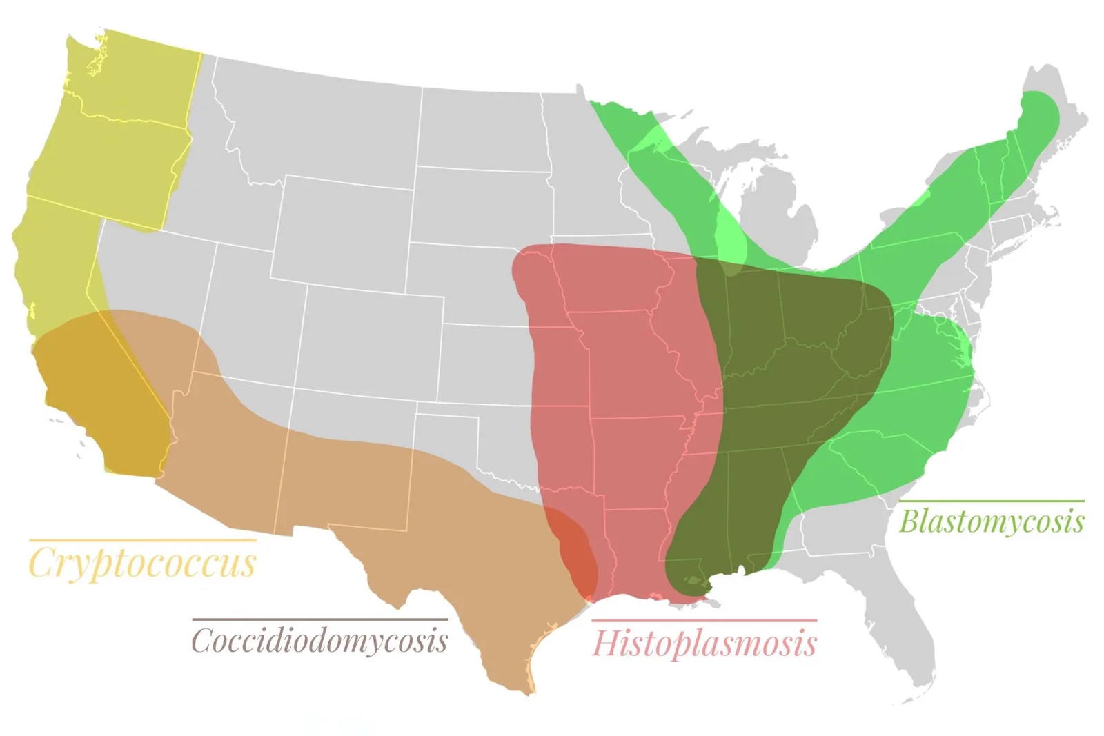
Long description
A self-published 2112×1408-pixel map titled 'Distribution of fungal pneumonia across USA', categorised on Wikimedia Commons under coccidioidomycosis, blastomycosis maps and maps of histoplasmosis.

| Infection | Main endemic regions | Typical exposure | Form seen in tissue |
|---|---|---|---|
| Histoplasmosis | Ohio, Mississippi and Missouri river valleys; Africa; parts of Asia and Latin America | Bird or bat droppings; soil, dust and construction or demolition sites | Small oval yeast (2 to 4 micrometres), often inside macrophages |
| Coccidioidomycosis | South-western United States; Mexico; Central and South America | Dust from arid soil; wind can carry spores over long distances | Spherules up to 75 micrometres, packed with 2 to 5 micrometre endospores [2] |
| Blastomycosis | Ohio and Mississippi river valleys; Great Lakes region; south-eastern United States; Africa and the Middle East | Moist soil, decaying vegetation, waterways; outbreaks near paper mills and rivers | Large thick-walled yeast (8 to 15 micrometres) with a single broad-based bud |
In the United States, histoplasmosis is found in the central and eastern states but is reportable in only 14 states, so true counts are uncertain. Seroprevalence studies suggest that 60% to 90% of people living in the most endemic parts of the Ohio, Mississippi and Missouri valleys are exposed during life, while the national estimate is 1 to 2 cases per 100,000 people. Histoplasmosis is also common elsewhere: worldwide more than 50 million people are thought to carry latent infection and about 500,000 new infections occur each year. Between 1938 and 2013, 105 outbreaks involving 2,850 people were reported across 26 states and Puerto Rico; exposure to birds, bats or their droppings contributed to 77% of those outbreaks, and workplace exposure to 41%.[1] [3]
Coccidioidomycosis is produced by Coccidioides immitis in California and by Coccidioides posadasii in other states and countries, the two being identical in clinical behaviour; the organism prefers arid soil. The Infectious Diseases Society of America estimates that about 150,000 infections occur in the United States each year: 50,000 cause illness that warrants medical attention, 10,000 to 20,000 are diagnosed and reported, 2,000 to 3,000 produce pulmonary sequelae, 600 to 1,000 spread beyond the lungs, and 160 are fatal. Reported cases from endemic states rose ten-fold between 1998 and 2011. Residence in or travel to an endemic area within the previous 1 to 2 months is a critical clue.[5]
Blastomycosis is reported at roughly 2 cases per 100,000 people per year in the United States, although true incidence is unknown because the infection is underdiagnosed and reported from only a few states; Wisconsin has the highest incidence. About 50% of exposed people have minimal or no symptoms, and roughly 70% of symptomatic cases occur in men. The range is expanding, partly with climate and environmental change, with cases now recorded beyond the traditional belts.[3]
All three are acquired from the environment, not from another person.
- Birds, bats and rotting vegetation enrich soil with nutrients that favour the mould; bats carry Histoplasma in the gut, and birds carry it on their feathers without being ill themselves. Birds tolerate high body temperatures, so they are unaffected.
- Spores become airborne when contaminated soil is disturbed. Intense construction, demolition, excavation, renovation of old buildings and caving are classic settings. Wind can carry Coccidioides arthroconidia over long distances.
- High inoculum or impaired immunity turns exposure into disease. For Histoplasma, fewer than 5% of people who inhale a low inoculum become ill, whereas up to 75% who inhale a high inoculum do.[1]
- Risks for severe or disseminated disease include advanced HIV infection, solid-organ and stem-cell transplantation, high-dose corticosteroids or TNF-alpha inhibitors, pregnancy (especially in the third trimester for coccidioidomycosis), African or Filipino ancestry and (for coccidioidomycosis) male sex and diabetes.
- Smoking and damaged lungs predispose to chronic pulmonary forms, particularly blastomycosis and histoplasmosis.
The germling then converts to the yeast form at body temperature. In histoplasmosis the yeast survives inside macrophages by blocking acidification of the phagosome, travels with trafficking macrophages and either is walled off in granulomas or disseminates. Coccidioides spherules rupture in tissue and release endospores that repeat the cycle locally. Blastomyces resists phagocytosis through its thick cell wall and spreads via bloodstream and lymphatics; an uncommon but distinctive feature is its pyogranulomatous inflammation, made of neutrophils followed by granuloma formation.
The sequence is the same for all three:
- Arthroconidia or conidia are inhaled and reach the alveoli.
- The fungus converts to the yeast or spherule form at 37 degrees Celsius.
- In healthy people, alveolar macrophages and neutrophils contain the infection and granulomas form within weeks.
- In heavy exposure or immunocompromise, replication outstrips control, producing pneumonia, cavitation and then dissemination.
This explains the clinical timing: incubation is about 3 to 17 days for histoplasmosis (most symptoms resolve within about 10 days), 7 to 21 days for coccidioidomycosis and 3 weeks to 3 months for blastomycosis.[1][2][3]
Typically a community-acquired pneumonia that does not respond to antibacterial treatment.
- Histoplasmosis: fever, chills, dry cough, headache, anterior non-pleuritic chest pain (thought to be enlarged hilar or mediastinal nodes), fatigue and myalgia; hilar or mediastinal lymphadenopathy with patchy or nodular infiltrates; rheumatological symptoms or erythema nodosum and erythema multiforme in about 5%, more often in women.[1]
- Coccidioidomycosis: 60% of infections cause no symptoms. When present, fever, cough, pleuritic chest pain, weight loss and marked fatigue are usual; eosinophilia and erythema nodosum or erythema multiforme ("desert rheumatism") are common. Chest imaging shows unilateral infiltrates, and hilar adenopathy raises suspicion for extrathoracic spread.[2]
- Blastomycosis: cough in 73%, fever in 54%, sputum in 50%, with weight loss, night sweats, chills and chest pain in a large minority. Chest radiographs most often show upper-lobe airspace consolidation involving more than one lobe; acute respiratory distress syndrome occurs in up to 10% and carries 50% to 90% mortality.[3]
- Histoplasmosis especially strikes damaged lungs: productive cough, weight loss, weakness, fatigue, fever, chest pain, haemoptysis and dyspnoea; cavitation occurs in about 30%, usually in the upper lobes. It mimics tuberculosis, sarcoidosis and malignancy.[1]
- Coccidioidomycosis presents as nodules, cavities and fibrocavitary disease; cavitary disease occurs in only about 8% of adults but more often in children. A thin-walled solitary cavity is typical.[2]
- Blastomycosis can resemble chronic lung infection or cancer with consolidation, mass lesions and cavitation.[3]
Histoplasmosis characteristically affects immunocompromised hosts: fever, weight loss, malaise and anorexia with hepatosplenomegaly and lymphadenopathy; pancytopenia and purpura occur with marrow infiltration; adrenal involvement can produce Addison's disease (fever, malaise, orthostatic hypotension, nausea and vomiting; CT shows enlarged adrenal glands with central necrosis), which is a distinctive feature. Pericarditis complicates about 6% and arthralgia or arthritis about 5% of symptomatic pulmonary cases. Central nervous system and fibrosing mediastinitis (rare) are further forms. Mortality in HIV-associated disease has been reported at 21% to 53%.[1] [3]
Coccidioidomycosis spreads in roughly 600 to 1,000 patients a year in the United States, usually within weeks to months of infection. Chronic skin ulceration or subcutaneous abscesses, vertebral osteomyelitis (which can mimic staphylococcal osteomyelitis, with psoas and epidural abscesses), synovial infection, and coccidioidal meningitis occur. Meningitis always needs treatment; in untreated cases it is fatal. Eosinophils in the cerebrospinal fluid are suggestive. The most lethal modern complications are hydrocephalus and cerebral vasculitis. Dissemination is more likely in immunosuppressed patients, pregnant women and those of African or Filipino ancestry.[5] [2]
Blastomycosis disseminates in 25% to 30% of patients after haematogenous spread, most often to skin (verrucous or ulcerative lesions with central clearing), bone (25% of extrapulmonary disease; lower spine and pelvis favoured, with sinus tracts) and, in 5% to 10% of extrapulmonary cases, the central nervous system. Genitourinary infection (prostatitis, orchitis, epididymitis) occurs. Immunosuppressed patients have mortality of about 30% and disseminated disease carries greater morbidity and mortality.[3] [2]
The clinical picture of all three overlaps with common and dangerous conditions:
- Tuberculosis and other mycobacterial disease (cavities, upper-lobe disease, weight loss, night sweats).
- Lung cancer and lymphoma (masses, cavities, nodules that do not respond to treatment).
- Sarcoidosis (hilar and mediastinal lymphadenopathy, diffuse reticulonodular infiltrates).
- Bacterial pneumonia that fails to respond to antibacterials.
- Other endemic and opportunistic fungi: Aspergillus (allergic, chronic and invasive forms), Cryptococcus (meningitis in immunocompromise), Mucorales (rhino-orbital disease in diabetes), Paracoccidioides, Sporothrix and Candida.
- Vasculitis and autoimmune disease when arthralgia, erythema nodosum and fever dominate in coccidioidomycosis.
A reliable exposure history and appropriate fungal tests are what separate them.
Figure 2Histoplasmosis (5414173501)
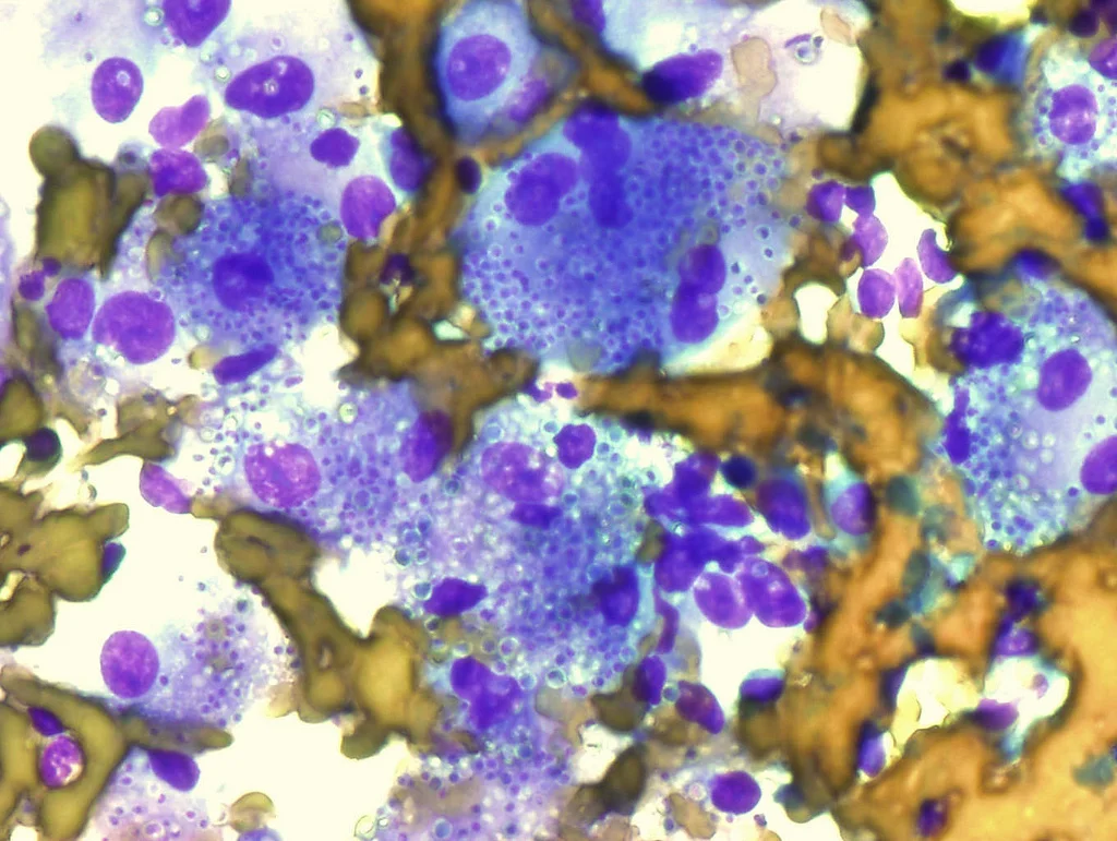
Long description
Macrophages in a lymph node containing numerous Histoplasma capsulatum, Diff-Quick stain.

- Histoplasma: 2 to 4 micrometre oval yeast with narrow-based budding, frequently inside macrophages or histiocytes.
- Coccidioides: spherules and endospores in tissue; culture colonies grow in 5 to 7 days as a white mould. [2]
- Blastomyces: 8 to 15 micrometre thick-walled yeast with a single broad-based bud; histology often shows pseudoepitheliomatous hyperplasia and pyogranulomatous inflammation. Culture may take 2 to 6 weeks. [3]
- Histoplasma urinary antigen (enzyme immunoassay): the preferred first test for suspected disease, especially severe or disseminated. Sensitivity 82% to 95%, specificity 97% to 99%; antigen can also be measured in serum and bronchoalveolar lavage fluid. Sensitivity falls to about 30% in subacute pulmonary disease. Cross-reactivity occurs with Blastomyces, Coccidioides, Paracoccidioides, Talaromyces, Sporothrix and Candida.[1] [2]
- Coccidioidal serology: infection is usually diagnosed this way. Precipitin (IgM) antibodies are detected in about 90% of patients in the first 3 weeks; complement-fixing (IgG) antibodies appear later and their titre reflects burden. Enzyme immunoassays are highly sensitive but less specific, so positive results should be confirmed. Serology may be negative in 15% of patients with culture-proven disease. Cross-reactivity with Histoplasma is well described.[2]
- Blastomyces urinary antigen: recommended alongside culture and histology when blastomycosis is suspected. Culture is highly specific (about 100%) but sensitivity is 66% to 86%; growth takes 2 to 6 weeks. Histology can be hard; the yeast must be sought carefully, often within histiocytes and abscesses. Serology is not recommended because it is insensitive and cross-reacts with other dimorphic fungi.[3]
A concise clinical-testing guideline covers all three endemic mycoses together for patients with community-acquired pneumonia; it recommends considering these infections when pneumonia fails to respond to empiric antibacterial therapy in a person who lives in or has travelled to an endemic area.[1]
- Histology detects fungi rapidly but needs skilled laboratory staff. Histoplasma is found in 9% to 50% of cases; fungal cultures have 40% to 74% sensitivity and grow in 5 to 6 weeks.[1]
- Serology in histoplasmosis: complement fixation, immunodiffusion and enzyme immunoassay have 54% to 95% sensitivity and 58% to 100% specificity. Immunodiffusion is more specific and is negative in fewer than 1% of residents of endemic areas; a complement fixation titre of 1:32 or a four-fold rise supports acute infection. Antibodies appear 3 to 6 weeks after infection and may be absent in severe immunocompromise.[1]
- Polymerase chain reaction: promising in histoplasmosis (86% to 98% sensitivity, near 100% specificity) and in research settings for coccidioidomycosis; standardisation is incomplete and commercial platforms are not universal.[1][2]
- Imaging: chest X-ray or CT for the lung forms; adrenal CT for suspected histoplasmosis; brain and spinal imaging when meningitic or osseous disease is suspected.
- CSF examination: essential when meningitis is suspected; coccidioidal meningitis may show eosinophils; cultures are often negative, so antigen or nucleic acid testing and travel history matter.
The updated IDSA histoplasmosis guideline uses severity categories rather than rigid scores:
- Mild acute pulmonary: treated or not treated depending on progress.
- Moderate acute pulmonary: individual decision with itraconazole favoured if treatment is given.
- Severe: requires lipid amphotericin B followed by itraconazole.
- Chronic pulmonary: always requires treatment.
- Asymptomatic nodules: in general not treated, except in high-risk patients.
There is no single "gold standard" clinical score. Each infection is diagnosed by combining an exposure history with compatible illness and at least one of:
- a positive fungal culture;
- characteristic tissue morphology on biopsy;
- a positive serological or antigen test in the correct clinical setting.
Staging is practical and clinical: localised pulmonary, chronic or cavitary pulmonary, disseminated, or meningeal. For coccidioidomycosis, a complement-fixing titre of 1:16 or higher is one of the markers that argues for treating primary pulmonary disease rather than observing; for severe histoplasmosis, the 2025 IDSA consensus recommends amphotericin B for all patients with severe acute pulmonary or disseminated disease.[4][5]
Clinical approach to a patient with a possible endemic mycosis
- Exposure and geographyResidence in or travel to an endemic region, plus soil, dust, construction or bird/bat-dropping exposure in the previous weeks to months
- Respiratory illness that does not respond to antibacterial treatmentCough, fever, chest pain, weight loss; cavities or nodules on imaging
- Question: Is the patient immunocompromised, pregnant or otherwise high risk?
Yes
- Treat symptoms while investigatingUse an antifungal when disease is severe or progressive; test broadly for dissemination
No
- Question: Which organism fits best?
Histoplasma
- Urinary Histoplasma antigen plus serologyPrefer antigen first; culture if needed (slow growth)
- Mild to moderate diseaseManage supportively; itraconazole if symptoms persist, progress or nodal enlargement occurs
- Severe diseaseLipid amphotericin B 3 mg/kg daily, then itraconazole
Coccidioides
- Serology (IgM then IgG); culture if possibleConfirm positive enzyme immunoassay with immunodiffusion
- Uncomplicated pneumoniaEducate, observe, recondition; treat if debilitating, extensive, diabetic or frail
- Meningitis, severe, bone or disseminated diseaseFluconazole 400 to 1,200 mg daily (meningitis lifelong); amphotericin B for severe or rapidly progressive disease
Blastomyces
- Urinary Blastomyces antigen plus culture and histologyAlert the laboratory to incubate for up to six weeks
- Mild to moderate diseaseItraconazole 200 mg three times daily for three days, then twice daily for 6 to 12 months
- Severe, disseminated or central nervous system diseaseLipid amphotericin B 3 to 5 mg/kg daily, then itraconazole
- Reassess and monitorRepeat imaging; therapeutic drug monitoring for itraconazole; continue or stop treatment based on clinical and radiological improvement
- Asymptomatic or calcified nodules: usually no treatment. In the recent IDSA assessment, treatment of asymptomatic nodules was suggested against (conditional recommendation, very low certainty). If a patient is highly immunocompromised, monitor closely and treat if disease appears. In pregnancy, avoid azoles in the first trimester and use lipid amphotericin if treatment is needed.[1][4]
- Mild acute pulmonary: many immunocompetent patients recover without antifungals. Consider treatment if illness is prolonged, imaging worsens, or hilar or mediastinal nodes enlarge.[1]
- Moderate acute pulmonary: an individual decision depending on duration and severity; when treatment is indicated, itraconazole is preferred: 200 mg three times daily for 3 days, then 200 mg twice daily for 6 to 12 weeks in adults; in children 5 mg/kg per dose (maximum 200 mg) three times daily for 3 days, then 5 mg/kg twice daily (maximum 400 mg daily) for 6 to 12 weeks. Therapeutic drug monitoring is recommended.[1][4]
- Severe pulmonary histoplasmosis (severe dyspnoea, hypoxaemia or acute respiratory distress syndrome): lipid amphotericin B 3 mg/kg daily intravenously, with consideration of concurrent methylprednisolone. Amphotericin is given for 1 to 2 weeks, followed by itraconazole 200 mg three times daily for 3 days, then 200 mg twice daily for at least 12 weeks. Children receive amphotericin for 1 to 2 weeks, then the itraconazole regimen above for at least 12 weeks.[1][4]
- Chronic cavitary pulmonary histoplasmosis: all patients should be treated with itraconazole. Adults receive 200 mg three times daily for 3 days, then 200 mg twice daily for at least 12 months (many clinicians use 18 to 24 months); children receive 5 mg/kg (maximum 200 mg) three times daily for 3 days, then 5 mg/kg twice daily (maximum 400 mg) for 12 to 24 months. Continue until clinical and radiological improvement; 10% to 20% relapse within a year of stopping. Drug monitoring two weeks after starting.[1] [3]
- Disseminated non-severe histoplasmosis: itraconazole as above for at least 12 months.
- Severe disseminated histoplasmosis: lipid amphotericin B 3 mg/kg daily for 1 to 2 weeks followed by itraconazole; for central nervous system disease use 5 mg/kg daily for 4 to 6 weeks before stepping down. Lifelong suppression with itraconazole 200 mg daily is advisable if immunosuppression cannot be reversed.[1][4]
- Therapeutic drug monitoring: random or trough serum itraconazole concentrations are usually checked after 1 to 2 weeks. An itraconazole level component between 1 and 4 mg/L (chromatographic assay) is suggested; a sub-therapeutic result is found in about 20% of patients and dose changes are common. Hydroxyitraconazole, an active metabolite, may also be measured.[1][4]
- Severe disease consensus (IDSA, 2025): amphotericin B is suggested initially for all patients with severe acute pulmonary or disseminated histoplasmosis. Liposomal amphotericin is preferred: 3 mg/kg daily for non-CNS disease and 5 mg/kg daily for CNS disease. Induction usually lasts 7 to 14 days for non-CNS disease and 4 to 6 weeks for CNS disease; shorter courses are reasonable with early clinical response or drug toxicity. The preferred post-induction azole is itraconazole (200 mg three times daily for 3 days, then 200 mg every 12 hours; suba-itraconazole 130 mg three times daily for 3 days, then 130 mg once or twice daily).[4] [1][3]
- Primary pulmonary disease: many patients need no antifungal. Education, observation and physical reconditioning are recommended for mild or resolving illness. Start treatment for significantly debilitating illness, extensive pulmonary involvement, concurrent diabetes, and for frail or older patients. (Some experts also treat patients of African or Filipino ancestry.) When treatment is begun in a non-pregnant adult, use an orally absorbed azole, for example fluconazole, at a daily dose of at least 400 mg.[5] An alternative practical dose range from StatPearls is fluconazole 400 to 1,200 mg daily.[2]
- When not to treat: asymptomatic pulmonary nodules and asymptomatic cavities should not receive routine antifungal therapy. Cavities that persist for more than 2 years, lie near the pleura or enlarge may be considered for surgical resection. Symptomatic chronic cavitary disease deserves an oral azole (fluconazole or itraconazole) for 3 to 6 months. Fibrocavitary disease is treated with an azole, for 12 months if there is sufficient response.[2][5]
- Pulmonary and soft-tissue dissemination: treat all extrapulmonary soft tissue disease. First-line oral azoles (fluconazole or itraconazole); use intravenous amphotericin B for azole failure, particularly synovitis. [5]
- Bone and joint disease: oral azole therapy is preferred unless disease is extensive, limb-threatening or threatening cord compression; in severe osseous disease start with amphotericin B and change to an azole for the long term.[5]
- Coccidioidal meningitis: fluconazole 400 to 1,200 mg daily, typically lifelong. Alternatives include itraconazole 200 mg two to four times daily, or intrathecal amphotericin B; higher doses or a change of azole are options for failure. Hydrocephalus is common and may need a shunt; vasculitis is the main life-threatening complication.[2][5]
- Severe, diffuse or rapidly progressive disease: amphotericin B until stabilised, followed by fluconazole. Liposomal amphotericin B 3 to 5 mg/kg daily is used in severe cases (including transplant recipients, in whom itraconazole drug interactions and AmB formulations matter). Prophylaxis with fluconazole 200 mg for 6 to 12 months is recommended for solid-organ transplant recipients in endemic areas without active disease. Donor-derived infection carries rapid onset and severe disease.[2][5]
- Pregnancy: symptomatic non-meningeal infection in the first trimester is treated with intravenous amphotericin B; after the first trimester an azole (fluconazole or itraconazole) is acceptable. Coccidioidal meningitis in pregnancy requires intrathecal amphotericin B in the first trimester, with azole therapy an option afterwards. For women with prior infection, the risk of reactivation in pregnancy is low and close serological follow-up is recommended. Treatment of pregnant women should be individualised with specialists. Breastfeeding is not recommended while taking azoles other than fluconazole.[2][5] [4]
- HIV infection: treat all HIV-infected patients with clinical evidence of disease if the CD4 count is under 250 cells/microlitre; above that, management is as for patients without HIV. Yearly serology and chest radiography screening are recommended in endemic areas. Antiretroviral therapy should not be delayed for fear of immune reconstitution inflammatory syndrome.[5]
- All patients are treated: although very mild pulmonary disease in immunocompetent hosts may be self-limited, antifungal therapy prevents extrapulmonary dissemination and is recommended for everyone. Treatment is recommended for all moderate-to-severe pneumonia, disseminated disease and immunocompromised patients.[3][6]
- Mild to moderate pulmonary or disseminated blastomycosis: oral itraconazole 200 mg three times daily for 3 days, then 200 mg twice daily for 6 to 12 months. Osteoarticular disease needs at least 12 months. Therapeutic drug monitoring is recommended after at least 2 weeks.[3]
- Moderate to severe or immunocompromised disease: lipid amphotericin B 3 to 5 mg/kg intravenously daily for 1 to 2 weeks or until improvement, then oral itraconazole (loading 200 mg three times daily for 3 days, then 200 mg twice daily for 12 months). In children, amphotericin deoxycholate 0.7 to 1.0 mg/kg daily or lipid amphotericin 3 to 5 mg/kg daily is followed by itraconazole 10 mg/kg daily (maximum 400 mg) for 12 months. If lipid amphotericin is unavailable, amphotericin B deoxycholate 0.7 to 1.0 mg/kg daily is used.[3]
- Central nervous system blastomycosis: lipid amphotericin B 5 mg/kg intravenously daily for 4 to 6 weeks, then an oral azole (fluconazole 800 mg daily, itraconazole 200 mg two or three times daily, or voriconazole 200 to 400 mg twice daily) for at least 12 months, until cerebrospinal fluid abnormalities resolve. If lipid amphotericin is unavailable, use amphotericin B deoxycholate 1.0 mg/kg daily.[3]
- Pregnancy: lipid amphotericin B 3 to 5 mg/kg intravenously daily is recommended; azoles are avoided because of teratogenicity. If the newborn shows infection, use amphotericin B deoxycholate 1.0 mg/kg daily.[3]
- Children with mild to moderate disease: itraconazole 10 mg/kg daily (maximum 400 mg) for 6 to 12 months. Therapeutic drug monitoring is recommended.[3]
- Immunosuppressed disease: initial lipid amphotericin 3 to 5 mg/kg daily, then itraconazole; lifelong suppressive itraconazole 200 mg daily may be required if immunosuppression cannot be reversed.[3] [1]
- Alternative azoles: posaconazole, isavuconazole, voriconazole and fluconazole are less well studied than itraconazole for blastomycosis and are reserved for intolerance, interactions or special sites.[3]
| Disease and severity | First-line | Severe or progressive disease | Typical duration |
|---|---|---|---|
| Histoplasmosis, mild to moderate acute pulmonary | Usually no antifungal; itraconazole if treated | Lipid amphotericin B 3 mg/kg daily, then itraconazole | 6 to 12 weeks (acute); at least 12 months if chronic |
| Histoplasmosis, chronic cavitary | Itraconazole 200 mg three times daily for 3 days, then twice daily | Same, with amphotericin if severe | At least 12 months (often 18 to 24) |
| Histoplasmosis, severe or disseminated | Lipid amphotericin B 3 mg/kg daily (5 mg/kg if CNS) | Itraconazole or suba-itraconazole step-down | Induction 7 to 14 days, or 4 to 6 weeks for CNS |
| Coccidioidomycosis, uncomplicated pneumonia | Observation, education and reconditioning | Fluconazole 400 mg daily or more | 3 months when treatment is given [2] |
| Coccidioidomycosis, chronic cavitary | Oral azole (fluconazole or itraconazole) | Amphotericin B if failure or severe osseous disease | 3 to 6 months; 12 months for fibrocavitary disease [2] |
| Coccidioidomycosis, meningitis | Fluconazole 400 to 1,200 mg daily | Intrathecal amphotericin B or alternative azoles | Lifelong |
| Blastomycosis, mild to moderate | Itraconazole 200 mg three times daily for 3 days, then twice daily | Lipid amphotericin B 3 to 5 mg/kg daily, then itraconazole | 6 to 12 months (at least 12 for bone) |
| Blastomycosis, severe, disseminated or CNS | Lipid amphotericin B (3 to 5 mg/kg daily, or 5 mg/kg for CNS) | Fluconazole, itraconazole or voriconazole step-down for CNS | 12 months; 4 to 6 weeks of amphotericin for CNS |
- Histoplasmosis: granulomas and calcified nodes; mediastinal granuloma; fibrosing mediastinitis (rare); pericarditis (about 6% of symptomatic pulmonary cases); arthralgia or arthritis (about 5%); relapse in 10% to 20% within a year of completing treatment for chronic pulmonary disease; disseminated disease with marrow, adrenal, gastrointestinal or central nervous system involvement.
- Coccidioidomycosis: persistent cavities; ruptured cavity with pyopneumothorax; pleural involvement; progressive fibrocavitary disease; dissemination to skin, bone and soft tissues; meningitis and its complications (hydrocephalus, infarction).
- Blastomycosis: relapse; dissemination to skin, bone, genitourinary tract and central nervous system; respiratory distress syndrome with very high mortality.
- Histoplasmosis carries an excellent prognosis in immunocompetent patients, with survival above 95%; mortality in HIV-associated disease is much higher (reported 21% to 53%).
- Coccidioidomycosis is fatal in about 160 of the roughly 150,000 annual United States infections; disseminated disease outside endemic-area practice is still often recognised late.
- Blastomycosis responds to treatment in about 80% to 95% of immunocompetent patients; immunosuppressed patients have mortality of about 30%, and acute respiratory distress syndrome carries 50% to 90% mortality. [3]
- Avoid disturbing soil in endemic areas; dampen contaminated soil before excavation and use fit-tested respiratory protection and protective clothing in high-risk work.
- Recognise outbreaks linked to construction, demolition and bird or bat roosts, and report them to public health.
- For coccidioidomycosis in pregnancy and transplant recipients, primary prevention (such as fluconazole 200 mg for 6 to 12 months after transplantation in endemic areas) is used; in HIV, prophylaxis against histoplasmosis is tailored to local risk.
- Reducing dust, using wet-cleaning rather than dry sweeping, and educating outdoor and laboratory workers are the principal exposure-control measures.
- No vaccine is available for any of the three infections.
- Pregnancy: coccidioidomycosis may worsen in the third trimester and is a concern in endemic regions; pregnancy changes antifungal choice (lipid amphotericin is preferred in the first trimester for histoplasmosis and for severe coccidioidomycosis). HIV-infected pregnant women with histoplasmosis are a high-risk group.
- Children: doses and durations differ (see the management sections); children more often have disseminated or severe disease and lower body mass, so drug monitoring matters.
- Immunocompromised hosts: transplant, HIV (CD4 counts 150 cells/microlitre or below for histoplasmosis risk), TNF inhibitors and other biologics increase the chance of dissemination. Diagnosis is harder because serology is weak; antigen testing and tissue diagnosis are more reliable.
- Coinfection and exposure: histoplasmosis can be reactivated years later in people who move away from endemic regions, particularly when immunosuppression is added.
- Where lipid amphotericin formulations are unavailable, amphotericin B deoxycholate at 0.7 to 1.0 mg/kg daily remains the standard fallback, with careful monitoring for nephrotoxicity and hypokalaemia. Modern regimens (lipid amphotericin, suba-itraconazole, posaconazole, voriconazole) may not be purchasable everywhere. [1]
- Fluconazole is inexpensive and widely available, which makes it the backbone of coccidioidomycosis management where laboratory testing is limited; itraconazole formulations vary in absorption and bioavailability between brands, and monitoring is often unavailable.
- Histoplasma urinary antigen and PCR tests are concentrated in reference laboratories. Where they are unavailable, diagnosis rests on microscopy of tissue, culture that may take weeks, and clinical and epidemiological judgement.
- For suspected disseminated histoplasmosis in people living with HIV in endemic regions, empirical antifungal therapy is often necessary, because delayed treatment is dangerous. Amphotericin B deoxycholate is commonly used for induction where liposomal amphotericin is unobtainable.
- Coccidioidomycosis and blastomycosis are geographically restricted, so every suspected case in a non-endemic country should prompt a detailed travel and residence history.
- Because the endemic mycoses mimic tuberculosis, HIV-associated mycobacterial disease or cancer, the absence of a response to anti-tuberculosis or antibacterial treatment should routinely prompt testing for fungi when exposure history fits.
| Fungus | Tissue form | Typical setting | Key treatment |
|---|---|---|---|
| Histoplasma | Small yeast in macrophages | Ohio, Mississippi and Missouri valleys | Itraconazole; amphotericin B for severe |
| Coccidioides | Spherules with endospores | Arid south-western USA and Latin America | Fluconazole (meningitis lifelong); amphotericin for severe |
| Blastomyces | Broad-based budding yeast | Ohio, Mississippi, Great Lakes | Itraconazole; amphotericin B for severe or CNS |
| Aspergillus | Septate hyphae branching at about 45 degrees | Ubiquitous in soil and dust | Voriconazole or isavuconazole for invasive; itraconazole for allergic and chronic |
| Candida | Budding yeast and pseudohyphae | Skin, mucosa, gut | Echinocandins for invasive candidiasis; fluconazole for mucocutaneous |
| Cryptococcus | Encapsulated yeast with narrow-based budding | Pigeon droppings worldwide | Amphotericin B plus flucytosine, then fluconazole |
| Mucorales | Broad ribbon-like hyphae with right-angle branches | Uncontrolled diabetes, acidosis, iron overload | Surgical debridement plus liposomal amphotericin B |
- The three endemic mycoses are dimorphic fungi acquired by inhaling conidia; they are not contagious from person to person.
- Geography and exposure history are the strongest clues; drug and diagnostic choices follow the organism.
- Histoplasma is the fungus of the river valleys and bird or bat droppings, with yeast inside macrophages.
- Coccidioides is the fungus of arid western regions, with spherules and endospores.
- Blastomyces is the fungus of the river valleys and lakes, with broad-based budding yeast and frequent skin or bone disease.
- Acute pulmonary disease may be self-limited, but chronic cavitary and disseminated forms require treatment.
- Antigen (urine) is the first test for Histoplasma and Blastomyces; serology is the main test for Coccidioides.
- Cultures can be slow (2 to 6 weeks) and need alerted laboratories.
- Cross-reactivity between the endemic fungi and with other dimorphic fungi complicates serological diagnosis.
- Severe disease in all three is treated first with lipid (or deoxycholate) amphotericin B, followed by an azole.
- Histoplasmosis and blastomycosis use itraconazole for mild or moderate disease and its step-down; coccidioidomycosis usually uses fluconazole.
- Coccidioidal meningitis needs lifelong fluconazole (or alternatives).
- Itraconazole requires therapeutic drug monitoring because 20% of patients have sub-therapeutic levels.
- Pregnancy, HIV and transplant status alter both diagnosis and drug choice.
- Low-resource settings rely more on deoxycholate amphotericin, fluconazole, microscopy and sound clinical judgement.
Answer each one in your head first, then open the answer.
A previously healthy man has a chronic cough, haemoptysis and upper-lobe cavities after years of working in demolition. Which fungus is most likely, and what tissue finding confirms it?
Show answerHide answer
Histoplasmosis is most likely (river-valley exposure with high dust and demolition). Biopsy or bronchoalveolar lavage showing small oval yeast inside macrophages with narrow-based budding confirms it. Tuberculosis and lung cancer remain important alternatives.
A patient from Arizona has fever, eosinophilia and erythema nodosum, and serology turns positive. What common nuclear marker confirms the diagnosis?
Show answerHide answer
No single marker is definitive. Precipitin (IgM) antibodies usually appear in the first 3 weeks; later a positive complement-fixing (IgG) antibody with a titre of 1:16 or more supports disease. Confirm an enzyme immunoassay result with immunodiffusion, because cross-reactivity and false positives occur.
A patient with disseminated blastomycosis to bone and severe immunosuppression: what is initial treatment, how long is the azole phase, and why is monitoring needed?
Show answerHide answer
Start with lipid amphotericin B 3 to 5 mg/kg daily for 1 to 2 weeks, then step down to oral itraconazole 200 mg three times daily for 3 days, then 200 mg twice daily for 12 months (longer for bone). Bone involvement needs at least 12 months. Therapeutic drug monitoring after at least 2 weeks is needed because itraconazole absorption is erratic.
A pregnant patient in her first trimester has severe coccidioidomycosis: what treatment should be used?
Show answerHide answer
Intravenous amphotericin B (liposomal amphotericin B, 3 to 5 mg/kg daily) is recommended in the first trimester for non-meningeal disease, and intrathecal amphotericin B for meningitis. Azoles may be considered after the first trimester once the risks have been discussed. Breastfeeding on azoles other than fluconazole is not recommended.
What differentiates all three endemic mycoses from tuberculosis and lung cancer from a practical point of view?
Show answerHide answer
They are not cured by antibacterial or anti-tuberculosis drugs. The combination of an endemic-area exposure, a non-resolving pneumonia or cavitary lesion, and an appropriate fungal test (antigen, serology, biopsy or culture) makes the diagnosis. Alerting the laboratory to extend culture incubation is essential.
Endemic mycoses including aspergillosis; pneumonia; tuberculosis; meningitis; HIV infection (opportunistic infections); chronic obstructive pulmonary disease and structural lung disease; osteomyelitis; adrenal insufficiency; infectious skin diseases.
- Jenks JD, Koirala J. Histoplasmosis. StatPearls [Internet]. Treasure Island (FL): StatPearls Publishing; 2026 Jan. Updated 2026 Jul 15.. Accessed .
- Akram SM, Koirala J. Coccidioidomycosis. StatPearls [Internet]. Treasure Island (FL): StatPearls Publishing; 2023 Jan. Updated 2023 Feb 25.. Accessed .
- Jenks JD, Gillespie E. Blastomycosis. StatPearls [Internet]. Treasure Island (FL): StatPearls Publishing; 2026 Jan. Updated 2026 Jun 8.. Accessed .
- Arnold SR, Spec A, Ardura MI, Stover KR, Baddley JW, et al. Histoplasmosis in Adults, Children, and Pregnant Individuals. Infectious Diseases Society of America; updated 2025-2026 clinical practice guideline update on histoplasmosis. Accessed .
- Galgiani JN, Ampel NM, Blair JE, Catanzaro A, Geertsma F, Hoover SE, et al. 2016 Infectious Diseases Society of America (IDSA) Clinical Practice Guideline for the Treatment of Coccidioidomycosis. Clinical Infectious Diseases 2016;63(6):e112-e146. Accessed .
- Chapman SW, Dismukes WE, Proia LA, Bradsher RW, Pappas PG, Threlkeld MG, Kauffman CA. Clinical practice guidelines for the management of blastomycosis: 2008 update by the Infectious Diseases Society of America. Clinical Infectious Diseases 2008;46(12):1801-1812. Accessed .
Figure credits
Educational content for medical students and doctors. It is not a substitute for clinical judgement or local guidelines.