Malaria
Malaria caused by Plasmodium parasites: transmission, species, clinical features, severe disease, diagnosis, treatment and prevention.
- Malaria is a mosquito-borne infection caused by Plasmodium parasites. A female Anopheles mosquito injects sporozoites into the blood; the parasites first develop in the liver, then multiply in red blood cells.
- Five species cause almost all human disease: P. falciparum (the most deadly and the main cause of severe malaria), P. vivax and P. ovale (which relapse from dormant liver stages), P. malariae (quartan fever), and P. knowlesi (a zoonotic species in Southeast Asia).
- Typical illness is fever with chills or rigors, headache, body aches and gastrointestinal symptoms, anaemia and an enlarged spleen. Severe malaria includes cerebral malaria, severe anaemia, acidosis, hypoglycaemia, acute kidney injury, ARDS and other organ failure, most often with P. falciparum.
- Thick and thin blood films are the diagnostic gold standard, with rapid tests used when microscopy is not immediately available. Child-sized travellers aside, always check for parasitaemia again after treatment.
- Severe malaria is treated with intravenous artesunate, then a complete oral regimen. Uncomplicated P. falciparum is treated with an artemisinin-based combination therapy. Hypnozoites of P. vivax and P. ovale need primaquine or tafenoquine after a G6PD test. Travellers use bite avoidance plus chemoprophylaxis chosen by destination and traveller factors.
By the end of this page you should be able to:
- Describe the life cycle of Plasmodium in the mosquito and in humans, including the hypnozoites of P. vivax and P. ovale.
- Compare the five malaria species by incubation, fever pattern, red-cell preference and risk of severe or relapsing disease.
- Recognise uncomplicated and severe malaria, including the criteria for severe disease.
- Plan the diagnosis of a febrile traveller or resident, including repeat blood films and parasitaemia thresholds.
- Choose and dose first-line treatment for severe malaria, uncomplicated malaria by species, and anti-relapse therapy, including safety checks.
- Advise a traveller on bite avoidance and select an appropriate chemoprophylactic drug and timing.
- State the clinical use of sickle-cell trait in relation to malaria.
DefinitionMalaria
Malaria is an acute and sometimes chronic febrile illness caused by protozoan parasites of the genus Plasmodium, passed on when an infected female Anopheles mosquito feeds on a person. It is a disease of red blood cells and the liver, and it becomes life-threatening when organ failure or severe metabolic disturbance develops.
Uncomplicated malaria is febrile illness with parasites in the blood but without evidence of serious organ involvement. Severe (complicated) malaria is present when there is any of the criteria listed below. The two forms guide different treatments: severe malaria demands urgent intravenous therapy and hospital care.
Malaria is not a single organism. The species that infect humans differ in geography, red-cell preference, incubation, fever pattern, ability to relapse, and risk of death. That is why identifying the species on the blood film changes management.
Malaria remains one of the most important infectious diseases worldwide. It is spread by female Anopheles mosquitoes in tropical and subtropical regions. In countries where malaria is not endemic, such as the United States, nearly all cases are imported in travellers, migrants or visitors, and malaria is treated as an emergency whenever a febrile person has recently been in an endemic area.
The incubation period is short enough that travel history in the past 12 months always matters. P. falciparum tends to produce illness after the shortest incubation and P. malariae after the longest, while P. vivax and P. ovale can reactivate from the liver long after travel. A returned traveller should remind their clinicians about any travel to malaria-endemic areas, because infection after prophylaxis can appear weeks or months later, particularly with P. vivax and P. ovale. This long delay reduces clinical suspicion and is a common cause of missed diagnosis.
Children, pregnant people, non-immune travellers and those without functioning spleen are at highest risk of severe disease. Residents of endemic areas develop partial immunity that reduces illness but not infection, so a positive smear in an ill person from an endemic area does not by itself prove malaria is the cause of that illness.
The five main human malaria parasites are Plasmodium falciparum, P. vivax, P. ovale (now often split into P. ovale curtisi and P. ovale wallikeri), P. malariae and P. knowlesi. Transmission occurs when an infected female Anopheles mosquito feeds on a person. P. knowlesi is a zoonosis usually acquired from macaque monkeys in Southeast Asia.
Other routes are uncommon but important: transfusion of infected blood, needle-sharing, mother-to-child transmission around delivery, and possibly organ transplantation. No vaccine is used in most non-endemic travel practice, and antimalarial chemoprophylaxis is highly effective when taken exactly as prescribed.
Risk factors for infection include travel to or residence in an endemic area, missing or incomplete chemoprophylaxis, late-evening and night-time outdoor exposure without bite protection, and blood transfusion in an endemic country. Risk factors for severe malaria include non-immune status, pregnancy, very young age, splenectomy, sickle-cell disease (not trait), low body weight, and delay in treatment.
When an infected mosquito feeds, it injects sporozoites into the skin and bloodstream. Within hours these reach the liver and invade hepatocytes. For 5 to 16 days (depending on species) the parasites multiply silently in the liver without causing symptoms. When liver cells rupture, thousands of merozoites spill into the blood. This liver stage is why a person can be infected but not yet ill, and why no drug acting only on blood stages prevents infection unless it persists in the plasma.
In P. vivax and P. ovale some parasites stay dormant in the liver as hypnozoites instead of developing immediately. These can reactivate weeks, months or even years later, release merozoites into the blood, and cause a relapse. This is why anti-relapse treatment is a separate step from treating the acute attack.
Merozoites invade erythrocytes. Inside the red cell the parasite develops from ring form to trophozoite to schizont, then the cell ruptures and releases more merozoites. Haemozoin pigment and other parasite products dump into the plasma at rupture and stimulate macrophages to release cytokines such as tumour necrosis factor-alpha. These cytokines produce fever and rigors and contribute to severe disease.
The interval between ruptures differs by species, giving the characteristic fever patterns. In P. vivax and P. ovale the erythrocytic cycle is usually 48 hours, so fever classically occurs every other day (tertian fever). P. vivax prefers reticulocytes and can invade young red cells; P. falciparum can parasitise red cells of all ages. P. malariae has roughly a 72-hour cycle and classically causes quartan fever. P. knowlesi has a 24-hour cycle, so fever can occur daily, and it can multiply to high densities quickly.
P. falciparum-infected erythrocytes, particularly those containing mature trophozoites, adhere to the endothelium of small vessels and do not circulate freely. This sequestration can occur in the brain, kidneys, lungs and placenta and is believed to be a factor in cerebral malaria and other organ failures. Non-falciparum species rarely sequester and therefore rarely cause the same fulminant presentation, although all species can produce significant illness.
| Species | Incubation | Fever pattern | Key features |
|---|---|---|---|
| P. falciparum | Shortest of the species; symptoms usually 10-15 days after the bite [5] | Irregular or continuous | Most deaths; severe malaria, cerebral disease, high parasitaemia; sequestration; no relapses from liver |
| P. vivax | About 10-15 days, longer after prophylaxis [5] | Tertian (every other day) once established | Relapses from hypnozoites; dormant liver stages; prefers reticulocytes; common in Asia and the Americas |
| P. ovale | About 10-15 days [5] | Tertian | Relapses from hypnozoites; milder course; West and Central Africa |
| P. malariae | Longest of the species | Quartan (every third day) | Low-level chronic parasitaemia; can persist for years; nephrotic syndrome in chronic infection |
| P. knowlesi | Short | Quotidian (daily) | Zoonotic from macaques; Southeast Asia; rapid rise in parasitaemia; can cause severe disease |
Clinical pearlOne blood stage versus another
The fever comes in steps because whole populations of parasites rupture red cells more or less together. An infection that looks "irregular" early may still settle into a tertian or quartan pattern as synchrony develops.
The most common presentation is a non-specific flu-like illness. Early symptoms include fever, chills or rigors, sweats, headache, muscle and joint aches, and tiredness. Nausea, vomiting and diarrhoea often occur. In countries where malaria is rare, these may be mistaken for influenza or another common infection, especially if history of travel was not taken or is not forthcoming.
Ask systematically about travel in the last 12 months, destination, rural or urban exposure, night-time outdoor activity, bed-net and repellent use, and chemoprophylaxis (which drug, when started, missed doses). Ask about fever spikes, rigors, dark urine (haemoglobinuria), shortness of breath, confusion, jaundice, and bleeding.
Physical findings may include elevated temperature, sweating, weakness, an enlarged spleen and sometimes an enlarged liver, mild jaundice, tachypnoea and tachycardia. In repeated or chronic infection the spleen can become massively enlarged.
A raised temperature on its own is not enough, and a normal temperature does not exclude malaria. Because malaria can deteriorate over hours, anyone with suspected malaria and systemic symptoms needs rapid evaluation for severity.
Severe malaria is a medical emergency. It develops when infection is complicated by organ failure or metabolic abnormality, usually after delay. The criteria for severe malaria used in the United States (CDC) are any of the following:
- High parasitaemia (at least 5 percent of red blood cells infected) [1] ...
- Severe anaemia (haemoglobin under 7 g/dL) [1]
- Impaired consciousness or coma
- Seizures
- Circulatory collapse or shock
- Pulmonary oedema or ARDS
- Acidosis
- Acute kidney injury or markedly reduced urine output
- Abnormal bleeding or disseminated intravascular coagulation (DIC)
- Jaundice accompanied by at least one other severe feature
Severe malaria is usually P. falciparum, but it can occur with other species, especially P. knowlesi. Cerebral malaria presents with altered consciousness, seizures or coma and carries high mortality. P. falciparum-infected red cells sequester in cerebral vessels, and this is believed to be a major factor in cerebral malaria. Neurological deficits can persist after recovery, particularly in children.
Severe anaemia arises from haemolysis plus dyserythropoiesis and can dominate in young children with repeated or inadequately treated infections. Acidosis reflects heavy parasitaemia, tissue hypoxia and lactate accumulation. Acute kidney injury ranges from reversible reductions in urine output to haemoglobinuric renal failure in "blackwater fever", a condition linked to massive intravascular haemolysis. Hypoglycaemia occurs in severe disease and is worsened by quinine treatment. ARDS and pulmonary oedema can develop during or after treatment as fluid and inflammation shift. Hypoglycaemia deserves explicit checking in every severe case.
Jaundice alone, without another severe feature, does not qualify as severe malaria in the US criteria; jaundice accompanied by another criterion does. Spleen rupture, although rare, can occur in P. vivax.
Malaria in pregnancy is especially dangerous. P. falciparum in pregnancy is linked to severe disease in the mother and to miscarriage, premature delivery or low-birth-weight infants, because infected red cells sequester in the placenta. Pregnant patients with malaria need prompt specialist-managed treatment. Artemether-lumefantrine can be used in all trimesters of pregnancy when needed, and chloroquine is used for uncomplicated malaria in some settings, while primaquine and tafenoquine are not given during pregnancy. [3][4]
The differential depends on geography and on whether the patient is a traveller or a resident. Fever with a mosquito exposure should also bring dengue, chikungunya, Zika, typhoid and paratyphoid, leptospirosis, rickettsial infection, influenza and other respiratory viruses, viral haemorrhagic fever in the correct setting, bacterial sepsis and meningitis into consideration. A malaria smear is simple enough that it should be done early rather than after a broad work-up.
| Condition | Points towards it rather than malaria |
|---|---|
| Dengue and chikungunya | Arboviral exposure, rash, marked thrombocytopenia, no parasites on smear |
| Typhoid | Enteric fever, rose spots, constipation or diarrhoea, positive blood culture |
| Leptospirosis | Jaundice, conjunctival suffusion, renal impairment, outdoor freshwater exposure |
| Influenza and COVID-19 | Respiratory symptoms, winter setting, negative thick film |
| Bacterial sepsis | Source of bacterial infection, no parasites on repeated films |
| Meningitis | Neck stiffness, photophobia, altered consciousness without parasitaemia |
A blood film also helps distinguish malaria from other causes of anaemia or splenomegaly, such as sickle-cell disease, thalassaemia, visceral leishmaniasis or lymphoma.
Figure 1Malaria falciparum ring forms
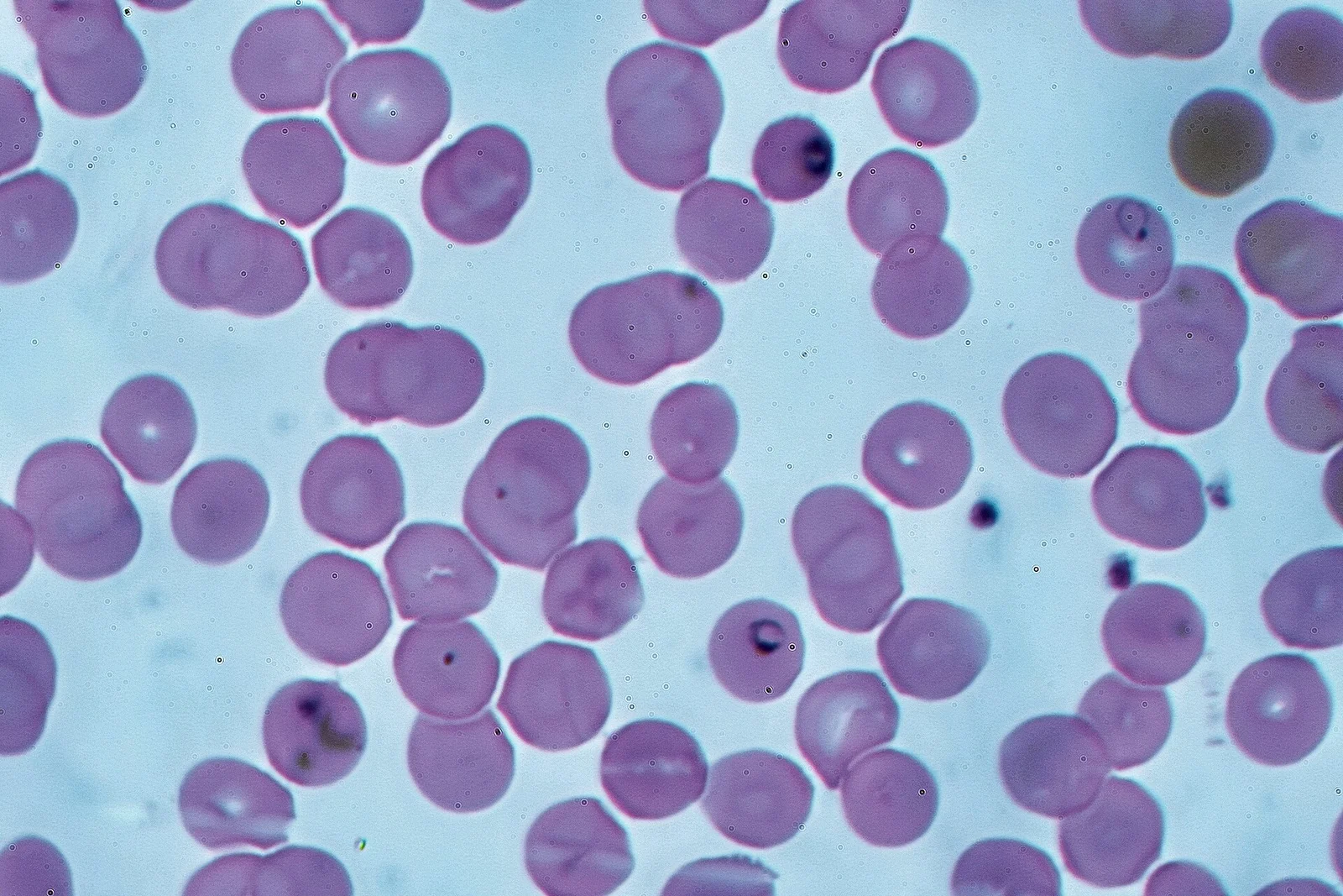
Long description
Ring forms of Plasmodium falciparum in red blood cells (Giemsa stain).

Thick and thin blood films are the diagnostic gold standard. The thick film concentrates red cells and is used to detect parasites at low density; the thin film fixes red-cell morphology and is used to identify the species and to estimate the percentage of parasitaemia. Anticoagulated EDTA blood is acceptable, but films should be made promptly because red-cell changes alter morphology over time.
Because non-immune travellers can be symptomatic with very low parasite densities, a single negative film does not exclude malaria. Repeat thick and thin films every 12 to 24 hours, for three sets, before deciding malaria is absent. Parasitaemia should be quantified as a percentage of red cells on every smear.
Antigen-detecting rapid tests give a result in 2 to 15 minutes and are valuable where reliable microscopy is not available, in urgent settings, and after hours. They detect malaria antigens rather than demonstrating parasites. Where one is used, every RDT should be followed by microscopy to confirm a positive result, to confirm the species and to quantify the percentage of infected red cells. Negative RDTs in a highly suspicious case should be backed up with repeated blood films. [2]
A complete blood count and routine chemistry panel are useful in every suspected or confirmed case. They detect severe anaemia, thrombocytopenia, hypoglycaemia, renal impairment, hyperbilirubinaemia and acid-base disturbance. Hypoglycaemia in particular must be checked in severe malaria. G6PD deficiency testing should be requested early in P. vivax or P. ovale evaluation so that anti-relapse treatment is not delayed. PCR is more sensitive than microscopy but is often too slow for emergency diagnosis; it is most useful for confirming species after diagnosis and for identifying mixed infections. [2]
No formal score is needed. The diagnosis is the demonstration of parasites on a blood film, RDT or PCR. Speciation matters because it directs treatment.
For travellers and residents in non-endemic areas, any febrile person with relevant travel history should be tested immediately, and treatment started for severe disease even before the film result if clinical suspicion is high and there is no rapid test.
Approach to a patient with suspected malaria
- Fever and travel history (last 12 months)Take destination, timing, prophylaxis and night exposure history
- Urgent thick and thin films plus RDTQuantify parasitaemia and identify species; repeat if negative
- Question: Any criterion for severe malaria?
Yes (or not yet known and very unwell)
- Start IV artesunate 2.4 mg/kg at 0, 12, 24 hGive 3 doses; repeat parasitaemia 4 h after the third dose
- Supportive careCorrect hypoglycaemia, fluid status, renal and respiratory failure
- Follow-on oral treatmentFull course beginning 8-24 h after last artesunate when parasitaemia is 1% or less
No (uncomplicated)
- Question: Species?
P. falciparum or unknown
- Artemisinin-based combination therapy, 5-day course with AL in the U.S.With food for absorption; avoid the drug used for prophylaxis
P. vivax or P. ovale
- Chloroquine where sensitive, or ACT, plus anti-relapse primaquine or tafenoquine after G6PD testAdd anti-relapse treatment
P. malariae or P. knowlesi
- Chloroquine or hydroxychloroquine if a mono-infection is confirmed, otherwise an ACTConfirm species, ideally by PCR
Intravenous artesunate is first-line for severe malaria of any species and from any endemic area. Dosing is 2.4 mg/kg intravenously, repeated at 0, 12 and 24 hours for the initial three doses. The percentage parasitaemia is rechecked at least 4 hours after the third dose. If parasitaemia is above 1 percent, intravenous artesunate continues once daily for up to 6 more days, to a total of 7 days. Once parasitaemia is 1 percent or less and oral medicines are tolerated, a full oral follow-on course is given beginning 8 to 24 hours after the last intravenous dose, even if intravenous artesunate was given for 7 days. The oral partner drug needs the longer half-life. [3]
If intravenous artesunate is not immediately available, give one of the oral options listed for uncomplicated disease while it is obtained, by mouth or by nasogastric tube after an antiemetic. Intravenous artesunate is preferred and oral therapy should be switched to intravenous artesunate as soon as it arrives.
Supportive care is as important as antimalarials. Treat hypoglycaemia with intravenous glucose. Be cautious with fluids: adults with severe malaria can develop pulmonary oedema, and fluid bolus strategies that work in bacterial sepsis are not automatically appropriate. Acute kidney injury may need dialysis. Severe anaemia may need transfusion. Seizures are treated with standard anticonvulsants. Acidosis usually improves with treatment and perfusion.
Artesunate is generally well tolerated. After recovery, watch for late post-treatment haemolysis, which can occur with intravenous artesunate, particularly in patients with high initial parasitaemia.
If severe malaria is accompanied by pregnancy, involve obstetric and critical-care teams early.
Always take or confirm the species first, and always give a full course. If a prophylaxis drug has already been taken, treat with a different drug.
Listed in order of preference (CDC):
- Artemether-lumefantrine, one tablet containing 20 mg artemether and 120 mg lumefantrine: 4 tablets per dose. Five-day course: a first dose and a second dose 8 hours later on day 1, then twice-daily dosing on days 2 to 5. The five-day course is now recommended for uncomplicated P. falciparum in the United States because of host and parasite factors. Take with a fatty meal or drink to maximise absorption. [3]
- Atovaquone-proguanil: 4 adult tablets once daily for 3 days (each adult tablet 250 mg atovaquone and 100 mg proguanil), with food. [3]
- Quinine sulfate 542 mg base (650 mg salt) by mouth three times daily for 7 days plus doxycycline 100 mg by mouth twice daily for 7 days; tetracycline is an alternative for adults. [3]
- Mefloquine only when other options cannot be used: 684 mg base (750 mg salt, 3 tablets) by mouth, then 456 mg base (500 mg salt, 2 tablets) 6 to 12 hours later. Mefloquine is contraindicated in epilepsy and neuropsychiatric disorders and is not recommended for infection acquired in Southeast Asia. [3]
Paediatric dosing by weight for artemether-lumefantrine is 1 tablet per dose for 5 to under 15 kg, 2 tablets for 15 to under 25 kg, 3 tablets for 25 to under 35 kg and 4 tablets for 35 kg or more, same schedule, not for infants under 5 kg unless other options are unavailable. Atovaquone-proguanil paediatric dosing and quinine plus clindamycin in younger children are given in the source tables and local protocols. [3]
Give both acute treatment and anti-relapse treatment.
Acute treatment if infection was acquired in a chloroquine-sensitive area:
- Chloroquine phosphate: 600 mg base (1,000 mg salt) by mouth once, then 300 mg base (500 mg salt) at 6, 24 and 48 hours. [3]
- Hydroxychloroquine: 620 mg base (800 mg salt) once, then 310 mg base (400 mg salt) at 6, 24 and 48 hours. [3]
In chloroquine-resistant areas (Papua New Guinea and Indonesia, and increasingly elsewhere) use artemether-lumefantrine or atovaquone-proguanil for acute treatment, in the same doses as above.
Paediatric chloroquine is 10 mg base/kg once, then 5 mg base/kg at 6, 24 and 48 hours, not exceeding the adult dose. Hydroxychloroquine paediatric dosing is 10 mg base/kg then 5 mg base/kg. [3]
Red flagG6PD before anti-relapse drugs
Primaquine and tafenoquine can cause life-threatening haemolysis in G6PD deficiency. Quantitative G6PD testing must be confirmed normal before giving either drug. In G6PD deficiency, alternative strategies such as prolonged weekly chloroquine for one year after acute infection are used instead, with specialist advice. [3]
- Primaquine: 30 mg base (52.6 mg salt, 2 tablets in the U.S.) by mouth once daily for 14 days. For patients weighing at least 70 kg the recommended total dose is 6 mg/kg divided into daily doses of 30 mg base. [3]
- Tafenoquine: 300 mg by mouth as a single dose, only for patients aged 16 years or older, and only when chloroquine or hydroxychloroquine was used for acute treatment because it has not been studied well with other partners. [3]
- Paediatric primaquine dose is 0.5 mg base/kg by mouth once daily for 14 days. [3]
- For intermediate G6PD activity, weekly primaquine 45 mg for 8 weeks may be used with close monitoring. [3]
- Tafenoquine or primaquine is used while breastfeeding only if the infant has normal G6PD activity.
Primaquine or tafenoquine is taken with food to reduce stomach upset. If G6PD activity is very low, give no anti-relapse drug but consider chloroquine 300 mg base (500 mg salt) once weekly for one year from the acute infection to suppress relapse. Pregnant patients should not receive either hypnozoite drug; use weekly chloroquine 300 mg base during pregnancy and treat after delivery if G6PD is normal. [3]
Use chloroquine phosphate or hydroxychloroquine at the same doses as for P. vivax, or an ACT if a mono-infection has not been confirmed by PCR (these species can be confused with P. falciparum on smear). Additional options include quinine plus doxycycline or clindamycin, or mefloquine as a last resort.
Uncomplicated P. falciparum in pregnancy is treated with artemether-lumefantrine preferentially, or quinine plus clindamycin, or mefloquine only if no other option is available (avoid in the first trimester if possible). Chloroquine or hydroxychloroquine are options only if the infection was acquired in a chloroquine-sensitive area. P. vivax or P. ovale in pregnancy is treated with chloroquine or hydroxychloroquine acutely, with weekly chloroquine 300 mg base after the acute attack until delivery. Anti-relapse drugs are contraindicated in pregnancy. [3]
Uncomplicated malaria is curable in nearly everyone with prompt, correct treatment. Severe malaria can progress within hours and demands immediate therapy; cerebral malaria carries substantial mortality, especially in children, and survivors may have long-term neurological deficits. Repeated P. falciparum infections in young children cause severe anaemia. Chronic P. malariae infection can lead to nephrotic syndrome.
- Cerebral malaria with coma and seizures
- Severe anaemia and haemolysis
- Acute kidney injury and blackwater fever
- Acidosis and shock
- ARDS and pulmonary oedema
- Hypoglycaemia
- Disseminated intravascular coagulation and bleeding
- Splenic rupture (rare, especially P. vivax)
- Post-malaria neurological syndrome
- Fetal loss and low birth weight in pregnancy
- Nephrotic syndrome after chronic P. malariae infection
Prevention rests on avoiding bites from Anopheles mosquitoes, which bite mainly from dusk to dawn. Sleep under insecticide-treated nets; screen windows and doors; wear loose-fitting long sleeves, long trousers and socks; use repellents approved by health authorities (DEET, picaridin or IR3535) on exposed skin; and reduce breeding sites around dwellings. Bite avoidance is recommended for everyone in endemic areas and complements chemoprophylaxis for travellers.
Choice of prophylaxis depends on the destination (resistance patterns), duration of travel, budget, age, pregnancy status and comorbidities. The commonly recommended options and their adult doses and timings are:
| Drug | Regimen | Key cautions |
|---|---|---|
| Atovaquone-proguanil | 1 adult tablet daily (250/100 mg), starting 1-2 days before travel and for 7 days after leaving | Not in pregnancy or breastfeeding low-weight infants; avoid in severe renal impairment [4] |
| Doxycycline | 100 mg daily, starting 1-2 days before travel and for 4 weeks after leaving | Not in pregnancy or children under 8; photosensitivity [4] |
| Mefloquine | 228 mg base (250 mg salt) once weekly, starting 1-2 weeks before travel and for 4 weeks after leaving | Not with certain psychiatric or seizure disorders; resistance in Southeast Asia [4] |
| Chloroquine | 300 mg base (500 mg salt) once weekly, starting 1-2 weeks before travel and for 4 weeks after leaving | Not in chloroquine-resistant areas [4] |
| Primaquine | 30 mg base daily, starting 1-2 days before travel and for 7 days after leaving | Requires normal G6PD; not in pregnancy or breastfeeding unless infant has normal G6PD [4] |
| Tafenoquine | 200 mg daily for 3 days before travel, weekly during travel, and 200 mg for one week after leaving | Adults only; requires normal G6PD [4] |
If prophylaxis fails or a traveller returns with fever, remember that prophylaxis can suppress symptoms, particularly for P. vivax, and that infection may appear after returning home.
WHO has recommended broad use of the RTS,S/AS01 malaria vaccine since October 2021, and a second vaccine, R21/Matrix-M, since October 2023, for children in regions with moderate to high P. falciparum transmission. These are given alongside, not instead of, bed nets and other malaria control measures. [5]
Sickle cell trait (HbAS) confers substantial protection against severe P. falciparum malaria in endemic areas, which explains its high prevalence in those regions through balanced polymorphism. Sickle cell disease, in contrast, is a risk factor for severe malaria. A traveller with sickle cell disease should have their malaria prevention and treatment planned carefully.
- Where microscopy is unavailable, use rapid tests but confirm results with microscopy as soon as possible; in their absence treat on strong clinical and epidemiological suspicion.
- Refer to national malaria guidelines (including WHO guidance) for the first-line ACT in that region, because resistance patterns differ.
- Intravenous artesunate is the preferred treatment for severe malaria; if unavailable, use the best available parenteral alternative (intravenous artemether or quinine) and transfer the patient.
- Oral therapy in a patient who cannot swallow can be given by nasogastric tube.
- G6PD testing may not be immediately available; if hypnozoite treatment must be delayed, use weekly chloroquine suppression and document the need for testing.
- Blood transfusion, dialysis and intensive care are scarce; prioritise early antimalarial treatment, glucose checks, and careful fluid management.
- In endemic areas, presumption often drives treatment when tests are unavailable; still record and review outcomes.
Children in endemic areas suffer repeated P. falciparum infections causing severe anaemia; infections should be diagnosed and treated promptly. Infants under 5 kg require alternative regimens. Pregnant people need urgent treatment and specialist involvement, and anti-relapse drugs are avoided. People with G6PD deficiency need alternatives to primaquine and tafenoquine. Travellers and non-immune visitors are at higher risk of severe disease from any species and should be treated urgently.
- Malaria is caused by Plasmodium parasites spread by female Anopheles mosquitoes, and always consider it in a febrile person with recent travel to an endemic area.
- P. falciparum is the most deadly species and causes most severe malaria through sequestration in organ vessels; P. vivax and P. ovale relapse from hypnozoites; P. malariae causes quartan fever and chronic low-level infection; P. knowlesi is zoonotic and can progress rapidly.
- Incubation is shortest for P. falciparum and longest for P. malariae, but P. vivax and P. ovale can relapse months later.
- Presentation is non-specific early fever with chills or rigors, headache, aches and gastrointestinal symptoms; severe features include cerebral disease, severe anaemia, acidosis, hypoglycaemia, acute kidney injury, ARDS and shock.
- Severe malaria is defined by parasitaemia at least 5 percent or any organ criterion.
- Thick and thin blood films are the diagnostic standard; repeat over up to 24-hour intervals for three sets if negative.
- Intravenous artesunate 2.4 mg/kg at 0, 12 and 24 hours, then guided by parasitaemia and oral follow-on, is first-line for severe malaria.
- Uncomplicated P. falciparum is treated with artemisinin-based combination therapy; uncomplicated non-falciparum malaria is treated with chloroquine where sensitive.
- P. vivax and P. ovale require anti-relapse treatment with primaquine or tafenoquine only after normal G6PD activity is confirmed.
- Travellers need bite avoidance plus region-appropriate chemoprophylaxis taken exactly as timed before and after travel.
- Sickle cell trait protects against severe P. falciparum malaria, while sickle cell disease is a risk factor.
Answer each one in your head first, then open the answer.
A 28-year-old returns from Ghana two weeks ago with fever, rigors and an enlarged spleen. Blood film shows 8 percent parasitaemia. Why is this severe malaria and what is the immediate treatment?
Show answerHide answer
It is severe malaria because parasitaemia is at or above 5 percent. Give intravenous artesunate 2.4 mg/kg at 0, 12 and 24 hours, check parasitaemia 4 hours after the third dose, and give a full oral follow-on course when parasitaemia falls to 1 percent or less and the patient can swallow.
Why do P. vivax infections relapse months after the original attack, and what must be checked before giving the drug used to prevent relapse?
Show answerHide answer
Dormant liver stages (hypnozoites) reactivate and release merozoites. Anti-relapse treatment with primaquine or tafenoquine can cause haemolysis in G6PD deficiency, so quantitative G6PD activity must be confirmed normal first.
A traveller leaves for a malaria-endemic area in two days and wants a daily tablet started as late as possible. Which prophylaxis options fit, and when should the first dose be taken?
Show answerHide answer
Atovaquone-proguanil (start 1-2 days before travel, continue 7 days after returning), doxycycline (start 1-2 days before travel, continue 4 weeks after), or primaquine or tafenoquine if G6PD is normal. Chloroquine and mefloquine need 1-2 weeks and are not suited to last-minute travel.
A febrile patient had one negative thick film 4 hours after admission. Can malaria be excluded?
Show answerHide answer
No. In non-immune patients parasites may be below detectable density initially. Repeat thick and thin blood films every 12 to 24 hours and do three sets before excluding malaria. [2]
Explain in one sentence why sickle cell trait is common in parts of Africa despite causing disease when homozygous.
Show answerHide answer
Heterozygotes have a marked survival advantage against severe P. falciparum malaria, so the trait persists despite the cost of sickle cell disease in homozygotes.
Sickle cell disease; haemolytic anaemias; acute kidney injury; transfusion reactions. Adult and paediatric dosing details should be checked against current national formularies and the CDC treatment tables.
- Centers for Disease Control and Prevention (CDC), National Center for Emerging and Zoonotic Infectious Diseases. Clinical Features of Malaria. CDC. Clinical guidance for malaria diagnosis and treatment in the U.S. Reviewed 20 Mar 2024.. Accessed .
- Centers for Disease Control and Prevention (CDC), National Center for Emerging and Zoonotic Infectious Diseases. Clinical Testing and Diagnosis for Malaria. CDC. Clinical guidance for malaria diagnosis and treatment in the U.S. Updated 20 Mar 2024.. Accessed .
- Centers for Disease Control and Prevention (CDC), National Center for Emerging and Zoonotic Infectious Diseases. Malaria in the United States: Treatment Tables. CDC. Clinical treatment tables for malaria. Updated 17 Aug 2026.. Accessed .
- Centers for Disease Control and Prevention (CDC), National Center for Emerging and Zoonotic Infectious Diseases. Choosing a Drug to Prevent Malaria. CDC. Guidance for healthcare providers on malaria chemoprophylaxis for travellers. Updated 18 Jul 2024.. Accessed .
- World Health Organization (WHO). Malaria. WHO. Malaria fact sheet, 4 Dec 2025 (WHO reference data and vaccine recommendations).. Accessed .
Figure credits
Educational content for medical students and doctors. It is not a substitute for clinical judgement or local guidelines.