Tuberculosis
Airborne infection with Mycobacterium tuberculosis: why most infections stay latent, how active disease presents in lung and elsewhere, how it is diagnosed.
- Tuberculosis (TB) is an airborne infection caused by Mycobacterium tuberculosis. It most often affects the lungs, but it can attack almost any organ.
- Only a minority of infected people become ill: an estimated quarter of the world's population carries the bacterium, yet about 5 to 10% of those infected develop active disease in their lifetime. [2]
- Disease emerges when the immune system fails to contain the bacillus, either soon after infection or years later through reactivation, especially with HIV or immunosuppression. [2] [8]
- Active pulmonary TB is treatable and curable: about 85% of people who develop TB disease can be treated successfully with standard drug regimens, if diagnosis is timely. [2]
- Latent infection causes no symptoms and is not contagious, but it can be detected and treated to prevent later disease. [2] [5]
By the end of this page you should be able to:
- Explain how M. tuberculosis travels from one person's lungs to another's and why only some infections become disease.
- Recognise the symptom patterns of pulmonary, pleural, miliary, meningeal, spinal, lymph node, genitourinary and abdominal TB.
- Use sputum smear, nucleic acid amplification (Xpert MTB/RIF), culture and chest imaging to diagnose TB, and interpret tuberculin skin tests and interferon-gamma release assays with their limits.
- Plan first-line treatment for drug-susceptible pulmonary TB in adults and children, and describe the shorter 4-month regimens and where they apply.
- Manage drug adverse effects, monitor therapy, treat latent infection and trace contacts.
- Time antiretroviral therapy in TB/HIV coinfection and state the place of BCG.
DefinitionTuberculosis (TB)
TB is a disease caused by bacteria of the Mycobacterium tuberculosis complex, most often M. tuberculosis itself. It is usually an infection of the lungs (pulmonary TB), but the bacillus can settle in almost any organ (extrapulmonary TB). Infection without symptoms or illness is called latent tuberculosis infection (LTBI); once the bacterium multiplies and causes symptoms, it is active TB disease.
A useful distinction runs through the whole topic:
- Latent TB infection: the person is infected, feels well, is not infectious, and has a positive test for infection (tuberculin skin test or interferon-gamma release assay). The bacterium is contained by granulomas. [2]
- Active TB disease: the person is symptomatic (and usually infectious if the lungs are involved), with bacteria recoverable from sputum or affected tissue. [2]
The causative organisms are slow-growing, robust, acid-fast bacilli. M. tuberculosis complex also includes closely related species such as M. bovis, which is acquired mainly through unpasteurised milk and is a minor contributor to human disease in most settings. [2]
TB is one of the world's great infectious killers. In 2024, an estimated 10.7 million people fell ill with TB, of whom 5.8 million were men, 3.7 million women and 1.2 million children. A total of 1.23 million people died of TB, including 150 000 among people living with HIV. Globally, TB is the leading cause of death from a single infectious agent and among the top ten causes of death. [1]
About a quarter of the world's population is estimated to have been infected with TB bacteria, yet only about 5 to 10% of those infected develop disease in their lifetimes; babies and children are at higher risk of progression once infected. [1] [2] Global efforts since 2000 are estimated to have saved 83 million lives. [1]
Geography matters. More than 80% of cases and deaths occur in low- and middle-income countries, and 87% of new cases occur in the 30 high-burden countries. The largest share of new cases in 2024 was in the South-East Asia Region (34%), followed by the Western Pacific (27%) and the African Region (25%). Eight countries accounted for about two-thirds of the global total: India (25%), Indonesia (10%), the Philippines (6.8%), China (6.5%), Pakistan (6.3%), Nigeria (4.8%), the Democratic Republic of the Congo (3.9%) and Bangladesh (3.6%). [1] [2] (The imaging review, using 2022 figures, noted South-East Asia 46%, Africa 23% and the Western Pacific 18%.) [2]
Certain conditions magnify risk. People living with HIV are 14 to 18 times more likely to develop TB disease than people without HIV, and TB is the leading cause of death among people with HIV. [2] [8] In 2024, about 150 000 people died of HIV-associated TB and only 61% of people living with HIV who developed TB received antiretroviral therapy. [1] Attributable cases in 2024 included an estimated 0.97 million from undernutrition, 0.93 million from diabetes, 0.74 million from alcohol use disorders, 0.70 million from smoking and 0.57 million from HIV. [1]
TB spreads through the air. People with infectious pulmonary TB release tiny droplet nuclei, 1 to 5 micrometres across, when they cough, sneeze, speak or sing. These particles are small enough to stay suspended and to be inhaled into the alveoli of a new host. [2] Larger droplets fall to the ground; it is the fine nuclei that matter. Close, prolonged contact in crowded or poorly ventilated spaces (households, shelters, prisons, health facilities) drives transmission. [1] [2]
Higher exposure risk is carried by household contacts of a person with infectious TB, people born in or frequently travelling to countries where TB is common, residents of homeless shelters, prisons and nursing homes, health care workers, and infants, children and adolescents exposed to adults at increased risk. [10]
Once infected, progression is governed by the balance between bacillus and host immunity:
- HIV infection, especially with low CD4 counts, the strongest risk factor. [8]
- Immunosuppressive therapy such as tumour necrosis factor-alpha antagonists, systemic corticosteroids equivalent to or greater than 15 mg of prednisone daily, or therapy after organ transplantation. [10]
- Diabetes mellitus, silicosis, chronic renal failure, leukaemia and head, neck or lung cancer. [10]
- Malnutrition and low body weight (under 90% of ideal body weight). [10]
- Substance use, including injection drug use and heavy alcohol use, and tobacco use. [10]
- Recent infection (within the preceding 2 years) and infection in children under 5 years. [10]
- Age extremes, and gastrectomy or jejunoileal bypass. [10]
Patients with TB-HIV coinfection have higher rates of extrapulmonary and disseminated disease and of reactivation of latent infection. Among people with TB who have HIV, 25% present with extrapulmonary TB, most often lymph node disease. [2]
In most people, inhaled bacilli are engulfed and destroyed by alveolar macrophages; the immune response walls off survivors in granulomas, and the person has latent infection without illness. [2] In a minority, the immune response fails, the bacillus multiplies, macrophages die, and infection spreads by lymphatics and blood to the lung and other organs, producing active disease. [2]
The course of infection is classically described in stages:
- Implantation of the bacillus in an alveolus, usually in a well-ventilated part of the lung.
- Lymphatic spread to hilar nodes, forming the primary (Ghon) complex: a subpleural Ghon focus plus involved hilar lymph nodes. This is the hallmark of infection in a person who has not previously met the organism; it can occur in adults as well as in children. [2]
- Haematogenous spread to many organs, most often without symptoms. In some this produces early severe disease (meningitis or miliary TB). [2]
- Healing with fibrosis and calcification, producing the Ranke complex, or progression to active disease. [2]
Primary TB is disease that develops within a year of infection. Postprimary (secondary) TB is reactivation of a quiescent focus, usually more than a year later, or reinfection with a new strain. Reactivation is more likely with HIV and other risk factors; reinfection matters more in high-incidence settings. [2]
On these grounds, 5 to 15% of people initially infected progress to active disease. [2] Patients with latent infection who do progress usually do so within the first 2 years, or later if risk factors such as immunosuppression appear. [2] Latent infection is not infectious and is usually asymptomatic; active disease is symptomatic and, when pulmonary, infectious. [2]
In HIV-negative people who are infected, the lifetime risk of progression is conventionally given as about 10% without other risk factors. In people with HIV who are not on antiretroviral therapy, the risk rises to roughly 7 to 10% each year. [8]
The lesion is a granuloma with central caseous necrosis, surrounded by macrophages and lymphocytes; Langhans giant cells (multi-nucleated cells with nuclei arranged in a horseshoe) are typical. Lesions may heal with fibrous scarring and calcification, or soften and cavitate, releasing bacilli into airways. Histology showing caseating granulomas with acid-fast bacilli in tissue is strongly supportive of TB, especially in extrapulmonary disease where cultures are often negative. [2]
Pulmonary TB often begins insidiously. Symptoms are common to other chest diseases, so TB is missed unless it is considered: in many settings it is underdiagnosed for months. [10]
Symptoms include a persistent cough, haemoptysis, chest pain, evening fever, night sweats, fatigue and weight loss; dyspnoea develops when disease is extensive. [2] On examination findings are often absent except with extensive consolidation; reduced breath sounds or signs of a pleural effusion may be present. [2] In immunocompromised patients, particularly with advanced HIV, presentation can be atypical (lower-lobe infiltrates, hilar lymphadenopathy, or even a normal chest film despite active disease). [8]
TB is called the great mimic: presentation depends on the organ involved.
- Pleural TB. A pleural effusion, often unilateral, that develops 3 to 7 months after infection and is seen in about a quarter of primary cases; effusion is ten times more common in high-incidence settings than in low-incidence ones, and more common with HIV. Concomitant parenchymal disease is found in 20 to 50% of cases. Pleural fluid alone is often culture-negative; biopsy helps. [2]
- Lymph node TB. The most common extrapulmonary form. Cervical (scrofula) and hilar or mediastinal nodes present as painless, firm, slowly enlarging lymph nodes that may soften and form sinuses. Hilar or paratracheal lymphadenopathy is usually unilateral and right-sided; bilateral in about a third. Radiographic lymphadenopathy is seen in up to 96% of children and 43% of adults with TB. [2]
- Miliary TB. Haematogenous dissemination with innumerable tiny (typically 1 to 3 mm) nodules distributed randomly through both lungs and often other organs. Fever, weight loss, hepatosplenomegaly and pancytopenia are common; it was once mainly a childhood disease and is now increasingly seen in severely immunocompromised adults. [2]
- Meningeal TB. A subacute meningitis with headache, fever, neck stiffness, cranial nerve palsies and altered consciousness. It demands urgent treatment and specialist care. [8]
- Spinal (Pott) disease. Chronic back pain, local tenderness, kyphotic deformity, cold abscess and possible cord compression with neurological deficits. The thoracic spine is commonly involved, often with disc space narrowing and paravertebral shadowing.
- Genitourinary TB. Often silent: sterile pyuria, haematuria, dysuria, renal colic, epididymo-orchitis or salpingitis, and infertility in either sex. It is usually a reactivation disease.
- Abdominal TB. Peritoneal TB (ascites, abdominal pain, weight loss), intestinal TB (obstruction, malabsorption), and hepatosplenomegaly. Can occur without pulmonary disease.
- Cardiac TB. Pericardial involvement is the most frequent cardiac form and carries a mortality of roughly 40%. [2] Cardiac TB is rare overall (up to 2% of immunocompetent people with TB) but much more common with HIV. [2]
| Condition | Features that point away from or towards TB |
|---|---|
| Community-acquired pneumonia | Acute onset, lobar consolidation, responds to ordinary antibiotics within days; TB cough and systemic symptoms persist |
| Lung cancer | Older smoker, haemoptysis, mass with irregular margins, hilar lymphadenopathy; can cavitate; biopsy separates them |
| Sarcoidosis | Bilateral hilar lymphadenopathy, non-caseating granulomas, often no fever or weight loss |
| Non-tuberculous mycobacterial disease | Different species, different setting (older women with bronchiectasis, for example); culture identifies species |
| Fungal infection (histoplasmosis, aspergillosis) | Geographic exposure, aspergilloma in a pre-existing cavity, positive fungal tests |
| Lymphoma | Generalised lymphadenopathy, night sweats and weight loss; biopsy shows lymphoma, not caseation |
| Other extrapulmonary infection | Pleural TB can look like bacterial empyema; spinal TB like pyogenic spondylodiscitis; genitourinary TB like chronic urinary infection. Culture, biopsy and PCR settle these |
A person with TB often looks like someone with "just a cough", which is why TB has to be actively looked for in anyone with a persistent cough and risk factors. [10]
Approach to suspected tuberculosis
- Symptoms or risk factors suggest TBPersistent cough, haemoptysis, fever, night sweats, weight loss; HIV, immunosuppression, travel or contact history
- Test for infection and diseaseSputum smear, nucleic acid amplification, culture; chest radiograph; HIV test; TST or IGRA as indicated
- Question: Bacteriology positive or strong clinical case?
Yes
- Start treatment and isolateFirst-line 2HRZE/4HR or an eligible shorter regimen; respiratory isolation; drug susceptibility pending
- Trace and test contactsOffer preventive therapy to those with latent infection
No or uncertain
- TST or IGRA positive, well and asymptomatic?Consider latent TB treatment after active disease is excluded
- Repeat or broaden testsCT, bronchoscopy, biopsy for extrapulmonary or paucibacillary disease; discuss with TB expert
The aim is three things: detect TB, identify the site and burden, and define drug susceptibility.
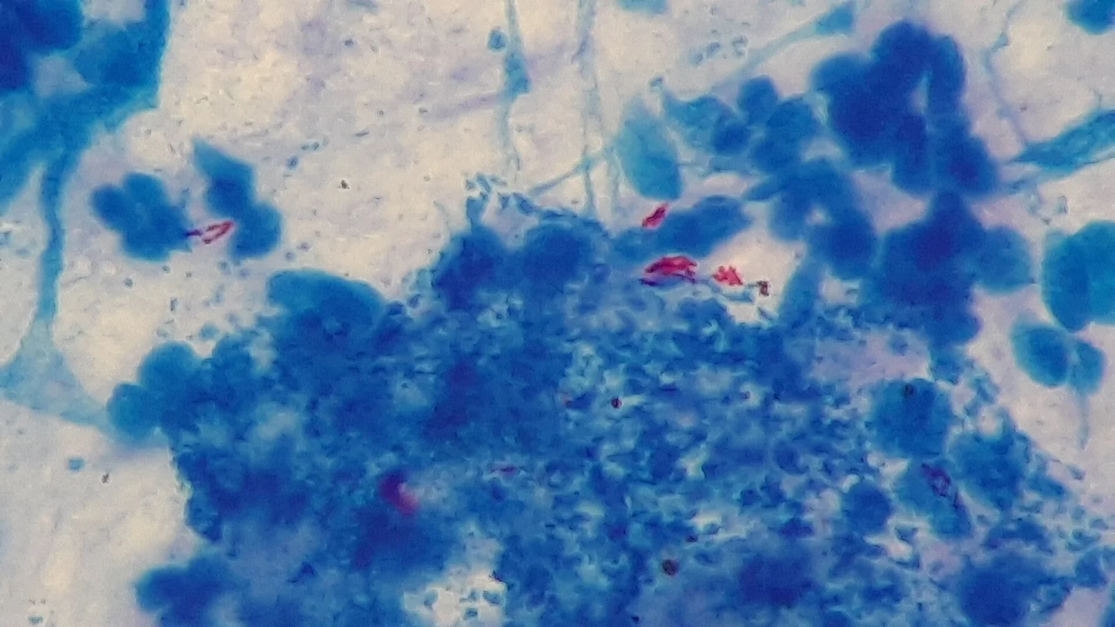
Figure 1Mycobacterium tuberculosis in Ziehl-Neelsen stained smear of sputum
Long description
Ziehl-Neelsen stained sputum smear: thin red acid-fast bacilli of Mycobacterium tuberculosis against a blue background of cells.

- Sputum smear microscopy for acid-fast bacilli gives a rapid answer (often same day) but is insensitive, especially in children, in HIV, and where disease is paucibacillary. [2]
- Nucleic acid amplification, most commonly Xpert MTB/RIF, detects M. tuberculosis and rifampicin resistance directly from sputum in under 2 hours and is the World Health Organization's recommended first test in anyone with symptoms of TB. [2] [1]
- Culture remains the gold standard microbiological test; it allows speciation and full drug susceptibility testing, but takes weeks. [2] [10]
- In extrapulmonary disease, sites are often paucibacillary and cultures frequently negative, so biopsy for histology and polymerase chain reaction is commonly needed. [2]
A diagnosis of TB can also be made clinically when bacteriology is negative, based on history, imaging, tuberculin or interferon-gamma results and clinical judgement. Worldwide in 2022, only about 63% of people diagnosed with pulmonary TB were bacteriologically confirmed, ranging from 56% in the Eastern Mediterranean Region to 79% in the Americas. [2]
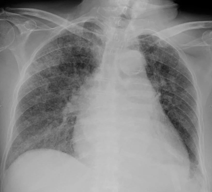
Figure 2Chest radiograph of miliary tuberculosis 1
Long description
Chest radiograph of miliary tuberculosis with innumerable tiny nodules spread evenly through both lungs.

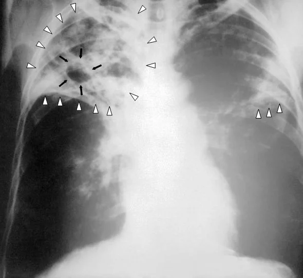
Figure 3Tuberculosis-x-ray-1
Long description
Chest radiograph of advanced pulmonary tuberculosis with a large cavitary lesion in the upper zone and surrounding infiltrate.

- Chest radiograph is central. Primary disease tends to produce lower- and middle-lobe consolidation and hilar or paratracheal lymphadenopathy; postprimary (reactivation) disease classically favours the upper lobes, with cavitation in up to 50% of cases, and nodules, tree-in-bud (centrilobular 2 to 4 mm branching opacities) and satellite nodules. A single tuberculoma occurs in about 5% of cases, and miliary disease shows innumerable 1 to 3 mm nodules in random distribution. Consolidation may take up to 2 years to clear. [2]
- Computed tomography characterises subtle or small lesions, lymph nodes (often larger than 2 cm with rim enhancement and low-density centres) and complications.
- Ultrasound helps guide pleural or nodal sampling and shows pleural thickening and loculation well.
Imaging cannot reliably distinguish active from latent or old disease by itself, so it must be paired with bacteriology and clinical assessment. [2]
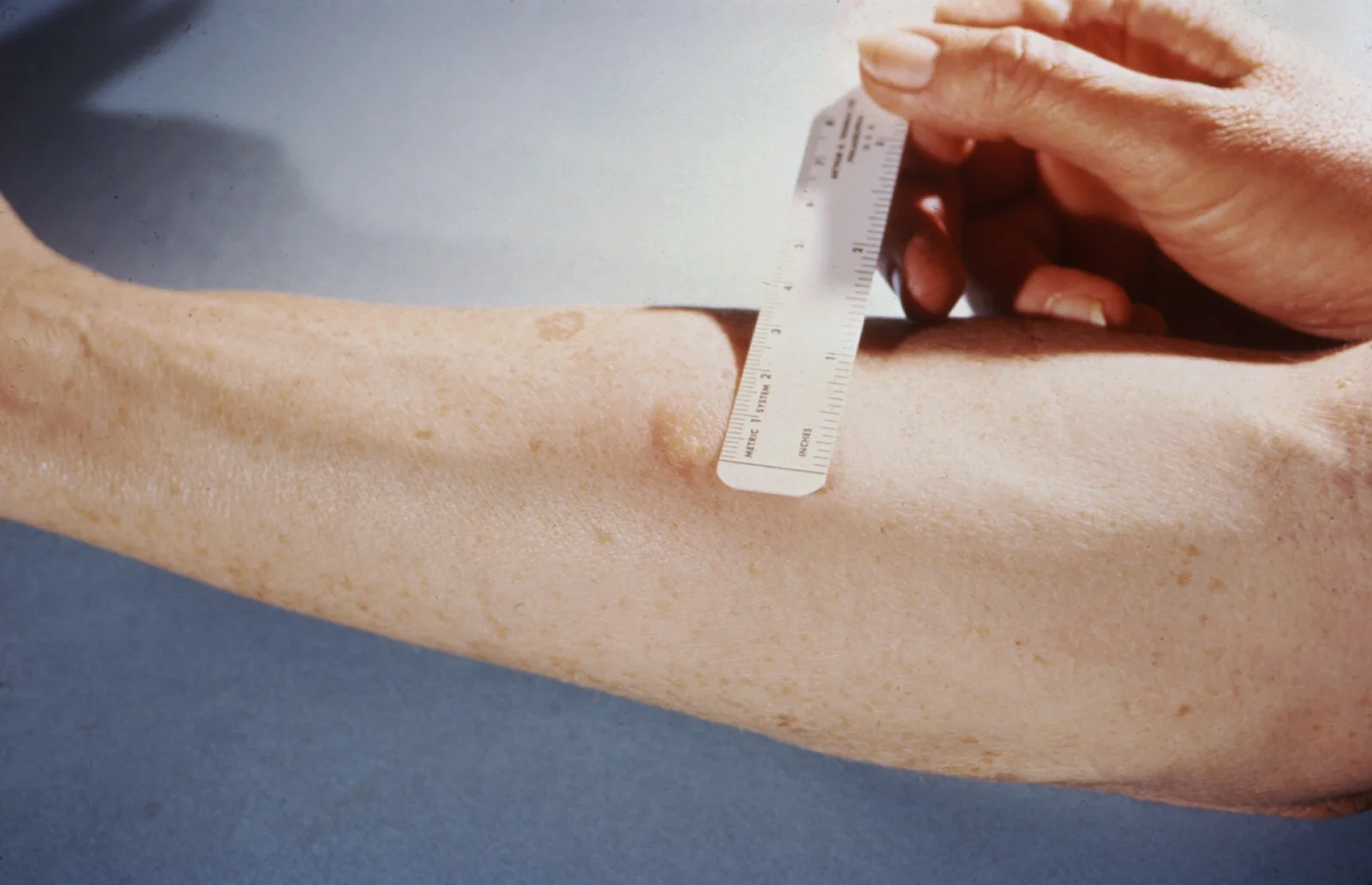
Figure 4Mantoux Test 48h
Long description
Reading a tuberculin skin test: a ruler held across the forearm to measure the width of the raised induration.

- Tuberculin skin test (TST, Mantoux). An intradermal injection of purified protein derivative; read at 48 to 72 hours by measuring induration. It becomes positive 2 to 8 weeks after infection. Induration thresholds: 5 mm or more is positive in people with HIV, recent contacts, immunosuppression (including corticosteroids at or above 15 mg prednisone daily or TNF-alpha antagonists), organ transplantation, and those with chest imaging suggesting prior TB; 10 mm or more in people born in high-incidence countries and other at-risk groups (children under 5, diabetes, silicosis, severe kidney disease, certain cancers, low body weight, people who use drugs or alcohol, congregate settings, laboratory workers); 15 mm or more in people with no known risk factors. [9]
- Interferon-gamma release assays (IGRAs) are blood tests using synthetic TB antigens; they are preferred for anyone who has had BCG vaccination because BCG does not cause a positive IGRA. [7] [9]
- Neither test can tell latent infection from active disease; both can be falsely negative in severe TB illness (including miliary disease and meningitis) and in HIV. [2] [9] Recent live virus vaccination (measles, rubella, varicella, yellow fever) can also suppress the TST. [9]
- BCG vaccination produces false-positive TST reactions that cannot reliably be distinguished from true infection; TST reactivity from BCG wanes over time and is boosted by repeated testing. [7]
All patients need testing for HIV and, where appropriate, renal and liver function and blood glucose or glycosylated haemoglobin, because diabetes and renal disease both raise TB risk and affect drug choice and dosing. [1] [10]
There is no single staging system for TB. For practical management, people fall into one of three states:
- Latent TB infection: positive TST or IGRA, no symptoms, normal (or old stable) imaging, and bacteriology negative. [2]
- Active TB disease: symptoms and/or imaging changes, with bacteriological confirmation where possible, or a confident clinical diagnosis. [2]
- Active TB disease, clinically diagnosed: started on treatment on strong clinical and epidemiological grounds when laboratory proof is unavailable. [2]
Severity is judged by site and extent rather than a score: cavitary or extensive pulmonary disease, positive smears after 2 months, meningitis, miliary or spinal disease, and immunosuppression each change the regimen and duration. Extrapulmonary TB outside a few "less severe" sites generally needs longer or specialist treatment. [3]
Because TB can kill quickly (miliary or meningeal TB especially), and because an infectious patient endangers others, the priorities are prompt diagnosis, isolation of infectious patients, treatment, and assessment of contacts. Baseline evaluation includes history, examination, imaging, bacteriology, drug susceptibility testing, HIV test and tests for the common comorbidities. [8] [10]
- Patients with suspected or confirmed infectious TB should be managed in appropriate respiratory isolation until infectiousness falls, under local public health rules. [1]
- Empirical treatment should not be delayed in a very sick patient when TB is strongly suspected. Drug susceptibility results then guide adjustment. [4]
The backbone is a six-month regimen: 2 months of isoniazid, rifampicin, pyrazinamide and ethambutol (2HRZE), followed by 4 months of isoniazid and rifampicin (4HR), given daily with direct observation. [3] [4]
- This 2HRZE/4HR regimen is the preferred regimen for adults and adolescents with newly diagnosed pulmonary TB. [4]
- A 9-month regimen (2HRZE/7HR) is used when there is cavitary disease and the culture at the end of 2 months is still positive with a drug-susceptible organism, when pyrazinamide was not given or the organism is pyrazinamide-resistant, or in HIV-positive patients who do not receive antiretroviral therapy. [4] [8]
- Shorter 4-month regimen for selected adults: 2 months of isoniazid, rifapentine, pyrazinamide and moxifloxacin, then 2 months and 1 week of isoniazid, rifapentine and moxifloxacin (17 weeks total). It is an option for pulmonary TB with organisms not known or suspected to be drug-resistant, in people aged 12 years and over who weigh at least 40 kg, with HIV only if the CD4 count is at least 100 cells per microlitre and antiretroviral therapy is planned or under way without known interactions, and with no contraindications. It is a conditional recommendation of moderate certainty. [3] [4] Dosing from the adapted guideline: isoniazid 300 mg daily for 17 weeks; rifapentine 1200 mg daily for 17 weeks; pyrazinamide by weight (1000 mg daily if 40 to under 55 kg, 1500 mg if 55 to 75 kg, 2000 mg if over 75 kg) for 8 weeks; moxifloxacin 400 mg daily for 17 weeks. Give on food and avoid milk, antacids and other cation-containing products. [3]
- Directly observed therapy (DOT) is the standard of care; video DOT (vDOT) is an acceptable equivalent. [4] [8]
- In the pivotal non-inferiority trial, the 4-month rifapentine-moxifloxacin regimen gave 84.6% cure against 85.4% for the 6-month standard, with grade 3 or higher adverse events of 18.7% versus 19.3%. [3]
Pyridoxine (vitamin B6), 25 to 50 mg daily, is given with isoniazid to everyone at risk of neuropathy (pregnancy, breastfeeding infants, HIV, diabetes, alcohol use, malnutrition, chronic renal failure, advanced age); if peripheral neuropathy occurs, increase to 100 mg daily. [4] [6]
Drugs are best taken together in a single dose. With DOT they may be given 5 days a week with the dose count adjusted. Bioavailability of all the drugs except rifapentine is greatest taken on an empty stomach. [4]
For children and adolescents aged 3 months to 16 years with non-severe TB (peripheral lymph node TB; intrathoracic lymph node TB without airway obstruction; uncomplicated pleural effusion; or paucibacillary, non-cavitary disease confined to one lobe, or without a miliary pattern), a strong recommendation supports a 4-month regimen: 2 months of isoniazid, rifampicin, pyrazinamide with or without ethambutol, then 2 months of isoniazid and rifampicin, rather than the standard 6 months. [3] Dosing is weight-based: isoniazid 10 to 15 mg per kg, rifampicin 10 to 20 mg per kg, pyrazinamide 30 to 40 mg per kg (target 35) and ethambutol 15 to 25 mg per kg (target 20) during the intensive phase. [3] Children who do not meet non-severe criteria, and those with severe extrapulmonary TB, should receive the 6-month regimen. [3] In the supporting trial of 1204 children with non-severe disease, treatment success was 96.9% with both the 4-month and 6-month regimens, and grade 3 or higher adverse events were 7.8% versus 8.0%. [3]
HIV treatment is critical to TB treatment success and must be planned alongside it. [8]
- Six months is the minimum duration for adults with HIV, even with culture-negative disease. If the patient is not on antiretroviral therapy, prolong treatment to 9 months (continuation phase 7 months). [4] [8]
- Prolong to 9 months when response is delayed (positive culture after 2 months). [8]
- The 4-month rifapentine-moxifloxacin regimen may be used with HIV if the CD4 count is at least 100 cells per microlitre and there are no significant drug interactions with antiretroviral therapy. [4] [8]
- Antiretroviral therapy timing: start within the first 2 weeks of TB therapy when the CD4 count is under 50 cells per cubic millimetre, and by 8 to 12 weeks when it is 50 or above. Exception: in TB meningitis, do not start antiretroviral therapy in the first 8 weeks. [8]
- Rifampicin and rifapentine have important and clinically significant interactions with antiretrovirals; rifabutin or adjusted regimens may be needed. [6] [8]
Tuberculosis of the central nervous system, bones and joints, miliary and pericardial TB were excluded from the shorter-regimen trials and are not treated with 4- or 6-month regimens in the same simplified way; they need longer, specialist-directed therapy, often with adjunctive management of complications such as spinal cord compression. Pregnancy, young children, malnourished patients and those with recurrence within the previous 2 years also require individualised plans. [3] [5]
- Multi-drug-resistant TB (MDR-TB) is caused by M. tuberculosis that does not respond to the two most effective first-line drugs, rifampicin and isoniazid. Rifampicin-resistant TB (RR-TB) is defined by resistance to rifampicin. [1]
- Resistance develops from inappropriate use: wrong prescription, poor-quality drugs, or stopping treatment early. [1]
- Drug-resistant TB requires different, usually more expensive drugs with more side effects; in 2024 only about 2 in 5 people with multidrug-resistant TB accessed treatment. [1]
- The World Health Organization prioritises a 6-month all-oral BPaLM/BPaL regimen (bedaquiline, pretomanid, linezolid, with or without moxifloxacin) for eligible patients with MDR or RR-TB; in 2024 about 34 000 people started on these shorter regimens, up from 5 653 in 2023. [1] [3]
- Dosing from the adapted guideline: BPaL uses bedaquiline 400 mg daily for 2 weeks then 200 mg three times weekly, pretomanid 200 mg daily, and linezolid 600 mg daily, all for 26 weeks; BPaLM adds moxifloxacin 400 mg daily for 26 weeks. [3]
- Every suspected case needs rapid bacteriological confirmation and drug susceptibility testing (molecular or culture) before or at the start of therapy; treatment is complex and should be supervised by specialists. [1] [2] [4]
- People exposed to MDR-TB may receive preventive treatment with levofloxacin. [1]
Treating latent infection prevents disease: in the United States, progression from untreated latent infection to disease accounts for about 80% of TB cases. [5] Treatment should be started only after active disease has been excluded. [5]
Short-course, rifamycin-based regimens are preferred over 6 or 9 months of isoniazid alone because they are effective, safer and better completed. [5]
- 3HP: isoniazid plus rifapentine once weekly for 3 months (12 doses), for people 2 years and older including people with HIV (when drug interactions allow). Not for children under 2, pregnancy, or presumed isoniazid- or rifampicin-resistant infection. Isoniazid adult dose 15 mg per kg up to 900 mg; rifapentine by weight (300 mg for 10 to 14.0 kg up to 900 mg for 50 kg or more). [5]
- 4R: rifampicin daily for 4 months, for adults and children who are HIV-negative and for isoniazid intolerance or exposure to isoniazid-resistant bacteria; 600 mg maximum daily. [5]
- 3HR: isoniazid and rifampicin daily for 3 months, for HIV-negative children and adults and some people with HIV on compatible antiretroviral therapy. [5]
- 6H or 9H: isoniazid monotherapy as an alternative when rifamycins cannot be used, daily or twice weekly (twice-weekly regimens must be given by directly observed therapy). [5]
Treatment for latent infection is estimated to be 90% effective at preventing disease. [9]
During therapy, monitor monthly for adherence, symptoms of disease, and adverse effects; baseline liver tests are considered for people with HIV, pregnancy or the immediate postpartum period, alcohol or injection drug use, liver disease or chronic liver risk. Stop the drug and seek care if jaundice, neurological symptoms, severe abdominal pain, or rash occurs. [5]
Contacts of a person with infectious TB, and people with recent infection or strong risk factors, usually receive preventive therapy. Those with a positive test after exposure to MDR-TB may receive levofloxacin-based prophylaxis. [1] [5]
| Drug | Common or serious effects | Key notes |
|---|---|---|
| Rifampicin / rifapentine | Orange discolouration of urine, tears and sweat (harmless but stains contact lenses); hepatitis; low platelets and bleeding; fever; skin rash; many drug interactions | Minor discoloration is expected. Withhold if symptomatic hepatitis or transaminases exceed 5 times the upper limit of normal when asymptomatic. [6] [5] |
| Isoniazid | Peripheral neuropathy; hepatitis; dizziness or mouth numbness; drug interactions (raises phenytoin levels) | Pyridoxine 25 to 50 mg daily (100 mg daily if neuropathy develops) for at-risk patients. [4] [6] |
| Pyrazinamide | Hepatitis; stomach upset, nausea; gout and raised uric acid; rash | Asymptomatic hyperuricaemia is expected. [6] |
| Ethambutol | Optic neuritis: blurred or altered vision and changed colour vision | Serious. Stop at once and report any visual symptoms. [6] |
| Any drug | Allergic rash; fever | Mild reactions can often be managed by adjusting timing and dose administration. [6] |
| Rifamycins | Decreased levels of oral contraceptives, warfarin and other anticoagulants, sulfonylureas and methadone; contraindicated with most non-nucleoside reverse transcriptase inhibitors and some integrase inhibitors | Assess all concurrent medicines; switch contraception advice to a barrier method. [6] |
| Rifabutin (when used) | Bone marrow suppression, anterior uveitis | Not routine first-line but appears where rifampicin interactions matter. [6] |
Monitoring: at least monthly, more often with risk factors. Clinical review for adherence and side effects each month; bacteriological review for pulmonary disease (smear and culture, ideally monthly until 2 consecutive negative cultures); chest imaging at 2 months and at completion if the baseline film was abnormal. [4] [6]
Untreated or late-treated TB, and even treated disease, can leave problems:
- Cavitation and haemoptysis, sometimes massive. A fungal ball (aspergilloma) may form in a healed cavity; Rasmussen aneurysm of a pulmonary artery branch beside a cavity can rupture. [2]
- Fibrothorax and residual pleural thickening after tuberculous empyema; empyema necessitans (extension to the chest wall) occurs in a minority and TB is its most common cause. [2]
- Bronchiectasis and airway stricture from endobronchial disease.
- Miliary dissemination, meningitis and Pott paraplegia, including cord compression.
- Pericardial TB, with a mortality of roughly 40%. [2]
- Immune reconstitution inflammatory syndrome (IRIS) after starting antiretroviral therapy in HIV-positive patients: paradoxical worsening despite effective therapy.
- Aspergilloma in old cavities, present on imaging as an air-crescent sign. [2]
With full treatment, almost all patients recover and are cured; about 85% of those who develop TB disease can be treated successfully with standard regimens when diagnosis is timely. [4] [2] Without treatment, TB can be fatal. Outcomes are worse with drug-resistant TB (because access and effective regimens are limited) and with miliary or meningeal disease; late relapse can occur, so a documented history of treatment is valuable if a patient is retested or reassessed years later. [1] [2]
- Early recognition and treatment benefit the patient and the community by curing the patient, reducing death and disability, reducing transmission, and preventing resistance. [4]
- Targeted testing for high-risk groups rather than universal testing, with a plan for evaluation and treatment of positives. [1] [10]
- TB preventive treatment (short-course regimens above) for latent infection. [1] [5]
- Infection control: airborne precautions, respirators, well-ventilated spaces and cough hygiene in health facilities and congregate settings; patients are advised to cough into tissues, wear masks and dispose of sputum safely. [1]
- BCG vaccination in countries where TB is common, protecting children from severe disease (see below).
- Reduce risk factors: HIV treatment, nutrition, diabetes control, alcohol and smoking cessation. [1]
- Children are more likely to progress once infected, can present with lymphadenopathy without adult-type cavitation, and are more prone to miliary and meningeal TB. Targeted investigations, child-appropriate dosing and shorter regimens for non-severe disease apply. [3] [2]
- People with HIV have higher progression, more atypical and extrapulmonary disease and worse imaging correlation with CD4 count; antiretroviral and anti-TB therapies must be coordinated. [8]
- Pregnancy: TB is treated; rifampicin, isoniazid and ethambutol are used, and pyrazinamide according to local guidance; the 3HP regimen is not for pregnant women. [5] Specialist input is advised.
- Older adults often have atypical, slower presentations and higher drug toxicity, so monitoring (including renal and hepatic function) is intensified. [6] [10]
- Immunosuppressed patients (steroids at 15 mg prednisone daily or more, TNF-alpha antagonists, transplantation) have blunted tests and higher risk; treatment decisions require an individualised plan and specialist advice. [9] [10]
- Diabetes raises the risk of TB several fold and worsens outcomes; glucose control is part of management. [1] [10]
- Long-term care and congregate settings need institutional screening and infection control. [10]
- All people with clinically active or presumed TB disease should be reported promptly to the local or state health department. [10]
- Suspected or confirmed infectious TB is managed with appropriate respiratory isolation, source case investigation and contact assessment by public health teams. [1] [10]
- Household and other close contacts are assessed for infection and disease; those with latent infection receive preventive therapy, and high-risk groups (young children, immunocompromised contacts) have lower thresholds for treatment. [5] [9]
- Post-treatment, patients should keep documentation of their diagnosis and regimen; most people who have ever had a positive test will remain positive, so repeat testing rarely helps. [4] [5]
- Directly observed therapy and adherence support are part of infection control as much as of treatment. [4] [8]
BCG is a live attenuated vaccine used where TB is common; it is not generally used in countries with low incidence (such as the United States), because of low exposure risk, variable protection against adult pulmonary TB, and false-positive tuberculin reactions. [7]
- BCG protects children against severe forms of TB disease, especially TB meningitis and miliary TB. [7]
- It is contraindicated in immunosuppressed people (including HIV) and in pregnancy; it may be considered for selected, continually exposed children who cannot be separated from an untreated or ineffectively treated infectious adult or from an adult with rifampicin- and isoniazid-resistant disease. [7]
- BCG produces false-positive TST reactions of unpredictable duration (boosted by repeated testing); TB blood tests (IGRA) are preferred for vaccinated people because BCG does not induce IGRA positivity. [7] [9]
People with HIV and TB need both diseases treated. In untreated HIV, the annual risk of progression from latent infection to disease is 7 to 10%, far above the roughly 10% lifetime risk without HIV. [8]
- CD4 counts guide both clinical and radiological patterns: with counts above 200 cells per cubic millimetre, chest disease looks like TB without HIV (upper-lobe infiltrates with or without cavitation); below 200 it may show lower-lobe infiltrates, hilar lymphadenopathy, or a normal film. [8]
- Six months is the minimum TB treatment in adults with HIV; extend to 9 months if antiretroviral therapy is not given or response is delayed. [4] [8]
- Antiretroviral therapy is usually started within the first 2 weeks when CD4 is under 50 cells per cubic millimetre, and by 8 to 12 weeks when it is 50 or above; not in the first 8 weeks with TB meningitis. [8]
- Drug-drug interactions between rifamycins and antiretrovirals are clinically significant; specialist and pharmacy input is essential. [6] [8]
Cross-check with the Pulmonary chapter: this topic is the infectious disease counterpart to the pulmonary chapter's discussion of pneumonia, lung abscess and pleural effusion; the imaging and microbiology sections here (cavitation, effusion, nodules) are the same findings encountered there, now read as TB.
TB remains concentrated where health systems are weakest, and the practical constraints change priorities:
- In 2024, more than 80% of cases and deaths were in low- and middle-income countries, and in 2022 only about 63% of people diagnosed with pulmonary TB were bacteriologically confirmed worldwide. [1] [2]
- Diagnosis may rest on clinical grounds and chest imaging (including ultrasound for effusions and guided sampling) when Xpert, culture and drug susceptibility testing are limited. [2]
- Sputum smear microscopy is inexpensive and rapid but misses paucibacillary disease; concentrate on specimens and, where possible, rapid molecular tests, which are more sensitive and faster. [2]
- Suboptimal adherence and interrupted supplies drive drug resistance; directly observed and community-based therapy protects treatment success. [4] [8]
- Rifampicin, isoniazid, pyrazinamide and ethambutol are cheap and widely available; rifapentine and moxifloxacin, and the MDR-TB regimens, may be difficult to obtain, so the 6-month standard often remains the realistic regimen. [3] [4]
- Infection-control basics (ventilation, separating coughing patients, simple masks) matter more where rooms are crowded. [1]
- Nutritional support, diabetes care and HIV treatment are themselves TB interventions. [1]
- Empyema and other complications can be managed with drainage under ultrasound guidance when surgery is unavailable. [2]
- TB is an airborne infection of Mycobacterium tuberculosis; a quarter of the world is infected, but only 5 to 10% develop disease, higher with immunosuppression.
- About 10.7 million people fell ill and 1.23 million died in 2024, mostly in low- and middle-income settings; HIV is the strongest risk factor for progression.
- Infection is contained in granulomas as latent TB infection (asymptomatic, non-infectious); failure of containment produces active disease, locally in the lung, disseminated (miliary, meningitis), or reactivated years later.
- The Ghon (primary) complex is a subpleural focus plus hilar nodes; healing with calcification forms the Ranke complex. Primary onset is within a year; postprimary reactivation usually after more than a year.
- Pulmonary symptoms are persistent cough, haemoptysis, chest pain, fever, night sweats, weight loss; examination is often normal.
- Extrapulmonary forms include pleural effusion, lymphadenitis, miliary, meningitis, spinal (Pott) disease, genitourinary and abdominal TB.
- Diagnosis rests on sputum smear (rapid, insensitive), Xpert MTB/RIF (fast, detects rifampicin resistance), culture (gold standard) and chest imaging; biopsy helps in paucibacillary disease. Latency and disease cannot be separated by TST or IGRA alone.
- TST is positive at 5 mm in high-risk or immunosuppressed people, 10 mm in at-risk groups and 15 mm in those without risk factors; IGRA is preferred with BCG vaccination. Both can be falsely negative in HIV and severe TB.
- Standard adult pulmonary treatment is 2 months of HRZE then 4 months of HR; a 4-month rifapentine-moxifloxacin regimen suits selected adults, and a 4-month regimen is strongly recommended for children with non-severe disease.
- Pyridoxine 25 to 50 mg daily accompanies isoniazid in at-risk groups; adverse effects of hepatotoxicity, neuropathy and optic neuritis need active monitoring.
- Drug-resistant TB (including MDR/RR-TB) is treated with newer all-oral regimens such as 6-month BPaLM/BPaL under specialist care.
- Latent infection should be treated (preferably rifamycin-based short courses) only after active disease is excluded; treatment is about 90% effective.
- Contacts should be assessed and offered preventive therapy; every case is notified, and directly observed therapy supports both individual and community benefit.
- HIV treatment timing (within 2 weeks if CD4 under 50, by 8 to 12 weeks otherwise; delay with TB meningitis) and rifamycin drug interactions are critical.
- BCG protects children against severe TB where the disease is common; it causes false-positive tuberculin tests and IGRA is preferred for the vaccinated.
Answer each one in your head first, then open the answer.
A 28-year-old contact of a person with smear-positive pulmonary TB has an indurated TST reaction of 12 mm, a normal chest radiograph and no symptoms. She has HIV with a CD4 count of 80 cells per microlitre and has just started antiretroviral therapy. What is the most likely interpretation and what should follow?
Show answerHide answer
She most likely has latent TB infection. With HIV, 5 mm or more is a positive TST, so 12 mm is positive; a normal, symptom-free chest film makes active disease unlikely. Because active disease must be excluded before starting preventive therapy, a thorough clinical assessment and chest imaging are done first. A 4-month rifampicin regimen or an isoniazid-based regimen can then be chosen with attention to antiretroviral drug interactions. Treatment is about 90% effective at preventing disease. Her contact history and positive test should also be documented so future testing is not repeated.
For adults with newly diagnosed, drug-susceptible pulmonary TB, what is the standard regimen and total duration?
Show answerHide answer
The standard is 2 months of isoniazid, rifampicin, pyrazinamide and ethambutol followed by 4 months of isoniazid and rifampicin: 6 months total, given daily under direct observation. Pyridoxine 25 to 50 mg daily is added for anyone at risk of neuropathy. A 9-month version (continuation for 7 months) is used with cavitary disease and persistently positive cultures at 2 months, pyrazinamide resistance or no pyrazinamide, and in people with HIV not taking antiretroviral therapy.
Which patients are eligible for the 4-month rifapentine-moxifloxacin regimen, and how is it dose?
Show answerHide answer
It is an option for pulmonary TB with organisms not known or suspected to be drug-resistant in people aged 12 or over weighing at least 40 kg, including people with HIV if CD4 is at least 100 cells per microlitre and antiretroviral therapy is compatible. Dosing is isoniazid 300 mg and rifapentine 1200 mg daily for 17 weeks, pyrazinamide by weight (1000 mg for 40 to under 55 kg, 1500 mg for 55 to 75 kg, 2000 mg if over 75 kg) for 8 weeks, and moxifloxacin 400 mg daily for 17 weeks, given with food and observed 5 of 7 days per week.
A 5-year-old immigrant has fever and weight loss. Chest imaging shows tiny, uniformly 2 mm nodules scattered randomly through both lungs, and abdominal imaging shows splenomegaly. What is the likely form of TB and what does it imply about management?
Show answerHide answer
This is miliary TB, from haematogenous dissemination. It tends to affect young children in endemic settings and immunocompromised adults and can involve many organs. It cannot be treated with the short 4-month regimens: it requires a full 6-month (or longer, specialist-guided) regimen, careful exclusion of meningitis, and stepped-up monitoring, because mortality is high. Nutritional support and evaluation for immunodeficiency, including HIV, are part of care.
Which first-line anti-TB drug is linked to optic neuritis, and what should the clinician do?
Show answerHide answer
Ethambutol. Any report of blurred, changed or colour vision during treatment should prompt stopping the drug and urgent ophthalmic and clinical assessment; it is a serious adverse effect and is the reason ethambutol is used with caution in young children. Hepatotoxicity from isoniazid, pyrazinamide and rifampicin, and peripheral neuropathy from isoniazid, are the other serious effects to watch for.
When should a patient with HIV and newly diagnosed TB start antiretroviral therapy?
Show answerHide answer
Start within the first 2 weeks of TB treatment if the CD4 count is under 50 cells per cubic millimetre, and by 8 to 12 weeks if it is 50 or above. The exception is TB meningitis, where antiretroviral therapy should not be started in the first 8 weeks. Rifamycin interactions must be reviewed first; the minimum total TB treatment duration in HIV is 6 months (9 months if the patient is not on antiretroviral therapy).
Why is a positive IGRA preferred over a positive TST in someone vaccinated with BCG?
Show answerHide answer
BCG commonly causes a false-positive tuberculin skin test that cannot be reliably distinguished from true infection, and repeated testing can boost (increase) the reaction. BCG does not produce a positive interferon-gamma release assay, so an IGRA better identifies true infection in BCG-vaccinated children and adults. Note that neither test separates latent from active disease, and both are unreliable in severe TB and advanced HIV.
- World Health Organization. Tuberculosis (fact sheet). World Health Organization, reviewed 24 March 2026. Accessed .
- Pattamapaspong N, Kanthawang T, Peh WCG, et al. Imaging of thoracic tuberculosis: pulmonary and extrapulmonary. BJR Open 2024;6(1):tzae031. doi:10.1093/bjro/tzae031. Accessed .
- Saukkonen JJ, Duarte R, Munsiff SS, et al. Updates on the Treatment of Drug-Susceptible and Drug-Resistant Tuberculosis: An Official ATS/CDC/ERS/IDSA Clinical Practice Guideline. Am J Respir Crit Care Med 2025;211(1):e15-e33. doi:10.1164/rccm.202410-2096ST. Accessed .
- Centers for Disease Control and Prevention (Division of Tuberculosis Elimination). Treatment for Drug-Susceptible Tuberculosis Disease. CDC, reviewed 17 April 2025. Accessed .
- Centers for Disease Control and Prevention (Division of Tuberculosis Elimination). Treatment for Latent Tuberculosis Infection. CDC, reviewed 17 April 2025. Accessed .
- Centers for Disease Control and Prevention (Division of Tuberculosis Elimination). Adverse Events During TB Treatment. CDC, reviewed 17 April 2025. Accessed .
- Centers for Disease Control and Prevention (Division of Tuberculosis Elimination). Bacille Calmette-Guerin (BCG) Vaccine for Tuberculosis. CDC, reviewed 31 January 2025. Accessed .
- Centers for Disease Control and Prevention (Division of Tuberculosis Elimination). Clinical Care for People with TB and HIV. CDC, reviewed 17 April 2025. Accessed .
- Centers for Disease Control and Prevention (Division of Tuberculosis Elimination). Clinical Testing Guidance for Tuberculosis: Tuberculin Skin Test. CDC, reviewed 31 January 2025. Accessed .
- Centers for Disease Control and Prevention (Division of Tuberculosis Elimination). Clinical Testing and Diagnosis for Tuberculosis. CDC, reviewed 17 April 2025. Accessed .
Figure credits
- Figure 1: Ajay Kumar Chaurasiya, via Wikimedia Commons, CC BY-SA 4.0, CC BY-SA 4.0. resized.
- Figure 2: Benjamín Herreros, Isabel Plaza, Rebeca García, Marta Chichón, Carmen Guerrero and Emilio Pintor, via Wikimedia Commons, CC BY 4.0, CC BY 4.0.
- Figure 3: Unknown authorUnknown author, via Wikimedia Commons, Public domain, Public domain.
- Figure 4: Transferred from en.wikipedia to Commons. This media comes from the Centers for Disease Control and Prevention's Public Health Image Library (PHIL), with identification number #3752. Note: Not all PHIL images are public domain; be sure to check copyright status and credit authors and content providers. العربية / Deutsch / English / македонски / slovenščina / +/−, via Wikimedia Commons, Public domain, Public domain. resized.
Educational content for medical students and doctors. It is not a substitute for clinical judgement or local guidelines.