Common adult orthopaedic injuries
How common adult orthopaedic injuries present and are managed: dislocations, fractures, sprains and tenosynovitis, with mechanism, imaging and treatment.
- Most adult musculoskeletal injuries are managed by the same few rules: protect the limb, document nerves and vessels, image the right joint, reduce or immobilise early, and involve the patient in decisions about surgery.
- Shoulder dislocation is reduced with a technique you know (a biomechanical method is preferred first), then imaged again and supported in a sling as needed for about a week; young men have the highest recurrence risk. [2]
- Rotator cuff tears are a major cause of shoulder pain; about 1% of adults seek primary care for a new shoulder pain episode each year, and roughly half of full-thickness tears are symptom-free. [3]
- Scaphoid fractures follow a fall on an outstretched hand, hurt in the anatomical snuffbox, and can look normal on the first X-ray; MRI is first-line imaging in many guidelines. [6][10]
- Hip fractures should be operated on the day of, or the day after, admission; a displaced intracapsular fracture is best treated with cemented arthroplasty. [1]
- Ottawa ankle rules decide when an X-ray is truly needed after an ankle injury, and functional bracing and early movement beat prolonged casting for an uncomplicated sprain. [4][5]
- Pain control follows simple rules: paracetamol regularly, opioids added by severity, and NSAIDs avoided in frail or older adults with fractures. [10]
By the end of this page you should be able to:
- Describe the mechanism and presentation of a primary traumatic anterior shoulder dislocation and its rotator cuff complications.
- Classify adult shoulder pathology and outline investigation and management of rotator cuff tears.
- Assess and manage clavicle, humerus, elbow and distal radius injuries, with timing of surgery. [7][10]
- Diagnose and manage scaphoid fractures, including when to use MRI, CT and casting. [6][10]
- Diagnose and treat de Quervain tenosynovitis. [9]
- Apply Ottawa rules, manage ankle sprains functionally, and advise on recovery. [4][5]
- Plan care for an adult with hip or femoral fracture, including surgery timing, anaesthesia and arthroplasty choice. [1]
- Recognise complications and arrange follow-up.
Common adult orthopaedic injuries are acute injuries of bones, joints and soft tissues of the limbs that present in adult practice, from sports fields to emergency departments. They range from dislocations and fractures to sprains, ligament ruptures and tenosynovitis. This page covers shoulder dislocation and rotator cuff tears, clavicle and humeral injuries, elbow and wrist injuries, scaphoid fracture, de Quervain tenosynovitis, hip and femoral injuries, and ankle sprain with the Ottawa rules. It assumes the adult skeleton: growth plates are closed and remodelling is limited, so acceptable deformity thresholds are tighter than in children. Open fractures, compartment syndrome and polytrauma follow the major trauma pathway; see Plain text: Long-bone fractures, fat embolism and dislocations (emergency medicine). Hip fracture is detailed below and follows the NICE pathway. [1][10]
Musculoskeletal injuries are among the commonest reasons for adult attendance. Shoulder pain affects around 70% of people in their lifetime and presents at an estimated incidence of 14.7 cases per 1000 patients per year. [3] About 1% of adults consult primary care for a new shoulder pain episode each year. [3]
Shoulder dislocation is the commonest major joint dislocation: incidence 12 to 26 per 100 000 person-years, with young men dislocating during sport and women more often at home. [2] Recurrence is common in the young: men aged 10 to 19 have a 29% recurrence rate and women the same age 19%; men under 40 carry a 13-fold increase in risk and male sex a 3-fold increase. [2]
Hip fractures are the commonest major fragility fractures of older adults and require coordinated multidisciplinary care. Scaphoid fractures account for roughly 60% to 70% of all carpal fractures and usually result from a fall onto an outstretched, dorsiflexed hand. [6] An ankle sprain is the commonest acute soft-tissue injury of the lower limb; ankle injuries are among the most common presenting problems in urgent care. [5]
Trauma mechanism explains the pattern: falls on the outstretched hand produce shoulder anterior dislocation, Colles and scaphoid fractures; direct blows and falls produce clavicle and humeral shaft fractures; rotational and inversion injuries produce ankle sprains. Rotator cuff tears arise from both degeneration and trauma. Risk factors include advanced age, diabetes, smoking, body mass index and osteoporosis; tears are seen more often in females and in the dominant arm. [3] Repetitive hyperextension of the wrist in sports such as gymnastics, tennis and diving predisposes to scaphoid stress fracture. [6] Football and jumping sports predispose to ankle inversion injuries, and previous sprain or chronic ankle instability predisposes to recurrence. [5] Alcohol use, poor bone quality and frailty increase the risk of hip fracture in older adults.
Shoulder dislocation. Most acute dislocations are anterior. The capsule and labrum are stripped (Bankart lesion), the humeral head may carry a compression fracture (Hill-Sachs) and the anterior glenoid rim may fracture. Reducing the joint early limits neurovascular stretch. Rotator cuff tears on reduction occur in more than half of dislocated shoulders, and in 98% of symptomatic shoulders in adults over 40. [2]
Rotator cuff. Chronic tendinopathy alters collagen organisation; repeated internal and external overload leads to partial- then full-thickness tear. Pro-inflammatory cytokines (including TNF-alpha and IL-1beta) are present in the subacromial bursa and correlate with pain. [3] Fatty infiltration of the cuff muscles (Goutallier grades) worsens prognosis and is a reason to refer. [3]
Scaphoid. The blood supply is retrograde from distal to proximal. A displaced or untreated fracture, particularly of the proximal pole, is liable to non-union and avascular necrosis; an ununited scaphoid can lead to scaphoid non-union advanced collapse (SNAC). [6]
Hip. A hip fracture is both a local fracture and a medical event: pain, immobility and surgery add to the risk of delirium, malnutrition and functional loss. Undisplaced intracapsular fractures are at risk of avascular necrosis of the femoral head. [1]
Ankle. An inversion injury stretches the lateral ligament complex, beginning with the anterior talofibular and progressing to the calcaneofibular ligament. Swelling and haemarthrosis follow. Functional treatment promotes ligament healing in a loaded, proprioceptive environment. [5]
Figure 1HipdisX
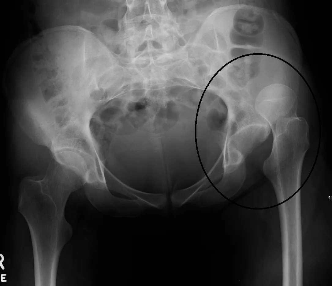
Long description
An X-ray showing a dislocated left hip after trauma, described on Commons as a superiorly dislocated hip.

Figure 2X-ray of scaphoid fracture
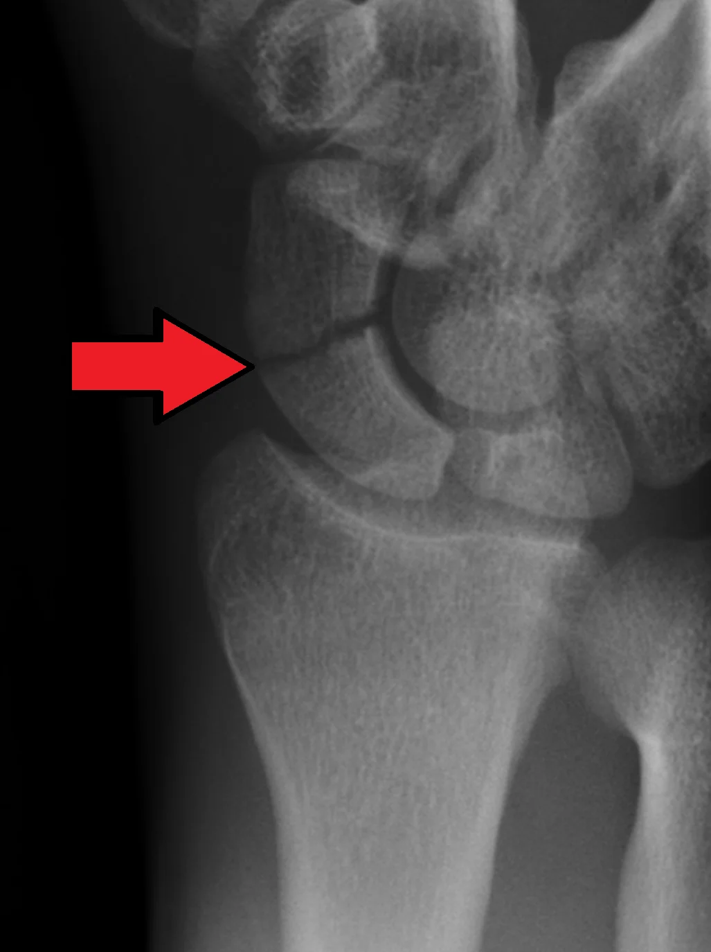
Long description
A wrist radiograph showing a fracture across the waist of the scaphoid bone, indicated by an arrow.

Figure 3Colles fracture
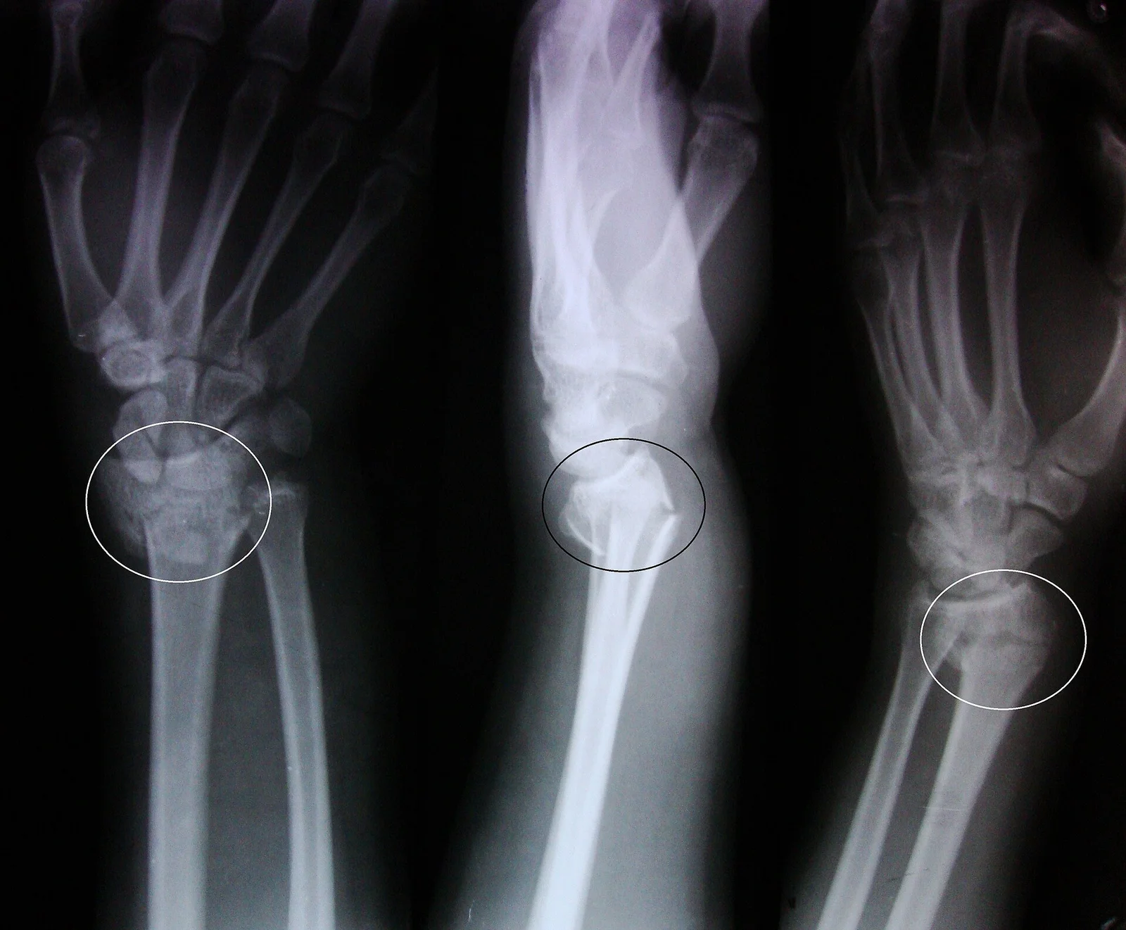
Long description
An X-ray image of a fractured radius showing the characteristic Colles' fracture with displacement and angulation of the distal end of the radius.

Figure 4Galeazzi-Fraktur Roentgen ap und seitlich - 002
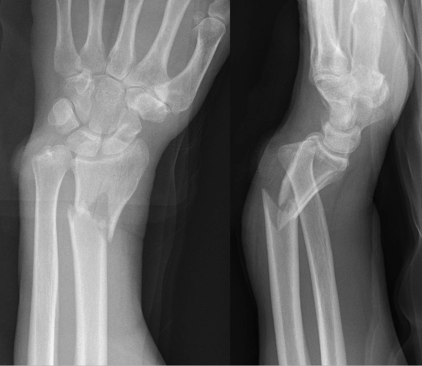
Long description
AP and lateral wrist/forearm radiographs of a Galeazzi fracture in which the lateral view clearly shows dorsal dislocation of the distal ulna.

Figure 5Monteggia LAT
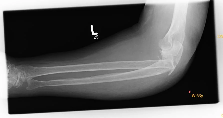
Long description
Lateral radiograph of the elbow of a 63-year-old woman showing a Monteggia fracture (fracture of the proximal ulna with dislocation of the radial head at the elbow).

Figure 6Shoulder dislocation, Y-projection before reduction
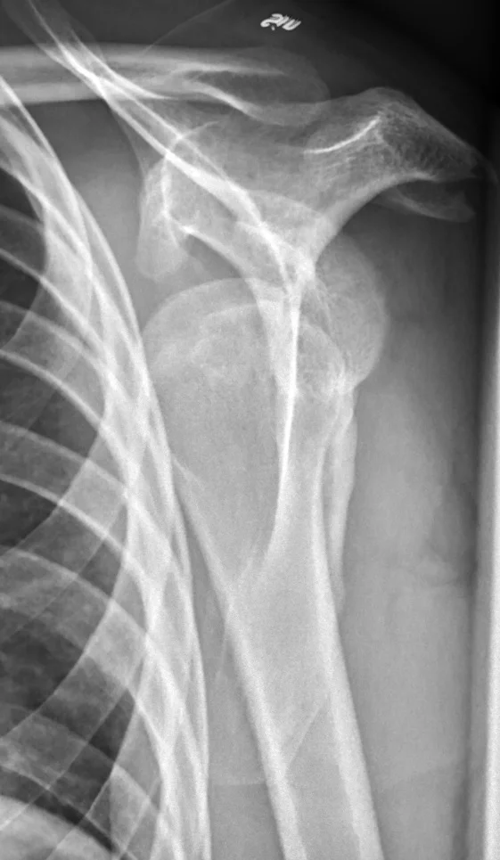
Long description
A Y-projection radiograph of the left shoulder of a 25-year-old male before reduction that is described on Commons as showing an anterior shoulder dislocation.

Figure 7AnterDisAP
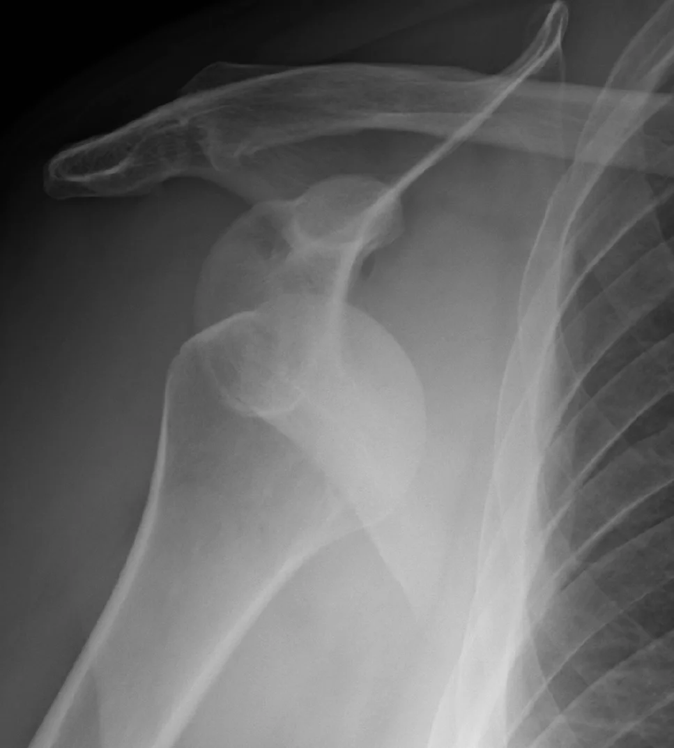
Long description
An AP X-ray described on Wikimedia Commons as 'Anterior dislocation of the right shoulder.'

Look, feel and move. Inspect deformity, swelling, bruising and skin (open versus closed). Palpate bony tenderness and compare both sides. Test active and passive range of motion and strength. Record a neurovascular examination: motor and sensory function in the relevant nerve territories and pulses, colour, warmth, capillary refill and temperature. Repeat after every manipulation, splint or position change. A pulseless deformed limb is realigned and splinted before imaging. Red flags are open fracture, compartment syndrome, pulselessness and multi-level injury.
Anterior dislocation presents with the arm slightly abducted and externally rotated, a squared-off shoulder and a palpable humeral head anteriorly. Elderly patients may present late with a "frozen" or painful shoulder; always consider rotator cuff tear and fracture. After reduction, test axillary nerve function (deltoid and lateral arm sensation) and check for cuff strength.
Rotator cuff tears present with pain, often at night and on overhead use; traumatic tears may follow a pop with weakness. Strength tests include Jobe (empty can) for supraspinatus, external rotation lag / lift-off (Gerber) for subscapularis, and drop arm. [3] Pseudoparalysis is an inability to hold the arm at 90 degrees of abduction or flexion. [3] Fatty infiltration, atrophy and deltoid bulk are noted. [3]
Clavicle fractures present with pain, deformity and a palpable step after a fall onto the shoulder or an outstretched hand. Most are mid-shaft. Neurovascular status and skin are checked. Proximal humerus fractures occur in older patients after a low-energy fall: pain, swelling and loss of shoulder movement. The greater tuberosity, surgical neck and four-part patterns matter because they change treatment. Humeral shaft fractures often allow the patient to cradle the arm; radial nerve function must be documented. [10]
Posterior dislocation follows a fall on the outstretched hand: shortened forearm and prominent olecranon, with gross valgus/varus instability. The terrible triad combines dislocation with radial head and coronoid fractures and is unstable. Olecranon fractures present with loss of active extension. Radial head fractures follow a fall on the hand with valgus load. [10] (See also Long-bone fractures, fat embolism and dislocations.)
Distal radius fractures (Colles, Smith, Barton) present with deformity, including the "dinner-fork" of a displaced Colles fracture. The wrist and distal radioulnar joint are assessed. Scaphoid fracture presents with radial wrist pain, tenderness in the anatomical snuffbox and reduced grip strength. [6] Focal snuffbox tenderness and pain on axial thumb compression raise suspicion. [6]
Hip fracture presents with a shortened, externally rotated leg and inability to bear weight after a fall, often with groin pain. Log-roll and careful skin and pressure-area checks are needed. Delirium, anaemia, volume depletion, electrolyte and diabetic problems are corrected pre-operatively. [1]
An ankle sprain presents with lateral ankle pain, swelling and bruising after an inversion injury. Examine the malleoli, the talar dome and the Achilles tendon. Test weight-bearing ability and record neurovascular findings. Ottawa rules decide imaging.
The table lists first-line and confirmatory tests. The exact cut-off for deciding imaging after ankle injury is the Ottawa ankle rule result; the cut-off for scaphoid first-line MRI is clinician suspicion after a careful examination. [6][10]
| Injury | First-line investigation | Confirmatory / special tests | What to look for |
|---|---|---|---|
| Shoulder dislocation | Radiographs in two orthogonal projections (AP and scapular Y) before and after reduction | CT if uncertainty; MRI arthrogram for soft tissues | Hill-Sachs, glenoid or tuberosity fracture; rotator cuff tear in those over 40 |
| Rotator cuff tear | Ultrasound or MRI | MRI (gold standard) for tear pattern, retraction, fatty infiltration | Partial versus full-thickness tear; muscle atrophy; Goutallier grade |
| Clavicle fracture | AP radiograph of clavicle/shoulder | CT for comminuted or medial fractures | Shortening and comminution |
| Proximal humerus fracture | Shoulder radiographs (AP, lateral, axillary) | CT for complex patterns | Head-split, tuberosity displacement |
| Elbow (dislocation / terrible triad) | AP and lateral elbow radiographs | CT for occult coronoid or radial head fracture | Joint congruence, fractures |
| Distal radius fracture | Wrist radiographs (AP and lateral) | CT for intra-articular step | Articular congruity, dorsal/volar tilt |
| Scaphoid fracture | Radiographs; MRI first-line for suspected occult fracture | CT for union or complex anatomy | Snuffbox region; proximal pole |
| Hip fracture | Pelvis and hip radiographs | MRI if X-ray negative but suspicion remains; CT alternative | Intracapsular versus extracapsular pattern |
| Ankle sprain | Ottawa rules decide X-ray | Radiographs if rule positive; MRI/ultrasound for ligament or syndesmosis if needed | Fracture, ligament disruption |
| Rule / cut-off | Statement |
|---|---|
| Ottawa ankle and foot rules | An ankle X-ray is indicated when there is pain in the malleolar zone plus bone tenderness at the posterior edge or tip of either malleolus, or inability to bear weight both immediately and in the emergency department; a mid-foot X-ray is indicated for pain in the mid-foot zone plus bone tenderness at the base of the fifth metatarsal or the navicular, or inability to bear weight. |
| Scaphoid imaging | MRI is recommended as first-line imaging for suspected scaphoid fracture after a thorough clinical examination. [10] |
| Hip fracture if X-ray negative | Offer MRI if a hip fracture is suspected despite negative X-rays of adequate standard; if MRI is unavailable within 24 hours or contraindicated, consider CT. [1] |
| Distal radius surgery timing | Surgery, when needed, within 72 hours for intra-articular and within 7 days for extra-articular fractures. [10] |
| Ankle fracture surgery timing | Consider operating on the day of injury or the next day. [10] |
A painful swollen limb after trauma raises a wide differential. The separating features are mechanism, distribution, joint involvement and imaging. [6]
| Presentation | Likely injury | Feature that separates it |
|---|---|---|
| Painful, empty shoulder after trauma, arm abducted and externally rotated | Anterior shoulder dislocation | Squared shoulder, humeral head palpable anteriorly; X-ray confirms; reduction relieves pain |
| Shoulder pain at night, weakness on overhead use, positive Jobe or lift-off | Rotator cuff tear | Strength tests positive; ultrasound or MRI shows tendon discontinuity |
| Shoulder stiffness after immobilisation or corticosteroid injection | Adhesive capsulitis | Global restriction of both active and passive range; normal imaging |
| Clavicle step after a fall onto the shoulder | Clavicle fracture | Tender bony step; fracture line on radiograph |
| Exquisitely tender shoulder in an older adult after a low-energy fall | Proximal humerus fracture | Deformity and loss of active movement; fracture on X-ray |
| Pain, shortening and radial nerve palsy after humeral trauma | Humeral shaft fracture | Mid-shaft tenderness; radial nerve deficit |
| Swollen painful elbow after a fall, olecranon prominent | Elbow dislocation or olecranon fracture | Loss of active extension; abnormal radiographs |
| Wrist deformity after a fall (dinner-fork) | Distal radius (Colles) fracture | Dorsal displacement and tilt on lateral X-ray |
| Volar wrist deformity with a fall on a flexed wrist | Smith fracture | Volar displacement and tilt |
| Radial wrist pain after a fall on an outstretched hand with normal-looking X-ray | Scaphoid fracture | Snuffbox tenderness, pain on axial thumb loading; MRI confirms |
| Thumb/finger radially-sided tendon pain on grasp and thumb extension | de Quervain tenosynovitis | Pain over the first dorsal compartment; positive Finkelstein test |
| Shortened externally rotated leg after a fall in an older adult | Hip fracture | Inability to bear weight; fracture on radiograph or MRI |
| Lateral ankle pain and swelling after inversion | Ankle sprain (or malleolar fracture) | Ottawa rules; inability to bear weight or bony tenderness point to fracture |
Management follows the simple order: life before limb, limb before bone. Resuscitate, examine, image, temporise, then decide. The table in the diagnosis section summarises mechanism and investigation; this section adds management by injury.
Align the patient, give analgesia and image. Choose a reduction technique you know well; a biomechanical (scapular) method is advised first. [2] If the first attempt fails, image for complications, attempt again under procedural sedation and analgesia, and resort to open reduction rarely. [2] After reduction, a sling as needed for one week; then early restoration of rotator cuff function, coordination and proprioception. [2] Consider surgery by shared decision for age under 40, contact athletes, or significant bone loss; a meta-analysis of early Bankart repair versus non-operative care reported 8.9% versus 49% recurrence and 90% versus 62% return to sport at two years. [2] Concomitant rotator cuff tears and iatrogenic fractures are common, particularly in women over 40, so re-examine carefully. [2]
Diagnose with ultrasound or MRI; MR arthrography is the most sensitive and specific method, while ultrasound and MRI have comparable accuracy. [3] Initial treatment is structured physiotherapy targeting the deltoid and cuff, analgesia and activity modification. [3] Oral paracetamol and NSAIDs give short-term relief; corticosteroid injection can help short-term pain but carries a signal of tendon harm with repeated use (steroid-injected patients developed tears more often, with mean follow-up 39 months). [3] Hyaluronic acid and platelet-rich plasma are alternatives with some short-term benefit. [3] Refer promptly for traumatic or massive tears, pseudoparalysis, significant functional loss, tendon retraction, delamination, fatty infiltration or failure after a conservative trial. [3]
Most mid-shaft fractures are managed conservatively in a sling, with analgesia and early pendular movement. A systematic review and meta-analysis of displaced mid-shaft fractures treated non-surgically reported a pooled DASH score of 16.1 at three months, 9.9 at six months and 6.3 at one year, non-union in about 14% and secondary surgery in about 13% at one year. [8] Operative fixation (plating) is considered for markedly displaced or shortened fractures, open injuries, skin tenting, neurovascular compromise or floating shoulder. A CT may be needed for medial or comminuted patterns. [7][8]
Proximal humerus: undisplaced and most low-energy displaced fractures are managed non-operatively; consider surgery for open injury, skin tenting, vascular injury, fracture-dislocation or head split. [10] Humeral shaft: most isolated fractures unite in a functional brace; displaced, open, pathological and failed conservative cases are fixed (plate preferred, nail selected). Radial nerve function must be documented before and after any manipulation. [10]
Posterior dislocation is reduced under analgesia or sedation (traction in line with the forearm) and tested for stability through range; a terrible triad needs surgical fixation and ligament repair. Olecranon fractures needing anatomic reduction are fixed. Radial head fractures are mostly managed according to displacement and articular step; excision or arthroplasty is reserved for complex or old cases.
Reduce under Bier's block if dorsally displaced; do not use nitrous oxide alone. [10] Manipulation and a cast is acceptable in many adults. [10] K-wire fixation is offered when the articular surface is not fractured or can be reduced closed; open reduction and internal fixation is considered if closed reduction of the joint surface fails. [10] Timing: intra-articular fractures within 72 hours, extra-articular within 7 days; re-displacement within 72 hours of the decision to operate. [10] Geriatric fractures are mostly non-operative. [7] Emergency priorities follow the Long-bone fractures pathway (open fractures, compartment syndrome).
Suspect with snuffbox tenderness after a fall on an outstretched hand. [6] Initial X-rays are often negative, so MRI is recommended as first-line imaging after a thorough examination; CT helps union assessment. [6][10] For undisplaced waist fractures, cast immobilisation is the primary treatment; surgical fixation (percutaneous screw) for displaced, proximal pole, delayed or non-union. [6] Prolonged immobilisation (thumb spica) is typical and clinical follow-up is needed because non-union and avascular necrosis may develop months later. [6]
Diagnose by pain over the first dorsal compartment, painful gripping and a positive Finkelstein test. Initial management is rest, splinting (thumb spica) and NSAIDs. Corticosteroid injection into the tendon sheath gives good benefit and ultrasound guidance increases efficacy. [9] Persistent symptoms after injection, or recurrent triggering, is an indication for surgical release. [9]
Hip fracture: operate on the day of, or the day after, admission after correcting reversible medical issues (anaemia, anticoagulation, volume depletion, electrolytes, diabetes, heart failure, arrhythmia or ischaemia, infection). [1] Give paracetamol 6-hourly and additional opioids as needed; NSAIDs are not recommended. [1] Aim to allow unrestricted weight-bearing immediately after surgery. [1] Displaced intracapsular fractures are treated with cemented arthroplasty (total hip replacement or hemiarthroplasty), with total replacement preferred where the patient walked independently outdoors with no more than a stick, is expected to live and function beyond two years, and is fit. [1] Subtrochanteric fractures are nailed; trochanteric fractures above and including the lesser trochanter are treated preferentially with extramedullary implants such as a sliding hip screw. [1] Use MRI or CT if radiographs are normal but clinical suspicion persists (MRI first; CT if MRI unavailable within 24 hours or contraindicated). [1] Mobilise from the day after surgery with orthogeriatric multidisciplinary care, falls and bone health assessment. [1]
Apply Ottawa rules to decide radiographs. [4][10] Functional treatment (early movement, bracing or taping, physiotherapy) consistently produces faster recovery and lower recurrence than prolonged immobilisation. [5] RICE in the acute phase; consider NSAIDs (avoid in frail or older adults). [10] Advise immediate unrestricted weight-bearing as tolerated for stable unimalleolar ankle fractures and orthopaedic review within two weeks if stability is uncertain. [10]
- Shoulder dislocation: iatrogenic fracture (women over 40, greater tuberosity fracture), axillary nerve injury, rotator cuff tear, recurrence. [2]
- Rotator cuff: progression of tear, fatty infiltration, sleep disturbance, recurrent shoulder pain; surgery carries re-tear risk. [3]
- Clavicle: non-union (~14%) and secondary surgery (~13%) at one year after non-operative treatment of displaced mid-shaft fractures; malunion and hardware failure after fixation. [8]
- Scaphoid: non-union, avascular necrosis of the proximal pole and SNAC wrist. [6]
- Hip: delirium, infection, venous thrombosis, pneumonia, pressure ulcers, failure of fixation or arthroplasty; falls and further fragility fractures.
- Ankle: chronic ankle instability, recurrent sprain, missed fracture, stiffness.
Most soft-tissue injuries recover well with functional rehabilitation. Displaced mid-shaft clavicle treated non-operatively shows substantial recovery by three months with further improvement to one year. [8] Rotator cuff repair relieves pain but re-tear is possible; recovery depends on tear size and fatty infiltration. Hip fracture carries significant mortality and dependence in older adults despite good surgery. Scaphoid fractures unite with adequate immobilisation, but delay worsens outcome. Ankle sprains rarely need surgery; the risk is recurrence and chronic instability. [5]
- Fall-prevention and bone health programs in older adults, including osteoporosis assessment after hip fracture. [1]
- Sports technique and conditioning, especially wrist and ankle proprioception.
- Early reduction and appropriate immobilisation to prevent recurrence and complications.
- Safeguarding awareness for injuries inconsistent with the history.
- Older adults: avoid NSAIDs for pain in fractures; cognitive and functional status guide management; comorbidities must be optimised before hip surgery. [1][10]
- Pregnancy: trauma principles as standard; minimise radiation; individual surgical and drug decisions with obstetric input.
- Children: different pathways apply (remodelling, growth plate injury); see Plain text: Common paediatric orthopaedic injuries.
- Document neurovascular status, realign, splint and transfer; a pulseless limb must not wait for imaging.
- Use clinical rules (Ottawa) to avoid unnecessary radiographs. [4][10]
- A thumb spica cast for scaphoid and a functional brace for ankle sprain are low-cost effective options. [6][5]
- Hip fracture care needs surgery and orthogeriatric support; arrange transfer where unavailable.
Approach to an adult with a painful, injured limb
- Primary survey and resuscitationAirway, breathing, circulation, disability, exposure; control bleeding; give analgesia
- Limb examinationDeformity, skin, swelling; tendon and joint status
- Neurovascular assessmentMotor, sensation, pulses, colour, warmth, capillary refill; document before and after any manipulation
- Realignment and splintGentle in-line traction; splint; repeat neurovascular check
- ImagingX-rays of the bone and adjacent joints in two planes; CT if uncertainty; MRI for occult hip or scaphoid
- Question: Does the limb have circulation, sensation and viable soft tissues?
No (or unsure)
- Treat as vascular emergencySenior orthopaedic and vascular input; revascularise promptly; fasciotomy if compartment syndrome
Yes
- Injury-specific planDislocation: reduce and image again; fracture: nail/plate/fixator/cast; sprain: Ottawa and functional rehab; tenosynovitis: splint and injection
- Admit or fracture-clinic follow-up; Falls and bone health for hip fracture
- Adults have limited remodelling; treat reductions, alignment and displacement with adult thresholds.
- Document neurovascular status before and after every manipulation or splint, and after position changes.
- Shoulder dislocation: image in two views before and after reduction; ultrasound cannot replace radiographs; recurrence is highest in young men. [2]
- Rotator cuff tears: MRI is the gold standard for pattern and retraction; ultrasound is cheaper and dynamic; refer for trauma, weakness or functional limitation. [3]
- About half of full-thickness rotator cuff tears are symptom-free; age is the main risk factor. [3]
- Scaphoid fractures often have normal first X-rays; MRI is recommended first-line imaging and management depends on displacement. [6][10]
- de Quervain tenosynovitis responds well to splinting and corticosteroid injection; ultrasound guidance improves efficacy. [9]
- Hip fractures need surgery within one to two days; displaced intracapsular fractures are treated with cemented arthroplasty. [1]
- Ottawa rules safely select ankle radiographs; functional bracing and movement beat prolonged immobilisation for sprains. [4][5]
- Pain in adult fractures: paracetamol regularly, morphine by severity, avoid NSAIDs in frail or older adults. [1][10]
- Low-resource care still begins with examination and documentation; reduce only after neurovascular checks.
Answer each one in your head first, then open the answer.
A 24-year-old man has an anterior shoulder dislocation reduced in the emergency department. Which imaging and aftercare does the guideline recommend, and what is his long-term recurrence risk?
Show answerHide answer
Radiographs in at least two projections are needed before and after reduction, with CT if uncertainty; ultrasound cannot replace radiographs. Use a sling as needed for one week, then early rotator cuff, coordination and proprioception work. Males under 40 have markedly higher recurrence (a 13-fold increase) and males overall 3-fold; a meta-analysis of early Bankart repair versus non-operative care reported 8.9% versus 49% recurrence. [2]
A 62-year-old woman has night shoulder pain and weak abduction. Jobe is positive. What imaging should you arrange and what are the conservative first steps?
Show answerHide answer
Arrange ultrasound or MRI; MRI is the gold standard for tear pattern, retraction and fatty infiltration, and MR arthrography is the most sensitive and specific. First steps are structured physiotherapy, analgesia and activity modification; consider corticosteroid injection cautiously because of a tendon-harm signal with repeated use. Refer if functional disability persists. [3]
An older adult falls and has groin pain and a shortened externally rotated leg. Radiographs are reported as normal but clinical suspicion remains high. What next and what are the surgical goals?
Show answerHide answer
Offer MRI (or CT if MRI is unavailable within 24 hours or contraindicated). Operate the day of, or the day after, admission after correcting reversible comorbidities. Aim for unrestricted weight-bearing immediately after surgery; a displaced intracapsular fracture receives cemented arthroplasty. [1]
A 30-year-old presents with wrist pain after a fall on an outstretched hand. X-rays are normal but the scaphoid region is tender. What first-line imaging and treatment?
Show answerHide answer
MRI is recommended as first-line imaging after a thorough examination; CT supports union assessment. For an undisplaced waist fracture, immobilisation in a thumb spica cast is typical, with surgery reserved for displaced, proximal or delayed cases. Warn the patient that non-union and avascular necrosis can appear months later. [6][10]
A 45-year-old has radial wrist pain worsened by gripping. Finkelstein test is positive. What is the diagnosis and stepwise management?
Show answerHide answer
de Quervain tenosynovitis. Start with splinting and NSAIDs, then a corticosteroid injection into the sheath, using ultrasound guidance when available for better efficacy. Persistent symptoms after one to two injections warrant surgical release. [9]
After an ankle inversion injury, how do you decide whether an X-ray is needed and how do you plan rehabilitation?
An adult has a dorsally displaced distal radius fracture. Within what time-frames should surgery occur, and what fixation choice does NICE suggest?
Show answerHide answer
If needed: intra-articular fractures within 72 hours; extra-articular within 7 days; re-displacement within 72 hours of the decision to operate. Offer K-wire fixation if no articular involvement or if the joint surface can be reduced closed; consider open reduction and internal fixation if closed reduction fails. Reduce under Bier's block if needed and do not use nitrous oxide alone. [10]
Common adult orthopaedic injuries (this page); long-bone fractures, fat embolism and dislocations (emergency medicine); common paediatric orthopaedic injuries; pelvic and hip fractures; neck and shoulder (rotator cuff) rehabilitation; hand and wrist trauma; ankle sprain and instability.
- National Institute for Health and Care Excellence. Hip fracture: management (NICE Clinical Guideline CG124). NICE clinical guideline CG124; published 22 June 2011; last updated 06 January 2023. Accessed .
- Van Gastel ML, Hekman KMC, Boon F, Nelen SD, Schep NWL, van der Meijden OAJ, Baden DN, Jonkergouw MAT, Huygen LE, Balemans ACJ, Griekepoor M, Derksen RJ, van den Bekerom MPJ. Clinical practice on treatment of primary traumatic anterior shoulder dislocation: a multidisciplinary guideline. Acta Orthopaedica 2026;97:531-537. doi:10.2340/17453674.2026.46434. Accessed .
- Adamus JP, Dywan K, Paszt Z, Dabrowska J, Tracz MJ, Legosz P, Struzik S. From Clinical Signs to Functional Recovery: A Practice-Oriented Review of Rotator Cuff Tear Management in Primary Care. Journal of Clinical Medicine 2026;15(17):6697. doi:10.3390/jcm15176697. Accessed .
- Gomes YE, Chau M, Banwell HA, Causby RS. Diagnostic accuracy of the Ottawa ankle rule to exclude fractures in acute ankle injuries in adults: a systematic review and meta-analysis. BMC Musculoskeletal Disorders 2022;23:885. doi:10.1186/s12891-022-05831-7. Accessed .
- Alharbi MM, Al Musaad M. Management of Ankle Sprains in Urgent Care: Adherence to Evidence-Based Guidelines. Cureus 2026;18(4):e106852. doi:10.7759/cureus.106852. Accessed .
- Gutierrez Y, Bonilla P, Franco E, Shaju R, Sepulveda A, Bonazza N, Schenck R, Ahmad A. Treatment and Return to Sport in Athletes With Scaphoid Fractures: A Narrative Review. Cureus 2026;18(9):e115689. doi:10.7759/cureus.115689. Accessed .
- Kirchberg T, Nolan R, Tomberg S. Emergency Medicine Management of Distal Radius and Ulna Fractures: Narrative Review. The western journal of emergency medicine 2026;27(4):1090-1104. doi:10.5811/westjem.52930. Accessed .
- Vaajala M, Tarkiainen J, Liukkonen R, Launonen AP, Mattila VM. Non-surgical treatment for displaced midshaft clavicle fractures: a systematic review and meta-analysis. BMC Musculoskeletal Disorders 2026;27:860. doi:10.1186/s12891-026-10208-1. Accessed .
- Urits I, Smoots D, Anantuni L, Bandi P, Bring K, Berger AA, Kassem H, Ngo AL, Abd-Elsayed A, Manchikanti L, Urman R, Kaye AD, Viswanath O. Injection Techniques for Common Chronic Pain Conditions of the Hand: A Comprehensive Review. Pain and Therapy 2020;9(1):129-142. doi:10.1007/s40122-020-00158-4. Accessed .
- National Institute for Health and Care Excellence. Fractures (non-complex): assessment and management (NICE Guideline NG38). NICE guideline NG38; published 17 February 2016; reviewed 23 June 2025. Accessed .
Figure credits
- Figure 1: James Heilman, MD, via Wikimedia Commons, CC BY-SA 3.0, CC BY-SA 3.0.
- Figure 2: Gilo1969, via Wikimedia Commons, CC BY 3.0, CC BY 3.0.
- Figure 3: Ashish j29, via Wikimedia Commons, CC BY 3.0, CC BY 3.0. resized.
- Figure 4: Hellerhoff, via Wikimedia Commons, CC BY-SA 3.0, CC BY-SA 3.0. resized.
- Figure 5: Pfeffer55, via Wikimedia Commons, CC BY-SA 4.0, CC BY-SA 4.0.
- Figure 6: Mikael Häggström, M.D., via Wikimedia Commons, CC0, CC0.
- Figure 7: James Heilman, MD, via Wikimedia Commons, CC BY-SA 4.0, CC BY-SA 4.0.
Educational content for medical students and doctors. It is not a substitute for clinical judgement or local guidelines.