Polymyositis, dermatomyositis and Sjogren syndrome
A clinician's guide to polymyositis, dermatomyositis and Sjogren syndrome: weakness, rashes, dryness, antibodies, malignancy screening and treatment.
- Polymyositis (PM) and dermatomyositis (DM) are idiopathic inflammatory myopathies: acquired, immune-mediated diseases of skeletal muscle that usually present with symmetric proximal weakness, often with a raised creatine kinase, and sometimes with characteristic skin findings.
- DM declares itself first in the skin: a heliotrope rash on the eyelids and Gottron papules over the knuckles are close to pathognomonic; PM has no rash.
- Both diseases are diagnosed under the 2017 EULAR/ACR classification criteria, supported by myositis-specific antibodies and muscle biopsy, and they always require exclusion of better explanations such as IBM, drug-induced myopathy and endocrine disease.
- DM, and to a lesser extent PM, are strongly associated with an underlying cancer in the three years before or after onset; every adult needs risk-stratified malignancy screening.
- Treatment rests on high-dose glucocorticoids plus an early steroid-sparing disease-modifying drug, with intravenous immunoglobulin, rituximab or cyclophosphamide for severe or refractory disease.
- Sjogren syndrome is a chronic autoimmune exocrinopathy: lymphocytic infiltration destroys salivary and lacrimal glands, producing dry eyes and dry mouth, with a female-to-male ratio up to 9 to 1 or more [5], and a lifetime non-Hodgkin lymphoma risk of roughly 2 to 9 percent [5]. [6]
- The 2016 ACR-EULAR criteria for primary Sjogren syndrome are built on five objective tests, of which anti-SSA (Ro) antibodies and a positive salivary gland biopsy carry the greatest weight, and dryness is managed symptomatically while systemic disease is treated with the minimum effective glucocorticoid dose plus steroid-sparing agents. [1]
By the end of this page you should be able to:
- Define polymyositis, dermatomyositis and Sjogren syndrome, and classify the inflammatory myopathies.
- Explain why proximal weakness occurs, why creatine kinase is raised, and how the DM skin lesions map to the underlying vasculopathy.
- Name the myositis-specific and myositis-associated antibodies and the cancer-risk information they carry.
- Apply the 2017 EULAR/ACR myositis criteria and the 2016 ACR-EULAR Sjogren criteria to a clinical vignette, and know the cut-offs and scores involved.
- Plan first-line and second-line treatment for an adult with active myositis or systemic Sjogren syndrome, with doses, route and monitoring.
- Screen for malignancy in an adult with IIM according to the 2023 international guideline, and say who needs basic versus enhanced screening.
- Recognise the complications, prognosis, and special-group considerations that alter management in pregnancy, childhood and older age.
This page covers two of the commoner questions in rheumatology and neurology clinics: a patient with progressive proximal weakness and a patient with relentless dryness. The two are linked by autoimmunity, by overlapping connective tissue disease, and by the need to think early about complications that are silent until they are dangerous. Read it alongside the broader adult myopathies and muscle disease topic for the bedside framework that places myositis among the other causes of weakness.
DefinitionPolymyositis and dermatomyositis
The idiopathic inflammatory myopathies (IIM, "myositis") are acquired, immune-mediated disorders in which skeletal muscle is infiltrated and destroyed by inflammatory cells, producing progressive, symmetric weakness that is usually proximal. Dermatomyositis adds characteristic skin disease, which can precede, accompany or follow the weakness. Polymyositis is the same process without skin lesions. [3]
The 2017 European League Against Rheumatism / American College of Rheumatology (EULAR/ACR) classification criteria divide IIM into major subgroups. [1] After a patient meets the criteria (probability of IIM at least 55 percent), a classification tree separates adults into polymyositis, dermatomyositis, clinically amyopathic dermatomyositis (DM skin disease without measurable weakness), inclusion body myositis (IBM) and, in children, juvenile dermatomyositis. [1] Immune-mediated necrotising myopathy (IMNM) is currently grouped with polymyositis in the tree because of small numbers; clinically it presents with very high creatine kinase levels, often after statins, and a paucity of inflammatory infiltrate on biopsy. [1]
DefinitionSjogren syndrome
Sjogren syndrome is a chronic systemic autoimmune disease in which lymphocytes infiltrate and damage exocrine glands, mainly the salivary and lacrimal glands, producing dry eyes (xerophthalmia) and dry mouth (xerostomia). It can occur alone (primary Sjogren) or alongside another connective tissue disease such as rheumatoid arthritis or systemic lupus erythematosus (secondary Sjogren, now often called Sjogren with overlap). [5]
Sjogren is overseen by rheumatologists with ophthalmologists and oral medicine specialists, and its spectrum runs from sicca symptoms through arthralgia, fatigue and systemic organ involvement to B-cell lymphoma. [5]
Myositis is rare. The British Society for Rheumatology (BSR) guideline states an incidence of up to 19 per 1,000,000 person-years in adults and 4 per 1,000,000 in children, with an estimated 10,000 adults with IIM in the United Kingdom. [3] Adults are most often affected in IIM; dermatomyositis occurs in children and adults, and polymyositis occurs almost exclusively in adults. [3] Every subtype except IBM is more common in women. [3]
No modifiable risk factor has been established for sporadic IIM. Infections, some drugs and malignancy are commonly cited triggers, and geography and ethnicity influence incidence. [3] There is a well-documented link with cancer: adult-onset IIM carries a raised malignancy incidence in the three years before and after onset, and dermatomyositis confers the highest risk among the subtypes. [4]
Sjogren syndrome is the most common systemic autoimmune disease and is overwhelmingly a disease of women. One systematic review estimated an incidence of 6.9 per 100,000 person-years and a prevalence of 60.8 per 100,000 from pooled studies, with a female-to-male ratio between 9 to 1 and 28 to 1; it is usually diagnosed between the fourth and sixth decades, and is rare in children. In a tertiary centre cohort, overlap with systemic lupus erythematosus, rheumatoid arthritis and systemic sclerosis occurred in about 15, 20 and 30 percent of patients respectively. [5]
Dermatomyositis is a humorally mediated microangiopathy. Antibody and complement (including the membrane attack complex) are deposited on small endomysial blood vessels, producing capillary injury, endothelial activation and ischaemia of the tissue they supply. Muscle fibres in the peripheral parts of the fascicle (perifascicular atrophy) are the most ischaemic, which is exactly the histological hallmark of the disease; the perivascular inflammation is predominantly CD4-positive T cells and B cells. [1] The same vascular process in the skin explains the characteristic rashes.
Polymyositis, in contrast, is a cell-mediated myocytotoxic process: CD8-positive T cells recognise and invade muscle fibres that abnormally express major histocompatibility complex class I (MHC-I) molecules, producing endomysial inflammation and fibre necrosis. [1] This mechanistic distinction matters at the bedside because it predicts the biopsy pattern and helps separate IMNM (necrosis with little inflammation) and inclusion body myositis (rimmed vacuoles and endomysial inflammation with poor steroid responsiveness) from true polymyositis. [1]
Sjogren syndrome is best understood as an autoimmune epithelitis. An environmental trigger (often a virus) in a genetically susceptible person, particularly with HLA class II alleles such as HLA-DQB1 and HLA-DQA1, disrupts the salivary and lacrimal gland epithelium. T cells, B cells and dendritic cells accumulate, germinal centre-like structures form, and the activated B lymphocytes produce large amounts of autoantibodies. [5] The resulting glandular hypofunction gives the sicca symptoms; systemic features (arthralgia, vasculitis, interstitial lung disease, renal tubular acidosis and neuropathy) are driven by the same B-cell hyperactivity, immune-complex deposition and cryoglobulinaemia. [5]
Both myositis subtypes present with painless, progressive, symmetric weakness that preferentially affects proximal muscle groups: difficulty rising from a chair, climbing stairs, brushing the hair, and, in more severe disease, lifting the head from the pillow. [3] Neck flexors are often unusually weak compared with extensors, and in the legs proximal muscles are weaker than distal ones; dysphagia is common and may be the presenting symptom. [1][3] Weakness is usually subacute, evolving over weeks to months; very rapid onset suggests either severe disease or an alternative (toxic, metabolic or neuropathic) process. Arthralgia, Raynaud phenomenon and a systemic feeling of being unwell may accompany the muscle picture. [3]
On examination, manual muscle testing shows symmetric proximal weakness; muscle tenderness is not usually prominent in IIM, which helps distinguish myositis from simple myalgia. Look for extra-muscular disease: skin in DM, lungs (basal inspiratory crackles), hands and joints, and the swallowing muscles. [3]
The skin findings of dermatomyositis are highly characteristic and often permit classification without a biopsy:
- Heliotrope rash: purple-lilac erythema over the upper eyelids, often with periorbital oedema. [1]
- Gottron papules: erythematous to violaceous, sometimes scaly papules over the extensor surfaces of joints, classically the knuckles, but also the elbows, knees, malleoli and toes. [1]
- Gottron sign: erythematous to violaceous macules over the same extensor surfaces, without being palpable. [1]
- Shawl sign and V-sign: erythematous patches on the back and shoulders and over the anterior chest and neck.
- Nail-fold changes: dilated, tortuous, or lost capillaries.
Clinical pearlExam tip
A rash that is photo-distributed and present over the knuckles rather than the interphalangeal joints (which is where psoriasis favours) is a strong pointer to dermatomyositis. In clinically amyopathic dermatomyositis, the skin lesions are present but weakness is absent or barely measurable; these patients still carry the malignancy and interstitial lung disease risks of DM. [1]
Sjogren syndrome presents with dryness. Xerophthalmia gives gritty, burning eyes, photophobia and reduced visual quality; xerostomia produces difficulty swallowing dry food, altered taste, oral ulceration and fissured lips. [5] In the Big Data Sjogren Project Consortium registry of over 6,000 patients, 92 percent had dry eyes and 94 percent had dry mouth. [5] Dryness is not confined to the eyes and mouth: vaginal dryness, dry cough (xerotrachea), and dry skin are common and easily overlooked. Fatigue (reported by 88 percent in one single-centre study) and diffuse pain (80 percent) are often the worst symptoms for the patient. [5]
Systemic involvement is common: in a multicentre study, 30 percent of patients had systemic manifestations at diagnosis and 39 percent had experienced them previously, and only 8 percent of patients in a Spanish registry had no systemic activity at all over a mean follow-up of six years. [5] The articular (56 percent), glandular (34 percent), pulmonary (15 percent) and cutaneous (13 percent) ESSDAI domains are the most frequently active. [5] Physical examination should record parotid and lacrimal gland swelling, palpable purpura (cutaneous vasculitis), synovitis, and pulmonary and neurological findings.
The wrong diagnosis here is expensive: treating inclusion body myositis or a statin myopathy with glucocorticoids does not help the patient, and missing an IBM means wasted months. Use age, symmetry, pattern of weakness and biopsy findings to separate the causes.
| Condition | Distinguishing feature |
|---|---|
| Inclusion body myositis (IBM) | Older men; asymmetric weakness with prominent finger flexors and quadriceps; rimmed vacuoles on biopsy; poor and transient response to immunosuppression. [1][3] |
| Immune-mediated necrotising myopathy (IMNM) | Very high creatine kinase; statin exposure; severe necrosis with little inflammatory infiltrate; anti-HMGCR or anti-SRP antibodies. [1][3] |
| Anti-synthetase syndrome | Fever, mechanic's hands, non-erosive arthritis, interstitial lung disease and anti-Jo-1 or other aminoacyl-tRNA synthetase antibodies. [3][4] |
| Statin-induced myalgia or myopathy | Onset after starting a statin; normal or modestly raised enzyme; resolves on withdrawal; no inflammatory infiltrate. [3] |
| Muscular dystrophy | Family history, limb-girdle or scapulohumeral distribution, chronic course, abnormal dystrophin or relevant gene, no response to steroids. [3] |
| Myasthenia gravis / Lambert-Eaton | Fatigable weakness, ocular/bulbar rather than proximal, normal creatine kinase, decrement on repetitive nerve stimulation. [3] |
| Hypothyroidism / endocrine myopathy | Slowed relaxation, raised thyroid-stimulating hormone, often hyperlipidaemia; weakness improves with replacement. [3] |
| Polymyalgia rheumatica | Older adults; severe shoulder and pelvic girdle pain and stiffness with near-normal strength and creatine kinase; dramatic response to low-dose glucocorticoids. [3] |
| Sensory neuronopathy (Sjogren) | Non-length-dependent numbness and sensory ataxia rather than weakness; anti-Ro serology; MRI or biopsy supportive. [5] |
Figure 1Dermatomyositis7
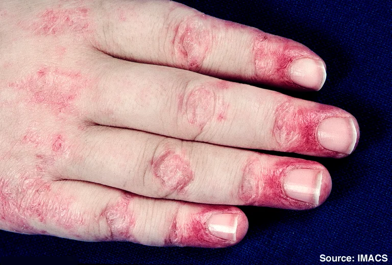
Long description
A clinical photograph shows Gottron's papules with secondary atrophy and telangiectasia in a girl with severe juvenile dermatomyositis, seen in association with prominent periungual erythema.

Figure 2Dermatomyositis9
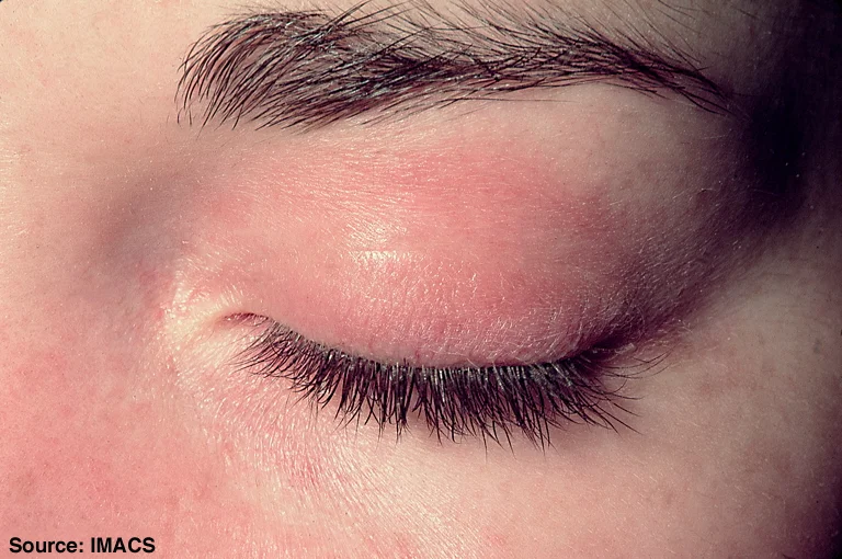
Long description
A clinical photograph of dermatomyositis showing confluent macular erythema confined to the upper eyelid with associated periorbital edema (heliotrope rash on the eyelids).

Creatine kinase (CK) is the central screening test: levels are raised in up to almost all patients with active polymyositis or dermatomyositis, and track disease activity and response. Lactate dehydrogenase, aspartate aminotransferase and alanine aminotransferase are also released from muscle and can be misleading if attributed to the liver. [1][3] CK is not raised in clinically amyopathic DM, which is why clinical judgement and biopsy still matter. Inflammatory markers are variable and non-specific. [3]
Electrophysiology and imaging play supporting roles. Electromyography shows a myopathic (short-duration, low-amplitude) pattern with spontaneous activity. Muscle magnetic resonance imaging identifies oedema, inflammation and fatty replacement, helps select a biopsy site, and monitors disease. [3] Pulmonary function testing (including diffusing capacity) screens for interstitial lung disease.
| Test | What it measures and typical values |
|---|---|
| Serum CK, LDH, AST, ALT | Muscle enzyme release; abnormal when above the laboratory upper limit of normal; the most abnormal value during the disease course is used in the IIM criteria. [1] |
| Myositis-specific autoantibody panel | Anti-Jo-1, anti-Mi-2, anti-TIF1-gamma, anti-NXP2, anti-MDA5, anti-SAE1, anti-HMGCR, anti-SRP and anti-synthetase antibodies (PL-7, PL-12, EJ, OJ). All adults with new-onset IIM should be tested. [3][4] |
| Myositis-associated antibodies | Anti-PM-Scl, anti-Ku, anti-RNP, anti-Ro/La (SSA/SSB). Associated with overlap, particularly Sjogren and systemic sclerosis. [3] |
| Anti-nuclear antibody (ANA), RF | Common in Sjogren; positivity for RF and cryoglobulins is frequent. [5] |
| Anti-SSA (Ro) / anti-SSB (La) | Anti-Ro in about 75 percent of primary Sjogren patients; anti-Ro/Ro52 is relatively specific. RF and anti-La occur in about 50 percent. Anti-Ro is the only serological item in the ACR-EULAR Sjogren criteria. [5][2] |
| Schirmer test | Lacrimal function; 5 mm per 5 minutes or less supports Sjogren classification. [2] |
| Ocular staining score (OSS) | Corneal and conjunctival damage; 5 or more on at least one eye (or van Bijsterveld score 4 or more) supports Sjogren classification. [2] |
| Unstimulated whole salivary flow rate | Less than 0.1 mL/min supports Sjogren classification. [2] |
| Labial salivary gland biopsy | Focus score: number of inflammatory foci of at least 50 cells per 4 mm2; a focus score of 1 or more supports Sjogren classification. [2] |
| Muscle biopsy | Diagnostic in atypical, antibody-negative or biopsy-pathway cases: endomysial CD8 inflammation in PM; perifascicular atrophy and perivascular inflammation in DM; necrosis with little inflammation in IMNM; rimmed vacuoles in IBM. [1] |
| Chest radiograph / CT / lung function | Screens for interstitial lung disease in IIM; ESSDAI and pulmonary domain in Sjogren. [3][5] |
Approach to suspected inflammatory myopathy or Sjogren syndrome
- Adult with proximal weakness or marked sicca symptomsHistory, examination, medication and family review
- Question: What is the leading problem?
Proximal weakness
- Check CK and muscle enzymesCK is usually raised in active PM/DM; normal CK suggests IBM, myasthenia or neuropathy
- Look for DM skin lesions, dysphagia, ILDHeliotrope rash or Gottron papules permit classification without biopsy
- Test MSA and MA panelsGuides phenotype, prognosis and malignancy risk
- Question: Biopsy needed?
No rash, no antibodies or atypical
- Muscle biopsyEndomysial vs perifascicular vs necrotic pattern identifies subtype
Typical DM rash
- Apply EULAR/ACR criteria without biopsyProbability 55 percent or more
- Risk-stratify for malignancyAll adults with new-onset IIM; basic or enhanced screening per 2023 guideline
- Start high-dose glucocorticoid plus steroid-sparing agentSee management section
Sicca (dryness)
- Confirm dryness with Schirmer and OSSAsk dry-mouth and dry-eye questions; ESSDAI for systemic activity
- Test ANA, RF, anti-SSA (Ro) and anti-SSB (La)Anti-Ro is the only serological criteria item
- Salivary gland biopsy if score below 4Focus score 1 or more gives 3 points
- Apply weighted 2016 ACR-EULAR scoreScore 4 or more out of 9
- Treat dryness first-line and systemic disease per 2019 EULAR frameworkGCs at minimum effective dose; GM-sparing agents second line
The criteria are built on 16 weighted variables across six domains: age of onset, muscle weakness, skin manifestations, other clinical manifestations, laboratory measurements and (when available) muscle biopsy features. [1] Each variable carries a score, with and without biopsy, and the total score converts into the probability of IIM. Skin manifestations carry high weights, so dermatomyositis can be classified from skin findings alone. [1]
Key cut-offs: a probability below 50 percent (score 5.3 or less without biopsy, 6.5 or less with biopsy) rules out IIM; 50 to less than 55 percent is "possible IIM"; at least 55 percent (score 5.5 or more without biopsy, 6.7 or more with biopsy) is "probable IIM" and is the recommended minimum to classify; a probability of 90 percent or more (score 7.5 or more without biopsy, 8.7 or more with biopsy) is "definite IIM", used when high specificity is required, such as in drug trials. [1] Remember that these are classification criteria for research and trial entry, not bedside diagnostic criteria. [1]
The target is anyone with at least one symptom of ocular or oral dryness, or suspected Sjogren from systemic features identified through the ESSDAI. [2] The diagnosis is supported when the weighted sum of five objective items is 4 or more. Anti-SSA (Ro) positivity and a labial salivary gland with focal lymphocytic sialadenitis and focus score 1 or more are the heavy items (3 points each). The light items (1 point each) are ocular staining score 5 or more on at least one eye (or van Bijsterveld score 4 or more), Schirmer test 5 mm per 5 minutes or less on at least one eye, and unstimulated whole salivary flow rate 0.1 mL/min or less. [2] Exclusion criteria (a pre-existing diagnosis of head/neck radiation damage, active hepatitis C infection with detectable virus, AIDS, sarcoidosis, amyloidosis, graft-versus-host disease or IgG4-related disease) remove the patient from the classification set because of overlapping tests. [2]
The aim is to induce remission quickly, reduce the steroid burden and prevent damage. Most recommendations come from the 2022 BSR guideline, which is one of the few rigorously produced IIM guidelines and is endorsed by NICE processes. [3]
Glucocorticoids. High-dose glucocorticoids should be used for active muscle inflammation at induction. [3] For adults, oral prednisolone at 0.5 to 1 mg per kg per day, usually 40 to 60 mg daily, is recommended, tapering according to clinical response. [3] Dose can be weaned when disease activity, assessed across all domains, has substantially improved, usually after about six weeks; available evidence does not define the rate of reduction, so taper in steps guided by strength and creatine kinase, and expect occasional flares during dose reduction. [3] Intravenous methylprednisolone may be considered when there are concerns about gastrointestinal absorption or when the disease is severe. [3]
Steroid-sparing agents. A disease-modifying anti-rheumatic drug should be used early to reduce muscle inflammation, induce remission and lower the cumulative steroid dose. [3] For adults there is no evidence that one agent is clearly first-line over another; methotrexate, azathioprine, tacrolimus, ciclosporin and mycophenolate mofetil are all to be considered. [3] The PROMETHEUS-type evidence is strongest for methotrexate and azathioprine in combination with glucocorticoids. [3] For children, the evidence is clearer: glucocorticoids plus methotrexate from the start, in preference to ciclosporin, with mycophenolate a good option for skin disease, and tighter initial control produces better long-term outcomes. [3]
Severe and refractory disease. Intravenous immunoglobulin should be considered for severe or refractory muscle inflammation. [3] For persistently active disease despite glucocorticoid and conventional agent therapy, rituximab is to be considered, especially in juvenile-onset disease, in antibody-positive patients and in those with a lower burden of damage; cyclophosphamide for severe or refractory disease; abatacept for refractory adult disease. [3] Intravenous cyclophosphamide, given in pulses, has fewer side effects than the oral route. [3] Anti-tumour necrosis factor agents and Janus kinase inhibitors currently have insufficient evidence for routine use, though emerging data are promising. [3]
Supportive care. A supervised exercise programme with specialist physiotherapy and occupational therapy improves function and quality of life and is safe at any disease activity. [3] Screen and treat bone health from diagnosis, regardless of glucocorticoid use. [3] Routinely assess for dysphagia and involve speech and language therapy; dysphagia is an indication for intravenous immunoglobulin. [3] Treat ILD alongside a respiratory physician, with high-dose steroids and ciclosporin or tacrolimus for rapidly progressive disease and mycophenolate, azathioprine, rituximab or cyclophosphamide for chronic disease. [3]
The 2019 EULAR recommendations, summarised in an up-to-date review, provide the framework: management is multidisciplinary, systemic immunomodulatory therapy is reserved for patients with active systemic disease (a clinical ESSDAI score of 1 or more), and treatment is tailored to organ-specific severity. [5] No systemic agent has a consistently robust evidence base, so many decisions are extrapolated from other autoimmune diseases. [5]
Dryness. First-line therapy for dryness is symptom-directed. Artificial tears and ocular gels or ointments for the eyes; for the mouth, non-pharmacological stimulation for mild dysfunction, muscarinic agonists (pilocarpine or cevimeline) for moderate dysfunction, and saliva substitutes for severe dysfunction. [5] Punctal plugs, topical ciclosporin eye drops and autologous serum drops are used for refractory eye disease. [5] Muscarinic agonists often help oral dryness and are limited by sweating and flushing. Fatigue and pain should be scored with specific tools and treated with analgesia, pacing and non-pharmacological measures. [5]
Systemic disease. Glucocorticoids should be used at the minimum dose and for the shortest time necessary to control systemic disease. [5] Immunosuppressive agents (leflunomide, methotrexate, azathioprine, mycophenolate, cyclophosphamide) are mainly used as glucocorticoid-sparing drugs, with no evidence favouring one over another. [5] B-cell targeted therapy (rituximab) may be considered in severe refractory systemic disease, particularly cryoglobulinaemic vasculitis; rituximab and belimumab combinations are under study, and tumour necrosis factor inhibitors have failed to show efficacy. [5] The organ-specific approach generally follows the sequential use of glucocorticoids, immunosuppressants and biologics. [5] Lymphoma is treated individually according to histological subtype and stage, directed by an oncologist. [5]
- Interstitial lung disease, especially in anti-synthetase and anti-MDA5 antibody-positive patients, is a major cause of morbidity and death; DM and PM on glucocorticoids plus DMARDs carry significant risk. [3][4]
- Dysphagia and aspiration pneumonia from pharyngeal muscle involvement. [3]
- Cardiac involvement (myocarditis, conduction abnormality, arrhythmia); use cardiac troponin I rather than troponin T for screening. [3]
- Malignancy, the leading cause of death in adult-onset IIM. [4]
- Micro- and macro-vascular disease (hypertension, diabetes, dyslipidaemia), calcinosis (children), and glucocorticoid-related osteoporosis. [3]
- Complete heart block in congenital Sjogren-related neonatal lupus is a separate but related issue (see special groups).
- B-cell non-Hodgkin lymphoma, usually MALT lymphoma of the salivary glands, occurs in about 2 to 9 percent of patients, four- to sevenfold above the general population risk, with particularly high risk in patients who have salivary gland enlargement, lymphadenopathy, purpura, peripheral neuropathy, Raynaud phenomenon, anti-Ro/La positivity, rheumatoid factor, monoclonal gammopathy, low C4 and cryoglobulinaemia. [5][6]
- Interstitial lung disease (cumulative incidence about 10 percent at one year and 20 percent at five years), renal tubular acidosis and glomerulonephritis, peripheral and central nervous system involvement, and cutaneous vasculitis. [5]
- Severe ocular complications (corneal ulceration, infection) and accelerated dental caries from xerostomia.
- Excess mortality, about 1.5-fold that of the general population, driven by vasculitis, ILD, hypocomplementaemia, cryoglobulinaemia, older age and male sex. [5]
Polymyositis and dermatomyositis are usually corticosteroid-responsive; survival is good in patients without major extra-muscular disease, though weakness may be chronic and the disease can relapse. The standardised mortality ratio is raised, and is particularly raised in myopathic (as opposed to amyopathic) DM and in anti-MDA5 disease with rapidly progressive interstitial lung disease. [3][4] Early steroid-sparing therapy, ILD surveillance and malignancy screening are the main modifiable influences on long-term outcome. [3][4]
Primary Sjogren syndrome is typically slowly progressive. Life expectancy is near normal in the absence of systemic features, but is reduced in those with vasculitis, interstitial lung disease, hypocomplementaemia or cryoglobulinaemia, in older and male patients, and in those who develop lymphoma. [5] Most patients never develop lymphoma (about 95 percent) but a substantial minority suffers years of fatigue, pain and functional limitation that are as disabling as the glandular dryness. [6] [6]
The 2023 international guideline risk-stratifies each adult patient against the IIM population (not the general population) on three dimensions: subtype, autoantibody status and clinical features. [4] High-risk features (dermatomyositis, anti-TIF1-gamma, anti-NXP2, onset after age 40, persistent high disease activity despite therapy, moderate or severe dysphagia, cutaneous necrosis or ulceration) place a patient in the high-risk group when two or more are present; IMNM, polymyositis, anti-HMGCR, anti-SAE1, anti-Mi2, anti-MDA5 and male sex are intermediate; anti-synthetase syndrome, overlap myositis, anti-Jo-1, anti-SRP, anti-PM-Scl/Ku/RNP/Ro-LA, Raynaud phenomenon, inflammatory arthropathy and ILD are low-risk features. [4] Standard-risk patients (those without high or moderate combinations) receive basic screening at diagnosis; moderate-risk receive basic and enhanced screening at diagnosis; high-risk patients receive basic and enhanced screening at diagnosis plus basic screening annually for three years (screening is not indicated in patients diagnosed more than three years after symptom onset). [4] High-risk patients in whom basic and enhanced screening have not found a cancer should be considered for 18F-FDG PET/CT (and, in anti-TIF1-gamma DM with onset over 40 years and another high-risk feature, PET/CT may be a single investigation), upper and lower gastrointestinal endoscopy, or nasoendoscopy in regions where nasopharyngeal carcinoma is common. [4]
Basic screening: a thorough history and examination, full blood count, liver function tests, ESR or plasma viscosity, C-reactive protein, serum protein electrophoresis with free light chains, urinalysis and plain chest radiograph, plus continuing age- and sex-appropriate national screening. [4] Enhanced screening adds CT of the neck, chest, abdomen and pelvis, and where not already performed, cervical screening, mammography, prostate-specific antigen, CA-125, pelvic or transvaginal ultrasound and faecal occult blood testing. [4] Clinicians should also remain alert to red flags regardless of risk category: unintentional weight loss, family history of cancer, smoking, unexplained fever and night sweats. [4] Routine malignancy screening is not warranted in juvenile IIM or verified IBM. [3][4]
No intervention prevents Sjogren syndrome. The practical preventive focus is on the complications of dryness (frequent hydration, sugar-free chewing gum, high-fluoride toothpaste, preservative-free artificial tears, humidified air) and on early detection of lymphoma (see complications and key points). [5] Hydroxychloroquine is often tried in Sjogren syndrome for fatigue, pain or hypergammaglobulinaemia despite limited trial evidence. [5]
Children. Juvenile dermatomyositis (onset 5 to 15 years) lacks the cancer association seen in adults; routine malignancy screening is not indicated. [3][4] Typical features include greater subcutaneous calcification (assess by examination and plain radiograph), vasculitis, shorter time-to-diagnosis (which improves outcome) and different autoantibody associations. [3] Therapy is glucocorticoids plus early methotrexate in most cases. [3] Children also accumulate organ damage earlier: ILD may be asymptomatic and routine pulmonary function testing, including diffusing capacity, should be used. [3]
Pregnancy. Polymyositis and dermatomyositis can first present or flare during pregnancy and postpartum; manage with maternal medicine specialists, plan conception in remission on compatible drugs, and monitor closely because complication rates (lower birth weight, pre-eclampsia, longer hospital stay) are higher. [3] Sjogren in pregnancy is marked by the risk of neonatal lupus from transplacental anti-Ro antibodies: complete heart block in the fetus, which is detected by fetal echocardiography and needs urgent specialist cardiology. [5]
Older adults. Onset of IIM after age 40 is a high-risk cancer feature; rising age also increases dermatomyositis incidence, which peaks in the 80s. Frailty, comorbidity and osteoporosis must be weighed when planning high-dose glucocorticoids, and bone health assessment is needed at diagnosis. [3][4] In Sjogren syndrome, older and male patients have worse survival; polypharmacy with anticholinergics can convert a mild disease into severe sicca. [5]
- Transthoracic echocardiography and a serum CK are the two tests that most change decisions; if neither is available, a clinical pattern (symmetric proximal weakness, rash) is sufficient to justify an empirical glucocorticoid and immunosuppressant trial.
- Dryness is managed with the cheapest evidence base: preservative-free lubricants (or boiled-then-cooled clean water as a stopgap, with infection precautions), chewing sugar-free gum, good hydration and high-fluoride toothpaste.
- Slit-lamp examination and a Schirmer test with filter paper need only a lamp, saline and a ruler.
- Myositis-specific antibody panels and labial salivary gland lip biopsies may be unavailable; in classification criteria, skin biopsy can substitute when DM skin is present. [1]
- Cancer screening therefore defaults to careful history and examination plus locally available tests; CT scanning is not a realistic option in many systems. [4]
- Polymyositis is inflammation without rash; dermatomyositis adds pathognomonic skin disease driven by a complement-mediated microangiopathy.
- The core muscle pattern is symmetric, proximal, painless weakness, often with dysphagia and a raised creatine kinase.
- Heliotrope rash and Gottron papules let the clinician classify a patient as DM without a muscle biopsy using the 2017 EULAR/ACR criteria.
- Anti-Jo-1, anti-Mi-2, anti-TIF1-gamma, anti-NXP2, anti-MDA5, anti-SAE1, anti-HMGCR and anti-SRP each predict phenotype, lung involvement and cancer risk.
- The EULAR/ACR criteria classify at a probability of 55 percent or more (score 5.5 or more without biopsy, 6.7 or more with biopsy) and definite at 90 percent or more (score 7.5 or more; 8.7 or more with biopsy).
- Every adult with new-onset IIM needs myositis autoantibody testing and malignancy risk-stratified screening.
- First-line adult therapy is oral prednisolone 0.5 to 1 mg per kg per day (usually 40-60 mg) plus an early steroid-sparing DMARD (methotrexate or azathioprine preferred). [3]
- Severe or refractory disease justifies IVIg, then rituximab or cyclophosphamide; IV cyclophosphamide has fewer side effects than oral.
- Sjogren syndrome is a B-cell-driven exocrinopathy with a 9 to 1 or greater female predominance; anti-Ro is present in about 75 percent of patients.
- The 2016 ACR-EULAR Sjogren criteria require a weighted score of 4 or more; anti-SSA(Ro) and a positive lip biopsy are weighted at 3 points each. [2]
- Dryness is treated first-line with lubricants, muscarinic agonists and saliva substitutes; systemic disease needs the minimum effective glucocorticoid dose plus steroid-sparing agents.
- Sjogren lymphoma risk is 2 to 9 percent lifetime, usually marginal zone (MALT) lymphoma of salivary glands.
Answer each one in your head first, then open the answer.
A 48-year-old woman has progressive difficulty climbing stairs and rising from chairs over three months, with a CK of 4,200 U/L. She has a lilac discolouration of both eyelids and papules over the knuckles. What is the diagnosis and what does it allow you to do?
Show answerHide answer
Dermatomyositis. The heliotrope rash and Gottron papules are pathognomonic skin findings, so the 2017 EULAR/ACR criteria can classify IIM from clinical features alone, without muscle biopsy. [1]
A patient has proximal weakness and a CK of 8,500 U/L. She started simvastatin four months ago and is anti-HMGCR-positive. How does her disease differ from typical polymyositis?
Show answerHide answer
She has immune-mediated necrotising myopathy. The creatine kinase is usually very high, statin exposure is common, biopsy shows necrosis with minimal inflammatory infiltrate, and anti-HMGCR confirms the subtype. It can overlap clinically with polymyositis but does not respond as reliably to glucocorticoids alone. [1][3]
A 55-year-old man with untreated dermatomyositis has developed moderate dysphagia and cutaneous ulceration. Which IIM-associated cancer risk category does he fall into and what screening should he have?
Show answerHide answer
High risk, because he has two high-risk features (dermatomyositis and cutaneous necrosis/ulceration) in addition to dysphagia. He should have basic and enhanced screening at diagnosis and basic screening annually for three years, with 18F-FDG PET/CT considered if no cancer is found; plus routine age- and sex-appropriate population screening. [4]
A 32-year-old woman reports dry eyes and dry mouth for a year. Schirmer is 3 mm in 5 minutes, OSS is 6 in one eye, anti-SSA (Ro) is positive, and she has no contraindications. What is her ACR-EULAR Sjogren score and classification?
Show answerHide answer
She scores 5 (anti-Ro 3 plus OSS 1 plus Schirmer 1), which meets the threshold of 4 or more, so she is classified as primary Sjogren syndrome under the current criteria. [2]
A 40-year-old woman with primary Sjogren syndrome has persistent salivary gland enlargement, lymphadenopathy, cryoglobulinaemia and low C4. What long-term complication is she at increased risk of?
First-line adult induction regimen for active polymyositis or dermatomyositis?
Show answerHide answer
Oral prednisolone 0.5 to 1 mg per kg per day (usually 40 to 60 mg daily), tapered according to response, started together with a steroid-sparing disease-modifying agent such as methotrexate or azathioprine. [3]
Plain-text follow-ups: adult myopathies and muscle disease; rheumatoid arthritis; systemic lupus erythematosus; systemic sclerosis; interstitial lung disease; peripheral neuropathy and Guillain-Barre syndrome; myasthenia gravis and Lambert-Eaton syndrome; acute rheumatic fever; cancer precursors and associations.
- Lundberg IE, Tjarnlund A, Bottai M, et al. EULAR/ACR classification criteria for adult and juvenile idiopathic inflammatory myopathies and their major subgroups. Ann Rheum Dis 2017;76:1955-1964. doi:10.1136/annrheumdis-2017-211468. Accessed .
- Shiboski CH, Shiboski SC, Seror R, et al. 2016 ACR-EULAR classification criteria for primary Sjogren's syndrome. Arthritis Rheumatol 2017;69:35-45. doi:10.1002/art.39859. Accessed .
- Oldroyd AGS, Lilleker JB, Amin T, et al. British Society for Rheumatology guideline on management of paediatric, adolescent and adult patients with idiopathic inflammatory myopathy. Rheumatology (Oxford) 2022;61:1760-1768. doi:10.1093/rheumatology/keac115. Accessed .
- Oldroyd AGS, Callen JP, Chinoy H, et al. International guideline for idiopathic inflammatory myopathy-associated cancer screening. Nat Rev Rheumatol 2023;19:805-817. doi:10.1038/s41584-023-01045-w. Accessed .
- Maleki-Fischbach M, Kastsianok L, Koslow M, Chan ED. Manifestations and management of Sjogren's disease. Arthritis Res Ther 2024;26:43. doi:10.1186/s13075-024-03262-4. Accessed .
- Fragkioudaki S, Mavragani CP, Moutsopoulos HM. Predicting the risk for lymphoma development in Sjogren syndrome: an easy tool for clinical use. Medicine (Baltimore) 2016;95:e3766. doi:10.1097/MD.0000000000003766. Accessed .
Figure credits
Educational content for medical students and doctors. It is not a substitute for clinical judgement or local guidelines.