Systemic lupus erythematosus
Chronic autoimmune multi-organ disease: diagnosis, complications and guideline-based treatment.
- Systemic lupus erythematosus (SLE) is a chronic autoimmune disease in which the immune system makes antibodies against the cell's own nuclear material, producing inflammation and damage in the skin, joints, kidneys, blood, nerves and other organs.
- It occurs worldwide but predominantly in women of childbearing age, with higher frequency and more severe disease in people of African, Asian and Hispanic ancestry.
- The 2019 EULAR/ACR classification criteria require a positive anti-nuclear antibody (ANA) at least once as an entry step, then a score of at least 10 from weighted clinical and immunological criteria.
- Hydroxychloroquine is recommended for all patients with lupus unless contraindicated, and glucocorticoids are used as bridging therapy at the lowest possible dose.
- Lupus nephritis is the most important organ-threatening complication; its treatment now favours upfront combination (triple) immunosuppression for active proliferative disease.
- With treatment, most patients live for decades, but damage accrues: the aims are remission or low disease activity, minimal steroids, and protection of the kidneys, heart and bones.
By the end of this page you should be able to:
- Define SLE and explain why ANA is the entry criterion for the 2019 classification criteria.
- Recognise the common clinical features by organ system and know the classic "DOPAMINE RASH" mnemonic for the older ACR criteria.
- Interpret ANA, anti-double-stranded DNA (anti-dsDNA), anti-Smith (Sm), complement and urinary findings, with the stated cut-offs.
- Classify a kidney biopsy by the ISN/RPS classes I to VI and know when a biopsy is indicated.
- Outline treatment following EULAR 2023 and the 2024 ACR lupus nephritis guideline, with doses for hydroxychloroquine and glucocorticoids.
- Counsel a patient about pregnancy, antiphospholipid syndrome and hydroxychloroquine.
- Distinguish SLE from drug-induced lupus and from other connective tissue diseases.
DefinitionSystemic lupus erythematosus
Systemic lupus erythematosus (SLE) is a chronic, relapsing and remitting, systemic autoimmune disease in which tolerance to self is lost and the immune system produces autoantibodies, notably against nuclear antigens. The resulting immune complexes and inflammation can damage almost any organ, with the kidneys, skin, joints, blood and nervous system most often affected.
SLE is a syndrome, not a single disease. Patients vary enormously in severity, and two patients may share almost no clinical feature. This is why classification criteria are tools for research recruitment, while the diagnosis remains a judgement made by a clinician who knows the patient.
The 2019 EULAR/ACR criteria [1] are the current standard. They require, as an obligatory entry criterion, positive anti-nuclear antibodies (ANA) at a titre of at least 1:80 on HEp-2 cells, or an equivalently sensitive test, at least once. ANA is highly sensitive for SLE (97.8% in meta-regression) but not specific, so it is used as a gate rather than as a scoring item. After the entry criterion, weighted criteria are scored from 10 clinical and immunological domains; a total of at least 10 points classifies SLE. Only the highest item within each domain is counted, and an item is counted only if there is no more likely alternative explanation. The criteria had a sensitivity of 96.1% and specificity of 93.4% in the validation cohort [1].
The weighted items in full are listed in the diagnosis section.
Older exam questions still use the 1997 American College of Rheumatology (ACR) criteria list, remembered by the mnemonic DOPAMINE RASH [7]:
- Discoid rash
- Oral (or nasal) ulcers
- Photosensitivity
- Arthritis, non-erosive
- Malar (butterfly) rash
- Immunological disorder (anti-dsDNA, anti-Smith, anti-phospholipid antibodies or a false positive syphilis test)
- NEurological disorder (seizures or psychosis)
- Renal disorder (proteinuria over 0.5 g/24 h or cellular casts)
- Antinuclear antibodies
- Serositis (pleuritis or pericarditis)
- Haematological disorder (haemolytic anaemia, leukopenia, lymphopenia or thrombocytopenia)
The 1997 criteria and the SLICC 2012 criteria are older alternatives still met in clinical practice; the SLICC set needs at least one clinical and one immunological item, or biopsy-proven lupus nephritis with a positive ANA or anti-dsDNA.
Clinicians also classify SLE by the organs involved: cutaneous lupus (acute, subacute, chronic/discoid), musculoskeletal, renal, haematological, neuropsychiatric, and so on. This organ-based view drives which treatment is chosen.
SLE is a worldwide disease with a striking female predominance. Women are affected about 9 times as often as men overall; in women aged 15 to 44 years the ratio can reach 13:1, while it falls to about 2:1 in children and in older adults [7]. Onset is usually in the childbearing years (15 to 44 years) [7]. Men are less often affected but tend to have more severe, faster-progressive disease and worse prognosis [7].
Frequency varies by geography and ancestry: people of African, Asian and Hispanic ancestry are affected more often and more severely than people of European ancestry, while SLE is relatively uncommon in most of Africa [7]. A 2022 global meta-analysis estimated an incidence of 5.14 (95% credible interval 1.4 to 15.13) new cases per 100,000 person-years and a prevalence of 43.7 (95% credible interval 15.87 to 108.92) cases per 100,000 people, with roughly 3.41 million people affected worldwide; women accounted for most of these, with a global prevalence of 78.73 per 100,000 in women versus 9.26 per 100,000 in men [6]. Mortality remains two to three times that of the general population [6]. In the United States, ancestry-related patterns are similar, with higher frequency and severity among Black, Hispanic, Asian and Indigenous peoples [7].
No single cause has been identified. The model is genetic susceptibility plus environmental trigger plus hormonal milieu.
- Sex and hormones. The female predominance and peak in reproductive years implicate oestrogen; puberty is when the female-male gap widens sharply.
- Ethnicity. As above, more frequent and more severe in non-European groups, suggesting polygenic risk.
- Genetics. Twin concordance is modest; many susceptibility loci have been identified, including HLA alleles and complement genes. A strong family history raises risk.
- Complement deficiency. Homozygous deficiency of C1q, C2 or C4 greatly increases the risk of SLE, which shows how important clearance of apoptotic debris is.
- Environmental triggers. UV light (both skin disease and systemic flares), Epstein-Barr virus infection, silica and some infections. Cigarette smoking is a risk factor.
- Drugs. Drug-induced lupus (below).
- Protective factors. Avoidance of UV and smoking; there is no vaccine.
The current model, simplified for first reading:
- Clearance of dying cells fails. Cells die by apoptosis daily; their nuclear material (DNA, RNA, proteins) must be cleared by the complement system and phagocytes. In SLE this clearance is inefficient, probably because of complement defects and other genetic factors.
- Nuclear material is presented to the immune system. Uncleared apoptotic debris exposes nuclear antigens. Plasmacytoid dendritic cells take up this material and release large amounts of type I interferon, the cytokine signature of SLE.
- Loss of tolerance and autoantibody production. B cells that recognise nuclear antigens (DNA, Smith, Ro, La, etc.) are activated, helped by CD4 T cells. Antibodies are produced against DNA and other nuclear components.
- Immune complexes form and deposit. Antibody plus antigen forms complexes that deposit in glomeruli, skin, joints and serosal surfaces, activating complement and attracting inflammatory cells - a type III hypersensitivity reaction.
- End-organ damage follows. In the kidney this produces lupus nephritis; in the skin the characteristic rashes; in joints non-erosive synovitis; in blood the cytopenias. Chronic low-grade inflammation also accelerates atherosclerosis, one of the reasons cardiovascular risk is increased.
This chain explains the clinical rule that symptoms relate to where immune complexes deposit: a nephritic sediment points to the glomerulus, while arthritis without erosion points to synovium without the cartilage destruction seen in rheumatoid arthritis.
Onset is often insidious. Constitutional symptoms - fatigue, arthralgia, low-grade fever, weight loss and myalgia - may precede specific organ findings by months or years. Median delay from first symptoms to diagnosis is about 2 years [3].
Arthralgia and non-erosive oligo- or polyarthritis of the small joints are among the commonest features. Importantly for examination, the joints often look surprisingly normal despite the patient's pain: SLE arthritis is usually non-deforming and non-erosive, unlike rheumatoid arthritis. Tenosynovitis and myositis can occur. Jaccoud arthropathy (reducible ulnar deviation) is an uncommon late feature.
- Acute cutaneous lupus: the classic malar or "butterfly" rash over cheeks and bridge of nose, characteristically sparing the nasolabial folds. A photosensitive maculopapular rash may be generalised.
- Subacute cutaneous lupus erythematosus (SCLE): annular or papulosquamous, photosensitive, healing without scar.
- Chronic (discoid) lupus (DLE): erythematous plaques with scale, follicular plugging and scarring. Discoid lupus can occur without systemic disease.
- Oral or nasal ulcers: often painless, especially on the hard palate.
- Non-scarring alopecia: diffuse hair thinning.
- Lupus panniculitis, bullous lupus and others are less common.
Lupus nephritis is the complication that determines prognosis. It is usually silent early on: a patient may feel well while the urine already shows proteinuria or active sediment. Nephrotic syndrome, hypertension and renal impairment develop if untreated.
Pleuritis and pericarditis are common and may present with chest pain. Small pleural and pericardial effusions are common. Shrinking lung syndrome (unexplained dyspnoea with raised diaphragms) is a rare cause.
A wide spectrum: headache, cognitive dysfunction, mood disorder, psychosis, seizures, stroke (especially in antiphospholipid syndrome), aseptic meningitis, movement disorders and mononeuritis multiplex. Attribution to SLE rather than to another cause requires careful clinical judgement.
Autoimmune haemolytic anaemia, leukopenia (under 4,000/mm3) and thrombocytopenia (under 100,000/mm3) are common. Anaemia of chronic disease and secondary antiphospholipid syndrome also occur. A positive Coombs test with haemolysis supports autoimmune haemolysis.
SLE carries a markedly increased risk of premature atherosclerosis and myocardial infarction, driven by inflammation, traditional risk factors and steroid exposure. Raynaud phenomenon and livedo reticularis are common. Secondary antiphospholipid syndrome causes venous and arterial thrombosis, pregnancy loss and thrombocytopenia.
Pleurisy, shrinking lung, diffuse alveolar haemorrhage (rare, life-threatening); abdominal pain from serositis, mesenteric vasculitis or pancreatitis; sicca symptoms; retinal vasculitis. SLE-associated interstitial lung disease is uncommon.
There is no single diagnostic sign, but examination should systematically cover:
- Vital signs: fever, hypertension, tachycardia.
- Skin: malar rash (sparing nasolabial folds), photosensitivity, discoid plaques, oral ulcers, alopecia; palpate for subcutaneous nodules.
- Joints: swelling, effusion, tenderness, range of motion; look for deformity without cartilage loss.
- Cardiorespiratory: pericardial rub, pleural rub, basal crackles.
- Abdomen: hepatosplenomegaly, ascites.
- Neurology: focal deficits, cognitive and mood assessment.
- Lymph nodes: generalised lymphadenopathy in active disease.
SLE mimics many conditions. Use the table to separate the common confounders. Key distinguishing features are the multi-organ pattern, positive ANA with SLE-specific antibodies, hypocomplementaemia and renal involvement.
| Condition | Feature that separates it from SLE |
|---|---|
| Drug-induced lupus (DIL) | Temporarily related to a drug; usually ANA-positive with less renal and central nervous system involvement; resolves after stopping the drug. Classic culprits include procainamide, hydralazine, some anti-tumour necrosis factor drugs and proton pump inhibitors [8]. |
| Rheumatoid arthritis | Symmetric erosive polyarthritis with morning stiffness; rheumatoid factor / anti-CCP; no lupus rash or renal disease. |
| Mixed connective tissue disease | High-titre anti-U1 ribonucleoprotein (RNP) antibodies, overlapping features of SLE, systemic sclerosis and myositis; Raynaud and swollen hands prominent. |
| Sjögren syndrome | Sicca (dry eyes and mouth) dominates; anti-Ro (SSA) / anti-La (SSB) antibodies; glandular biopsy shows focal lymphocytic sialadenitis. |
| Primary antiphospholipid syndrome | Thrombosis and pregnancy loss with persistent antiphospholipid antibodies but without the multisystem criteria of SLE. |
| Systemic sclerosis | Skin thickening / fibrosis, Raynaud, digital ulcers, anti-topoisomerase I (Scl-70) or anti-centromere antibodies; no fluctuating multisystem relapses. |
| Undifferentiated connective tissue disease | Some features of SLE, fewer than full criteria; often mild and stable for years. |
Figure 1Lupusfoto
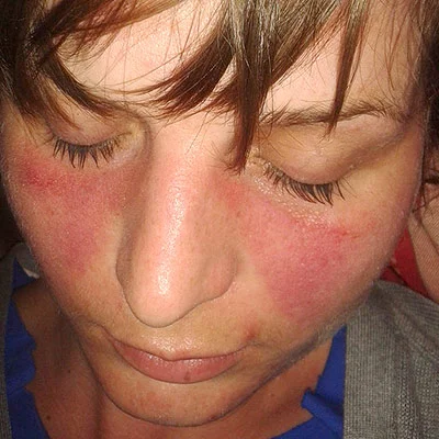
Long description
A clinical photograph of a child with butterfly (malar) erythema in systemic lupus erythematosus.

No single test diagnoses SLE. The pattern of results supports the clinical impression and monitors activity.
- Full blood count and blood film: anaemia, leukopenia, thrombocytopenia, haemolysis.
- Inflammatory markers (ESR and CRP): ESR is often raised in SLE; CRP is usually normal unless there is serositis or infection. A raised CRP therefore suggests infection or serositis, not uncomplicated activity.
- ANA by immunofluorescence on HEp-2 cells: positive in the large majority (more than 95%) of patients with SLE [7].
- Anti-dsDNA antibodies: highly specific for SLE when the assay is validated (at least 90% specificity against relevant disease controls) [1].
- Anti-Smith (Sm) antibodies: very specific for SLE.
- Complement (C3, C4, CH50): low in active disease, particularly in lupus nephritis; falling levels herald renal flares.
- Urinalysis and spot urine protein-to-creatinine ratio: screens for renal involvement; quantify proteinuria over 24 hours when indicated.
- Renal function (creatinine, estimated glomerular filtration rate, eGFR).
- Antiphospholipid antibodies: lupus anticoagulant, anticardiolipin (IgG/IgM/IgA) and anti-beta2-glycoprotein I (IgG/IgM/IgA), on two occasions at least 12 weeks apart.
- A positive ANA with negative anti-dsDNA and anti-Sm, normal complement and normal urine often means an ANA-positive person without SLE; children, family history and medications should be reviewed, and the test should be repeated if clinically indicated.
- Anti-dsDNA with low complement and rising proteinuria indicates active lupus nephritis until proven otherwise.
- Anti-cardiolipin or lupus anticoagulant identifies secondary antiphospholipid syndrome and thrombotic risk.
- A positive direct antiglobulin (Coombs) test with reticulocytosis, low haptoglobin and raised indirect bilirubin defines autoimmune haemolysis.
| Item | Stated cut-off or definition |
|---|---|
| ANA | At least 1:80 on HEp-2 cells (or equivalent), at least once [1]. |
| Fever | Temperature greater than 38.3 °C [1]. |
| Leukopenia | White blood cell count under 4,000/mm3 [1]. |
| Thrombocytopenia | Platelet count under 100,000/mm3 [1]. |
| Proteinuria | Greater than 0.5 g/24 h, or equivalent spot urine protein-to-creatinine ratio [1]. |
| Anti-dsDNA | Immunoassay with at least 90% specificity for SLE against relevant disease controls [1]. |
| Complement (C3/C4) | Below the lower limit of normal for the laboratory [1]. |
| Antiphospholipid antibodies | Anticardiolipin at medium or high titre (over 40 APL/GPL/MPL or above the 99th percentile), or positive anti-beta2-glycoprotein I, or positive lupus anticoagulant [1]. |
| Joint involvement | Synovitis of at least 2 joints, or tenderness in at least 2 joints with at least 30 minutes of morning stiffness [1]. |
| Lupus nephritis class by biopsy | Class III (focal) or IV (diffuse) nephritis scores 10 points on its own, which equals the classification threshold [1]. |
Use the 2019 EULAR/ACR criteria as a classification and as a prompt to look systematically for organ involvement, not as a yes/no diagnostic test. The SLICC 2012 and ACR 1997 criteria remain in wide use and in old exam questions.
Severity is better described by organ involvement and damage than by a single score. Practically:
- Mild disease: constitutional, musculoskeletal and skin features only, normal renal function and urine, no serious haematological or neuropsychiatric involvement.
- Moderate disease: persistent arthritis, widespread skin disease, leukopenia or thrombocytopenia without major organ threat.
- Severe / organ-threatening: lupus nephritis, neuropsychiatric SLE, severe haematological (autoimmune haemolytic anaemia or thrombocytopenia with bleeding), pulmonary or cardiac involvement. These groups require specialist management and usually cyclophosphamide, rituximab or biologics.
- Damage is tracked with a damage index (SLICC/ACR Damage Index) so that late complications (renal failure, cataract, avascular necrosis, premature atherosclerosis) are not mistaken for active disease.
Disease activity is monitored with tools such as SLEDAI or BILAG; low disease activity (LLDAS) or remission (DORIS criteria) is the treatment target.
- Life-threatening flares (lupus nephritis with renal impairment, neuropsychiatric SLE, diffuse alveolar haemorrhage, severe cytopenias) need urgent specialist care, high-dose intravenous methylprednisolone (125 to 1,000 mg/day for 1 to 3 days, with some guidelines using 250 to 1,000 mg/day [2][3]), and usually intravenous cyclophosphamide or rituximab.
- Infection must always be excluded before treating a flare: in an immunosuppressed patient with SLE, fever and raised CRP are more likely to be infection than inflammation.
- Renal flares should be biopsied promptly where feasible.
Sun protection (high-SPF sunscreen, protective clothing, avoidance of midday sun) is a genuine treatment, because UV light drives both skin and systemic disease [3]. Smoking cessation, regular exercise, weight control, attention to bone health, and treatment of cardiovascular risk factors (blood pressure, lipids, diabetes) are part of routine care. Tailoring drug choices, contraception and fertility counselling to the individual.
Pharmacological: the backbone (EULAR 2023 [3])
- Hydroxychloroquine for every patient with SLE, unless contraindicated, at a target dose of 5 mg/kg real body weight per day, individualised for flare risk and retinal toxicity [3]. Hydroxychloroquine reduces flares, thrombotic events and damage accrual, and improves survival. It should be continued long term, including in pregnancy (see below).
- Glucocorticoids are bridging therapy, not maintenance. Use the lowest dose that controls activity. pulses of intravenous methylprednisolone 125 to 1,000 mg/day for 1 to 3 days (some guidelines and most clinicians use 250 to 1,000 mg/day) can be given in moderate to severe disease [3][2]. Maintenance dose should be 5 mg/day or less (prednisone equivalent) and, if possible, withdrawn; at doses of 0.3 to 0.5 mg/kg/day after a pulse, genomic pathways are largely saturated, so the traditional 1 mg/kg regimen adds little [2]. Observational data support 5 mg/day or less over 7.5 mg/day for lower damage accrual [2].
- Prompt add-on of an immunomodulator in anyone not controlled on hydroxychloroquine (with or without glucocorticoids), or unable to reduce glucocorticoids to an acceptable chronic dose: methotrexate, azathioprine or mycophenolate, and/or biologicals (belimumab or anifrolumab), should be considered [3].
- Organ-threatening or life-threatening disease: intravenous cyclophosphamide, and rituximab in refractory disease [3].
- Severe haematological disease (e.g. immune thrombocytopenia): high-dose glucocorticoids, with or without intravenous immunoglobulin, rituximab or high-dose intravenous cyclophosphamide, then maintenance with rituximab, azathioprine, mycophenolate or cyclosporine [3].
- Active skin disease: topical glucocorticoids or calcineurin inhibitors, antimalarials, plus systemic steroids as needed; methotrexate, mycophenolate, anifrolumab or belimumab as second line [3].
- Neuropsychiatric disease attributed to SLE: glucocorticoids and immunosuppressants for inflammatory disease; antiplatelets or anticoagulation for atherothrombotic or antiphospholipid-related disease [3].
Screening is continuous: screen for proteinuria at least every 6 to 12 months in SLE without known kidney disease, or sooner during extra-renal flares [4]. Biopsy promptly when lupus nephritis is suspected, and conditionally when there is proteinuria over 0.5 g/g and/or impaired kidney function not otherwise explained [4].
Active proliferative (class III/IV, with or without V) disease is treated with anchor drugs (glucocorticoids plus mycophenolate or low-dose "EuroLupus" intravenous cyclophosphamide) and the ACR 2024 guideline [4] favours upfront triple immunosuppression: [3]
- pulse intravenous glucocorticoids 250 to 1,000 mg methylprednisolone daily for 1 to 3 days, then oral glucocorticoid at most 0.5 mg/kg/day (maximum 40 mg/day), tapered to at most 5 mg/day by 6 months, plus [2][4]
- mycophenolic acid analogue (MMF / MPA) plus belimumab, or mycophenolic acid analogue plus a calcineurin inhibitor (CNI), or EuroLupus low-dose cyclophosphamide plus belimumab (with MMF / MPA substituted for cyclophosphamide after the cyclophosphamide course).
An MMF-based regimen is favoured over a cyclophosphamide-based one. Where proteinuria is at least 3 g/g, choose a regimen containing MMF plus a CNI. Where extra-renal manifestations are prominent, choose a regimen containing belimumab [4]. If using cyclophosphamide, EuroLupus low-dose intravenous cyclophosphamide is preferred over high-dose monthly pulse or daily oral regimens [4]. People at high risk of renal failure (reduced GFR, cellular crescents, fibrinoid necrosis, or severe interstitial inflammation) may receive high-dose (NIH regimen) intravenous cyclophosphamide with pulse methylprednisolone [3].
Pure membranous (class V) disease with proteinuria at least 1 g/g is treated with the same pulse glucocorticoids then triple therapy (MPA plus a CNI). With proteinuria under 1 g/g, glucocorticoids and/or an immunosuppressant (MPA, azathioprine or a CNI) are used [4].
Non-responsive or refractory disease should first prompt a check of drug dose and adherence [4].
After renal response, treatment of lupus nephritis should continue for at least 3 years; patients initially treated with mycophenolate, with or without belimumab or a CNI, should remain on those drugs, and azathioprine or mycophenolate should replace cyclophosphamide after an initial cyclophosphamide course [3][4]. The ACR guideline sets a therapy duration of at least 3 to 5 years after complete renal response [4]. Therapy is titrated against proteinuria and kidney function trends, with complete renal response as the goal [4].
SLE with thrombotic APS is managed with long-term vitamin K antagonists after the first arterial or unprovoked venous thrombotic event; in patients with SLE without APS but with a high-risk antiphospholipid profile, low-dose aspirin (75 to 100 mg/day) should be considered [3]. In pregnancy this extends to heparin (below).
In patients achieving sustained remission, gradual tapering should be considered, withdrawing glucocorticoids first [3]. Glucocorticoid withdrawal may be possible after 12 months of quiescent disease and slow taper, although randomised data in SLE favour maintenance over abrupt stopping [2]. Non-pharmacological measures (sun protection, smoking cessation, healthy diet, exercise, bone health) [3] and immunisation against herpes zoster, human papillomavirus, influenza, COVID-19 and pneumococcus, plus monitoring of bone health, kidney protection, cardiovascular risk and malignancy screening [3] are recommended.
Any patient with suspected lupus nephritis, neuropsychiatric SLE, diffuse alveolar haemorrhage, severe cytopenias or life-threatening flare needs urgent specialist (rheumatology, nephrology, neurology, haematology) involvement. Early referral also improves outcomes in mild disease, because diagnostic delay is common [3].
- A basic work-up (full blood count, ESR, ANA if available, urinalysis, creatinine, complement if possible) still allows most diagnoses; clinical judgement carries more weight when tests are unavailable.
- Renal biopsy is not always available; heavy persistent proteinuria with active sediment in a patient with SLE is treated as proliferative nephritis if specialist advice supports it. A repeat biopsy to confirm response is often impossible, so creatinine and proteinuria trends become the guide.
- Hydroxychloroquine and oral prednisolone are inexpensive and form the backbone of treatment; methotrexate and azathioprine are widely available alternatives when mycophenolate or biologics are not.
- Biologics (belimumab, anifrolumab, rituximab) and voclosporin may not be accessible; the principle of minimising steroids and using the cheapest effective immunosuppressant remains.
- A region note: in East Africa SLE is under-recognised and often presents late with renal disease; maintaining a low threshold for ANA and urine testing in young women with joint, skin or haematological problems is good practice.
East Africa
In many East African settings hydroxychloroquine, prednisolone, azathioprine and methotrexate are available and form the workhorse regimen; biologics and voclosporin usually are not. Urine dipstick screening of young women with systemic symptoms is a cheap and useful habit.
- Lupus nephritis (classes I to VI, see below): the leading cause of serious morbidity; severe disease can progress to end-stage renal disease despite treatment [5].
- Secondary antiphospholipid syndrome: venous and arterial thrombosis, pregnancy loss, thrombocytopenia.
- Cardiovascular disease: accelerated atherosclerosis from the disease, its treatment and traditional risk factors.
- Neuropsychiatric SLE: stroke, seizures, psychosis.
- Infection: from the disease and its immunosuppression.
- Haematological: autoimmune haemolysis, thrombocytopenia.
- Bone disease: osteoporosis and avascular necrosis (especially from glucocorticoids).
- Malignancy: increased risk of non-Hodgkin lymphoma and cervical dysplasia (human papillomavirus screening).
- Pregnancy complications: pre-eclampsia, preterm birth, fetal loss, neonatal lupus.
Most patients now survive for decades, but damage accumulates and mortality remains two to three times that of the general population [6]. Renal involvement, early age at onset, high SLEDAI, cytopenias, low complement, antiphospholipid antibodies and high steroid exposure predict worse outcome. Good control of disease activity, early use of steroid-sparing agents and prompt renal treatment are the levers that improve it.
Renal disease develops in more than half of patients with SLE and is the first manifestation in 15 to 20% [5]. The 2003 International Society of Nephrology / Renal Pathology Society (ISN/RPS) classification is the framework [5]:
| Class | Name | Brief description |
|---|---|---|
| I | Minimal mesangial | Normal glomeruli by light microscopy; mesangial immune deposits on immunofluorescence. |
| II | Mesangial proliferative | Mesangial deposits with mesangial proliferation. |
| III | Focal | Proliferative / necrotising / crescentic lesions involving under 50% of glomeruli. |
| IV | Diffuse | Lesions involving at least 50% of glomeruli; subdivided into segmental (IV-S) and global (IV-G). |
| V | Membranous | Subepithelial immune deposits; often presents as nephrotic syndrome. |
| VI | Advanced sclerotic | 90% or more of glomeruli globally sclerosed, without residual activity. |
Classes III and IV (and combinations III + V or IV + V) are the proliferative, aggressive forms that need immunosuppression. Activity (A), chronicity (C) or both (A/C) are specified for III and IV.
A biopsy is generally indicated with an acute rise in creatinine, proteinuria over 500 mg/24 h or a protein-to-creatinine ratio over 0.5 g/g, haematuria at any level of proteinuria, or an active sediment with cellular casts [5]. Some patients with modest proteinuria have active nephritis on biopsy, so biopsy should be considered with new unexplained renal findings in SLE. [4]
- Sun protection is both prevention and treatment: high-SPF sunscreen, protective clothing, avoidance of peak UV.
- Cardiovascular risk factor control (smoking, hypertension, dyslipidaemia, diabetes) from diagnosis, because damage is partly irreversible.
- Bone protection: baseline bone mineral density when long-term steroids are planned; calcium and vitamin D; bisphosphonates where indicated; lowest glucocorticoid dose.
- Infection prevention: vaccinations (herpes zoster, human papillomavirus, influenza, COVID-19, pneumococcus), plus prophylaxis where local protocols require it [3].
- Malignancy screening: cervical cytology (increased dysplasia with immunosuppression and HPV), and site-specific screening.
- Renal screening: urinalysis at every visit; proteinuria every 6 to 12 months in patients without known renal disease [4].
- Retinal screening for hydroxychloroquine toxicity: follow local ophthalmology schedules; risk rises with dose (above 5 mg/kg/day), renal impairment and duration.
Pregnancy is possible and often successful in SLE, but requires planning. The principles are [9][10]:
- Conceive during inactive disease for at least 6 to 12 months; active lupus nephritis and severe organ disease are reasons to delay or avoid pregnancy [9][10].
- Continue or initiate hydroxychloroquine in pregnancy unless contraindicated; doses used are 400 mg/day or less [9].
- Low-dose aspirin from 12 weeks of gestation until delivery for all patients with SLE to reduce pre-eclampsia risk [10]. Low-dose aspirin 75 to 100 mg/day during pregnancy is also recommended for patients with SLE with a high-risk antiphospholipid profile without prior events [9].
- Azathioprine, cyclosporine and tacrolimus are pregnancy-compatible; methotrexate must be stopped 1 to 3 months and mycophenolate at least 6 weeks before conception; cyclophosphamide, leflunomide and warfarin are contraindicated or used only in exceptional circumstances [10].
- Antiphospholipid syndrome in pregnancy: prophylactic heparin plus low-dose aspirin for obstetric APS, therapeutic heparin for thrombotic APS [10]. A high-risk aPL profile without prior events is managed with low-dose aspirin alone [10].
- Antenatal monitoring: serial growth scans and fetal monitoring because of fetal growth restriction and stillbirth risk; anti-Ro/SSA and anti-La/SSB antibodies put the fetus at risk of congenital heart block and neonatal lupus, which is why antibody status is checked before or early in pregnancy [9].
- Pre-pregnancy counselling with both maternal-fetal medicine and rheumatology is recommended, with discussion of maternal and fetal risks [10].
Childhood-onset SLE tends to be more severe, with more frequent renal involvement [7]. Management mirrors the adult approach, but accrued damage and growth must be monitored; the ACR 2024 lupus nephritis guideline applies to adults and children, although steroid dosing in children is largely extrapolated from adult evidence.
Late-onset SLE often has less rash and more serositis and pulmonary disease, with a broader differential. Immunosuppressant dosing, renal function monitoring and drug interactions deserve extra care, and steroid-sparing strategies are emphasised.
Drug-induced lupus (DIL) is a lupus-like syndrome temporally related to drug exposure that usually improves after stopping the offending drug [8]. Systemic DIL is reported with anti-tumour necrosis factor drugs (the most common triggers in recent case series), but also with classic agents such as procainamide and hydralazine, and with other immune-modulating drugs [8]. Subacute cutaneous lupus is increasingly reported with proton pump inhibitors, and chemotherapeutic agents predominate among cutaneous cases [8]. Enhanced neutrophil extracellular trap formation induced by procainamide and hydralazine may be one mechanism [8]. There are no standard diagnostic criteria, so the diagnosis is clinical. Compared with idiopathic SLE, DIL usually has less renal and central nervous system involvement and fewer high-specificity antibodies; complement is usually normal. Stopping the drug is the treatment, with short courses of anti-inflammatories or steroids for symptoms.
Approach to suspected systemic lupus erythematosus
- Suspected SLEYoung woman with arthritis, rash, cytopenia, serositis, renal or neuro features
- ANA (HEp-2)Titre ≥1:80 or equivalent
- Question: Is the ANA positive?
No
- SLE unlikelyReconsider diagnosis; drug-induced, infection, other connective tissue disease
Yes
- Score 2019 EULAR/ACR criteriaClinical + immunological weighted criteria
- Question: Score ≥10?
No
- Not classified; re-evaluate if clinical suspicion remains
Yes
- Confirm SLE; organise baseline investigationsFBC, ESR/CRP, anti-dsDNA, anti-Sm, C3, C4, urinalysis, UPCR, renal function, aPL
- Start hydroxychloroquine 5 mg/kg/day real body weight
- If active disease, add glucocorticoid bridging + immunomodulator or biologic as neededMinimise and withdraw steroids
- Screen for renal involvementProteinuria every 6 to 12 months or with extra-renal flares; biopsy if proteinuria over 0.5 g/g
- Plan prevention and follow-upSun protection, bone health, cardiovascular risk, immunisation, pregnancy counselling
- SLE is a relapsing and remitting systemic autoimmune disease with autoantibodies against nuclear antigens; immune complex deposition drives organ damage.
- Female predominance is about 9:1, peaking in reproductive years; frequency and severity are greater in people of African, Asian and Hispanic ancestry.
- ANA positivity (at least 1:80) is the obligatory entry criterion for the 2019 EULAR/ACR classification criteria; a score of at least 10 classifies SLE.
- Anti-dsDNA and anti-Smith antibodies are the most specific serological findings; low C3 and C4 indicate active disease.
- Lupus nephritis occurs in more than half of patients and is usually silent early; biopsy is the gold standard and guides therapy.
- Hydroxychloroquine is recommended for all patients at 5 mg/kg real body weight per day. [3]
- Glucocorticoids are bridging therapy: pulse 125 to 1,000 mg/day intravenous methylprednisolone for severe disease, maintenance at most 5 mg/day prednisone equivalent.
- Active proliferative nephritis is now treated with upfront triple immunosuppression, with therapy continued for at least 3 to 5 years after complete response.
- High-risk antiphospholipid profiles are managed with low-dose aspirin 75 to 100 mg/day; thrombotic APS needs long-term anticoagulation.
- Pregnancy is usually successful if planned during remission; continue hydroxychloroquine (400 mg/day or less), give low-dose aspirin from 12 weeks, stop teratogenic drugs well before conception.
- Drug-induced lupus should be considered with a compatible drug and usually improves when the drug is stopped.
Answer each one in your head first, then open the answer.
A 24-year-old woman has arthritis of the hands, a malar rash sparing the nasolabial folds, oral ulcers and Raynaud phenomenon. ANA is 1:320. Anti-dsDNA is positive and C3 is low. What is the diagnosis and the most useful next investigation?
Show answerHide answer
Systemic lupus erythematosus. The next most useful investigation is urinalysis with a spot protein-to-creatinine ratio (and renal function), because lupus nephritis can be silent early and determines prognosis and treatment. A renal biopsy follows if proteinuria exceeds 0.5 g/g or kidney function is unexplained.
What is the minimum hydroxychloroquine dose rule and the glucocorticoid maintenance target in EULAR 2023?
Show answerHide answer
Hydroxychloroquine for all patients with SLE at 5 mg/kg real body weight per day, individualised for flare risk and retinal toxicity. Glucocorticoids as bridging therapy only, with maintenance minimised to at most 5 mg/day (prednisone equivalent) and withdrawn if possible; pulse intravenous methylprednisolone 125 to 1,000 mg/day for 1 to 3 days in moderate to severe disease.
A patient with active class IV lupus nephritis has proteinuria of 4 g/g and extra-renal manifestations. What regimen suits best?
Show answerHide answer
Upfront triple immunosuppression: pulse glucocorticoids (250 to 1,000 mg methylprednisolone for 1 to 3 days) then oral glucocorticoid 0.5 mg/kg/day or less (maximum 40 mg/day) with taper, plus mycophenolic acid analogue plus a calcineurin inhibitor for heavy proteinuria (at least 3 g/g) and belimumab for extra-renal manifestations. Therapy is continued for at least 3 to 5 years after complete renal response. [4]
When is kidney biopsy indicated in lupus nephritis, and what proteinuria threshold applies?
Show answerHide answer
Prompt biopsy when lupus nephritis is suspected; conditionally when there is proteinuria over 0.5 g/g and/or impaired kidney function not otherwise explained. Biopsy also confirms class (I to VI), which drives treatment and prognosis.
A woman with SLE wishes to become pregnant. Summarise the preconception and pregnancy plan.
Show answerHide answer
Plan conception during 6 to 12 months of inactive disease. Continue hydroxychloroquine (400 mg/day or less). Stop methotrexate 1 to 3 months and mycophenolate at least 6 weeks before conception; cyclophosphamide is avoided. Start low-dose aspirin from 12 weeks to delivery. Use azathioprine, cyclosporine or tacrolimus if immunosuppression is needed. Manage antiphospholipid syndrome with heparin appropriately, and check anti-Ro/SSA and anti-La/SSB status for fetal heart block risk.
How does drug-induced lupus differ from idiopathic SLE?
Show answerHide answer
Drug-induced lupus follows drug exposure and usually improves when the drug is stopped; it generally has less renal and central nervous system involvement, fewer highly specific antibodies and usually normal complement levels.
Systemic sclerosis, Sjögren syndrome, rheumatoid arthritis, mixed connective tissue disease, antiphospholipid syndrome, glomerulonephritis, cutaneous lupus, chronic kidney disease, pregnancy counselling.
- Aringer M, Costenbader KH, Daikh DI, et al. 2019 European League Against Rheumatism/American College of Rheumatology Classification Criteria for Systemic Lupus Erythematosus. Arthritis & Rheumatology 2019;71(9):1400–1412. doi:10.1002/art.40930. Accessed .
- Moysidou GS, Fanouriakis A. EULAR 2023 Recommendations for the Management of Systemic Lupus Erythematosus: One Step Forward. Mediterranean Journal of Rheumatology 2024;35(1):63–65. doi:10.31138/mjr.130124.erm. Accessed .
- Fanouriakis A, Kostopoulou M, Andersen J, et al. EULAR recommendations for the management of systemic lupus erythematosus: 2023 update. Annals of the Rheumatic Diseases 2024;83(1):15–29. doi:10.1136/ard-2023-224762. Accessed .
- Sammaritano LR, Askanase A, Bermas BL, et al. 2024 American College of Rheumatology (ACR) Guideline for the Screening, Treatment, and Management of Lupus Nephritis. Arthritis & Rheumatology 2025;77(9):1115–1135. doi:10.1002/art.43212 (summary in The Rheumatologist, 2 Dec 2025). Accessed .
- Giannico G, Fogo AB. Lupus Nephritis: Is the Kidney Biopsy Currently Necessary in the Management of Lupus Nephritis?. Clinical Journal of the American Society of Nephrology 2013;8(1):138–145. doi:10.2215/CJN.03400412. Accessed .
- Tian J, Zhang D, Yao X, et al. Global epidemiology of systemic lupus erythematosus. Annals of the Rheumatic Diseases 2023;82(3):351–356. doi:10.1136/ard-2022-223035. Accessed .
- Ameer MA, Chaudhry H, Mushtaq J, et al. An Overview of Systemic Lupus Erythematosus (SLE) Pathogenesis, Classification, and Management. Cureus 2022;14(10):e30330. doi:10.7759/cureus.30330. Accessed .
- He Y, Sawalha AH. Drug-induced lupus erythematosus: an update on drugs and mechanisms. Current Opinion in Rheumatology 2018;30(5):490–497. doi:10.1097/BOR.0000000000000522. Accessed .
- Gamba A, Zen M, Depascale R, et al. Modern Management of Pregnancy in Systemic Lupus Erythematosus: From Prenatal Counseling to Postpartum Support. Journal of Clinical Medicine 2024;13(12):3454. doi:10.3390/jcm13123454. Accessed .
- Silver R, Craigo S, Porter F, et al. Society for Maternal-Fetal Medicine Consult Series #64: Systemic lupus erythematosus in pregnancy. American Journal of Obstetrics and Gynecology (SMFM 2023, reaffirmed 2025). Accessed .
Figure credits
Educational content for medical students and doctors. It is not a substitute for clinical judgement or local guidelines.