Aphasias and cortical syndromes
Aphasia types and cortical syndromes (neglect, agnosia, apraxia): artery localisation, causes, assessment, imaging, and rehabilitation in stroke and brain.
- Aphasia is an acquired disorder of symbol use: spoken, written or signed language, not a problem with the muscles of speech, hearing or general intelligence.
- Most language is housed in a left hemisphere triangle formed by the Wernicke area, the adjacent inferior parietal lobe and the Broca area, so a single vessel, usually a branch of the middle cerebral artery, can destroy only part of it and produce a recognisable syndrome.
- The classic post-stroke syndromes differ in what is lost and what is spared: Broca is non-fluent with good comprehension and poor repetition; Wernicke is fluent with poor comprehension; conduction has fluent speech and good comprehension but strikingly poor repetition; global loses everything; transcortical types spare repetition.
- Neglect, agnosias and apraxia are also cortical or disconnection syndromes: they are not just “weakness” or “senility”, and they are diagnosed by bedside tests that do not depend on strength.
- Good recovery and communication are possible: treat the cause urgently, refer all suspected communication difficulties after stroke to speech and language therapy within 72 hours, and adapt all information to the person's language [6].
By the end of this page you should be able to:
- Localise a language deficit to the Wernicke, Broca or connecting pathways, and to a cerebral artery territory.
- Recognise Broca, Wernicke, conduction, global and transcortical aphasias, and describe what is preserved in each.
- Explain three other cortical syndromes — neglect, agnosia and apraxia — and how they are demonstrated at the bedside.
- Take a focused history and perform a focused examination for language and other cortical deficits.
- Order investigations in a sensible sequence, including brain imaging and speech-language assessment.
- Outline immediate, definitive and low-resource management, and name the common traps.
Figure 11605 Brocas and Wernickes Areas-02
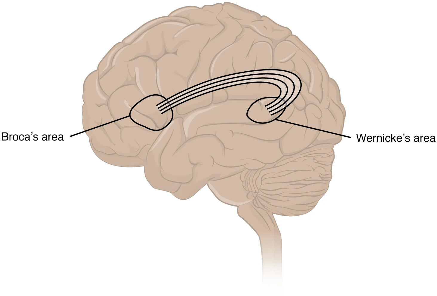
Long description
A diagram of the human brain circling Broca's and Wernicke's areas and showing the connections between them, from an OpenStax Anatomy & Physiology illustration hosted on Wikimedia Commons.

DefinitionAphasia
Aphasia is an acquired disorder of language: impaired comprehension, production or use of words and other symbols, resulting from damage to the language network in the dominant cerebral hemisphere. It is distinct from dysarthria, which is a motor problem of articulation, and from developmental language disorders.
In right-handed people and about two-thirds of left-handed people, language function resides in the left hemisphere; in the other third of left-handed people much of it resides in the right hemisphere [3]. Prosody — the rhythm and emphasis that add meaning to speech — is usually influenced by both hemispheres [3]. Language areas form a rough triangle: the posterosuperior temporal lobe (Wernicke area), the adjacent inferior parietal lobe, and the posteroinferior part of the frontal lobe just anterior to the motor cortex (Broca area), joined by subcortical pathways [3]. Damage to any part of this network, by infarct, haemorrhage, trauma or degeneration, interferes with some aspect of language use [3].
Two preliminary cautions matter for every later section. First, aphasia says something about where the lesion is, but not about the person's intellect; many people with aphasia retain reasoning in the non-verbal realm. Second, global medical crises often look like language problems: Wernicke aphasia can be mistaken for delirium, and a pure language disturbance has no fluctuating consciousness, hallucinations or inattention [3].
Broca aphasia was the first clearly localised language syndrome. It is non-fluent, with effortful, telegraphic, dysprosodic speech and good comprehension, but impaired repetition and agraphia. Wernicke aphasia is the fluent counterpart: normal rate, rhythm and grammar, but semantically empty speech with poor comprehension, anomia, and alexia and agraphia [3]. Conduction aphasia is defined by strikingly poor repetition in a person who is otherwise fluent with good comprehension [3]. Global aphasia is the most common type of aphasia, affecting comprehension and expression to varying degrees [1]. Transcortical motor aphasia resembles Broca aphasia, and transcortical sensory aphasia resembles Wernicke, but repetition is preserved in both because the perisylvian language core is spared and only the surrounding cortex is damaged [3].
The table is a clinical key, not a rigid label: the syndromes overlap, and careful description of the actual deficits is more precise than the name alone [3].
| Syndrome | Usual lesion | Speech pattern and impaired functions | Spared abilities |
|---|---|---|---|
| Broca (non-fluent, expressive) | Left frontal or frontoparietal area, including Broca area, usually a large lesion [3] | Anomia in oral and written language; slow, effortful speech; short phrases; impaired prosody and prepositions; impaired repetition; non-fluent agraphia | Comprehension relatively preserved; conceptualisation preserved |
| Wernicke (fluent, receptive) | Left temporoparietal area, including Wernicke area, usually a large lesion [3] | Fluent but semantically empty speech with paraphasias; poor auditory and written comprehension; impaired repetition; alexia; fluent agraphia | Rate, prosody and grammar may be preserved; often no hemiparesis |
| Conduction | Subcortical white matter under the superior temporal or inferior parietal lobe [3] | Prominent anomia and paraphasias; fluent speech; good comprehension; markedly impaired repetition; good reading comprehension; writing unaffected | Comprehension and spontaneous fluency |
| Global | Large left frontotemporoparietal lesion including both Broca and Wernicke areas [3] | Severe anomia; non-fluent, sparse output; poor comprehension; impaired repetition; alexia; agraphia | Little or nothing, although some automatic speech may remain |
| Transcortical motor | Left frontal lesion excluding Broca and Wernicke areas | Like Broca aphasia: non-fluent output, good comprehension | Repetition is strikingly preserved; articulation often unaffected |
| Transcortical sensory | Left temporoparietal lesion excluding Broca and Wernicke areas | Like Wernicke aphasia: fluent, empty speech, poor comprehension | Repetition preserved; may include echolalia |
| Anomic | Small lesion anywhere in the left language area [3] | Isolated difficulty naming objects, leading to circumlocution and empty speech; fluency, comprehension, repetition and reading largely preserved | Almost everything except naming |
Recovery is influenced by cause, lesion size and location, extent of impairment, and response to therapy, with age, education and general health playing a lesser part [3]. Children younger than 8 years often regain language function even after severe damage to either hemisphere; in older children and adults most recovery occurs within the first 3 months, with further improvement possible up to a year [3]. One practical consequence is that Broca aphasia tends to have better recovery than global aphasia, while global aphasia fares better than Wernicke aphasia [1][2]. A population-based study from Zurich reported 43 new aphasias per 100,000 residents per year after a first acute ischaemic stroke, mostly from cardioembolic causes; a Lund study reported 31 per 100,000 person-years and aphasia in 30 per cent of strokes [2].
Neglect is an acquired syndrome in which a person fails to attend to, act on or report stimuli on one side of the body or space, most often the left side, despite intact primary sensation and vision. It follows damage to the nondominant parietal cortex and networks, most often in the right middle cerebral artery territory, and extends beyond the bedside sensory examination. The patient may deny the deficit (anosognosia), ignore it (hemi-inattention) or both, even in the face of complete paralysis [5]. Neglect is not a "visual problem" alone; it affects personal space, peripersonal space and extrapersonal space.
An agnosia is inability to identify an object using one or more of the senses, despite intact peripheral perception. The damage is usually in the unimodal association cortex of the affected sense, from stroke, tumour, abscess or trauma, or in degenerative disease [5]. Visual agnosia has several forms: simultanagnosia, in which only one object can be seen at a time; prosopagnosia, the inability to recognise familiar faces, usually associated with bilateral inferotemporal or fusiform damage; and environmental agnosia, difficulty recognising familiar places [5]. Balint syndrome — simultanagnosia, optic ataxia and ocular apraxia — arises from bilateral dorsal occipitoparietal damage [5]. Auditory, tactile, gustatory and olfactory agnosias also occur [5].
Apraxia is the inability to execute a purposeful, previously learned motor task despite having the physical ability, willingness and understanding of the task, as a result of brain damage. It usually comes from parietal lesions or their connections, or from more diffuse degenerative damage [4]. Ideomotor apraxia is the most common form and shows as inability to imitate or perform a gesture to command. Ideational apraxia is inability to sequence a complex task; constructional apraxia is inability to draw, copy or construct; conceptual apraxia is the wrong use of tools [4]. Isolated apraxia is relatively uncommon [4]. A very important point for patients: people with apraxia usually do not recognise their deficit, and the patient's own account therefore cannot be trusted.
The artery table is a memory aid, not a measurement. Large or delayed lesions blur boundaries, and patients often have combinations rather than neat syndromes.
| Artery territory | Dominant hemisphere | Non-dominant hemisphere |
|---|---|---|
| Middle cerebral artery, superior division | Contralateral face and arm weakness greater than leg weakness; lower field homonymous hemianopia; Broca (expressive) aphasia; possible apraxia of speech | Contralateral weakness and lower field hemianopia; spatial inattention and neglect; anosognosia |
| Middle cerebral artery, inferior division | Contralateral upper field homonymous hemianopia; Wernicke (receptive) aphasia | Upper field hemianopia; visual-spatial and dressing apraxia; neglect emerging with large lesions |
| Anterior cerebral artery | Transcortical motor aphasia; contralateral leg weakness | Leg weakness; abulia; impaired executive function |
| Posterior cerebral artery | Visual cortex involvement; anomia and alexia can occur; transcortical or semantic impairment depending on extension | Hemi-inattention, visual agnosia and prosopagnosia with occipitotemporal involvement |
| Watershed (border-zone) between anterior, middle and posterior cerebral territories | Transcortical motor, sensory or mixed aphasia with relatively preserved repetition | Visual-spatial neglect, constructional deficits, dressing apraxia |
| Arcuate fasciculus disconnection (subcortical) | Conduction aphasia: disproportionate impairment of repetition with fluent speech and good comprehension | Conduction aphasia is usually dominant hemisphere; bilateral independence is rare |
Wernicke aphasia is most commonly caused by an acute ischaemic stroke affecting the posteroinferior division of the dominant middle cerebral artery [2]. That artery detail is why Wernicke aphasia often presents without hemiparesis, although visual pathway involvement gives a right visual field cut in many patients [3].
Aphasia is an acquired language loss; it is not developmental delay and it is not the language drift of childhood. If the problem wanes with time after a transient episode, consider transient ischaemic attack; post-stroke aphasia that persists at 24 hours is stroke until proven otherwise in routine practice. A convenient grouping by mechanism makes the differential wide enough to avoid premature closure.
| Mechanism or system | Examples | Typical cortical picture |
|---|---|---|
| Vascular: arterial (focal, acute) | Ischaemic stroke in MCA/ACA/PCA territory; intracerebral haemorrhage; TIA (resolved) | Broca, Wernicke, global or transcortical aphasia; neglect in right hemisphere lesions; visuospatial agnosias |
| Vascular: venous / dural sinus | Cerebral venous thrombosis; Sturge-Weber syndrome with cortical venous abnormalities | Acquired language regression; hemiparesis; neglect depending on site |
| Space-occupying lesions | Glioma, brain metastasis, abscess, subdural collection | Gradual aphasia; seizures; headache; papilloedema; constructional and semantic deficits |
| Trauma (traumatic brain injury) | Contusion, diffuse axonal injury, penetrating injury | Mixed fluent and non-fluent deficits; apraxia and agnosia from parietal injury; anosognosia |
| Neurodegenerative disease | Alzheimer disease (language early or late), primary progressive aphasia, vascular dementia (cognitive stepwise decline) | Primary progressive variants: non-fluent/agrammatic, semantic and logopenic aphasias; neglect and apraxia in later disease |
| Inflammatory / infectious | Herpes simplex encephalitis, brain abscess, chronic meningitis, autoimmune encephalitis | Acute cognitive and language upset; temporal lobe memory-semantic disturbance; anomia out of proportion |
| Metabolic / toxic / systemic | Hypoglycaemia, hepatic or renal failure, drug delirium, Wernicke thiamine deficiency (encephalopathy) | Delirium masquerading as Wernicke aphasia; global cognitive reduction; often reversible with treatment of cause |
| Seizure | Postictal aphasia and ictal aphasia | Transient; Todd-like language deficit after dominant hemisphere seizure; rapid improvement |
| Primary progressive aphasia spectrum (frontotemporal degeneration) | Non-fluent/agrammatic, semantic and logopenic variants | Gradual (not stroke), often with apraxia of speech; loss of word knowledge or grammar |
| Developmental / non-acquired mimics | Hearing loss, congenital or early brain injury producing different, often milder, speech organising | Usually not a sudden aphasia; if present from birth it belongs to a different framework |
- Any new or sudden change in language, comprehension, repetition, reading or writing is an acute neurological event until proven otherwise; call the stroke team. Aphasia onset is invisible to the patient.
- Neglect and anosognosia are associated with a higher falls and accident rate because the patient does not perceive half the environment; remove hazards and do not assume compliance.
- Suspected brain infection, suspected tumour or progressive aphasia needs urgent imaging and neurology or neurosurgical referral; never reassure on a "migraine aura" or "stress" label.
- Wernicke aphasia can be misdiagnosed as delirium, mania or psychosis: look for pure language impairment without clouding of consciousness [3].
- Dysphagia after stroke carries aspiration pneumonia risk: most major guidelines require an early swallow screen before any oral intake.
Ask what the person can understand and produce in each language they speak, including a second language, and ask whether the ability changed suddenly or gradually. Sudden onset points to vascular causes (TIA, stroke), stepwise progression to vascular cognitive impairment, and insidious progressive decline to neurodegeneration. Ask about vascular risk factors (hypertension, diabetes, atrial fibrillation, smoking), recent infection, head injury, seizure, neurosurgery and medications. Ask how the family communicates, whether the person is aware of the problem, and what the sudden language loss has cost them in terms of banking, driving, work and identity — this shapes therapy goals. When a second language, deaf sign or writing-only language is involved, ask directly; people are often assessed only in the language of the clinic, which can miss the deficit or exaggerate it.
Use the classic five-point mental status language screen [3]:
- Spontaneous speech for fluency, phrase length, effort, prosody, paraphasias and word-finding pauses.
- Naming objects, parts of objects, and actions; note circumlocutions.
- Repetition of single words and grammatically complex phrases.
- Comprehension with object naming, one-step and multistep commands, and yes-no questions of graded complexity.
- Reading and writing, including spontaneous writing, reading aloud, reading comprehension and writing to dictation.
Different deficit profiles then match the syndromes in the table above. Screen articulation too, because dysarthria and apraxia of speech often co-exist. In multilingual speakers check each relevant language; comprehension and production do not fail uniformly. Note automatic and rehearsed speech (for example, counting or prayers) and echolalia, which can be deceptively preserved. Bedside testing takes a few minutes but is the most valuable part of the neurological examination.
NICE defines visual inattention as an inability to orient towards and attend to stimuli on the side of the body affected by stroke. Screen with valid standardised assessment and behavioural observation that affects functional tasks such as mobility, dressing, eating and using a wheelchair [6]. Useful bedside tests include left-right line cancellation, circle or star cancellation, line bisection, copying and drawing, double simultaneous stimulation and examination of personal, peripersonal and extrapersonal space. Look for dressing apraxia and extinction as well as overt neglect. The Merck description reminds us that anosognosia can coexist with fully preserved denial of the paralysed limb [5].
Present common objects for identification by sight, touch and sound before concluding agnosia, because a primary sensory loss or an aphasia can imitate it [5]. For apraxia, test command imitation and use of common tools (such as a hammer or toothbrush) and drawing or copying; assess strength first, so that weakness is not mistaken for apraxia [4]. Observe swallowing, dressing, tool use and writing as functional screens; these are often more informative than isolated tests.
- Immediate brain imaging to separate haemorrhage from ischaemia and to look for mass, abscess or tumour; use CT first and MRI if the lesion is small, posterior or equivocal, and if vascular imaging is needed.
- Vascular and cardiac work-up guided by the clinical picture: carotid or intracranial imaging, ECG, rhythm monitoring and echocardiography when embolism is plausible, plus standard stroke risk studies.
- Metabolic screen (glucose, electrolytes, renal and liver function, infection markers, thyroid where indicated) to trap reversible causes.
- Formal speech and language therapy assessment within 72 hours of stroke onset, where a screening test is positive or there is clinical suspicion; NICE requires screening for communication difficulties within that window and referral of suspected cases to a speech and language therapist [6]. Assessment should analyse the impairment and its impact, including with the Western Aphasia Battery or similar tools, and should include the Barthel Index and National Institutes of Health Stroke Scale data in stroke patients [6].
- Cognitive, neglect and functional assessment using valid tools [6][8]; NICE recommends screening for cognitive impairment after stroke followed by detailed assessment before designing a treatment programme [6]. The ESO/EAN joint guideline found no evidence to support routine cognitive screening for everyone after stroke, but emphasised the importance of targeted assessment [8].
- Neuropsychological testing when deficits are subtle, the diagnosis is unclear, functional predictions are required, or behavioural symptoms are prominent [3][4][5].
- Disease-specific tests for suspected infection, tumour, vasculitis or degenerative disease (for example, EEG for seizure, cerebrospinal fluid when encephalitis or vasculitis is suspected, and advanced imaging when the diagnosis remains unclear).
Speech and language therapists, not the screening tool, make the detailed diagnosis. A screen is a sieve; the wording "passed" is often misleading, because a person with mild comprehension loss may score normally on a broad test.
Clinical approach to suspected aphasia or other cortical syndrome
- New or unexplained language, spatial or praxis deficitAphasia, neglect, visual agnosia, apraxia or constructional failure on testing
- Red flags for emergency?Acute onset, vascular risk, seizure, fever, signs of meningitis or cancer, altered consciousness
- Question: Should emergency brain imaging be performed now?
Yes
- CT head; consider MRI
- Identify haemorrhage, infarct, mass, abscess or venous thrombosis
- Follow local acute stroke and emergency pathway
No
- Arrange MRI or further imaging depending on suspected cause
- Bedside language, neglect, agnosia and praxis batterySpontaneous speech, naming, repetition, comprehension, reading and writing
- Question: Is repetition impaired out of proportion?
Yes, fluent with good comprehension
- Conduction aphasia; look for arcuate fasciculus or subcortical lesion
Yes, with non-fluent speech
- Broca or global aphasia depending on comprehension
No
- Transcortical or anomic pattern; consider watershed stroke or neurodegeneration
- Screen and referScreen communication within 72 hours of stroke onset and refer suspected cases to speech and language therapy; screen cognition and vision
- Cause-directed treatmentStroke care, revascularisation when indicated locally, infection treatment, tumour treatment, medication for seizure and vascular risk control
- Rehabilitation, family support and adaptationCommunication training, aphasia-friendly information, neglect scanning and prism glasses, occupational therapy for apraxia
Immediate management is cause directed. Where stroke is suspected, follow local acute stroke protocols for airway, aspiration prevention, glucose and vascular or neurosurgical pathways. Swallow before feeding. Treat seizures according to local neurology or emergency guidance. For the specific cortical deficits:
- Aphasia: Institute speech and language therapy early, because there is Cochrane evidence that speech and language therapy improves communication after stroke [3]. NICE requires services to offer at least 3 hours of rehabilitation a day on at least 5 days a week, including speech and language therapy, and to tailor all information to the person's communication and cognitive needs [6]. Make all written information aphasia friendly from the appointment letter onwards [6].
- Communication environment: Provide conversations with trained staff, community communication support groups, and training in slowed, supportive communication for family and ward staff, including gestures and props [6]. Consider computer-based word-finding programmes alongside face-to-face therapy [6], and assess whether the person needs a communication aid [6].
- Neglect: Screen functional impact and use task-focused interventions to redirect scanning to the neglected side, using bright page markings, sounds, repeated practice and prism glasses when indicated [6].
- Apraxia: There is no specific medical treatment. Physical and occupational therapy may improve function and safety modestly, but the mainstay is adaptation and the environment [4].
- Agnosia: Treat the cause if reversible, and use speech or occupational therapy to help the person compensate [5].
- Pharmacological treatment of language itself: No drug is established for aphasia after stroke. Current drug candidates are research only, including catecholaminergic agents and nootropics such as piracetam [1][2]. Cognitive impairment after stroke should be assessed and explained, and information provided to the person and family, even though treatment trials have not established benefit from cholinesterase inhibitors, memantine or cognitive rehabilitation [8].
Definitive long-term management is rehabilitation and adjustment, not "fixing the lesion". Provide speech and language therapy led by a specialist, with individualised therapy for specific impairments such as aphasia or dysarthria, and coaching for family and staff [6]. Review persisting communication difficulties at the 6-month and annual stroke reviews and refer back to speech and language therapy if beneficial [6]. The same AAN-affirmed 2026 guideline concerns comprehensive communication assessment after stroke, but the AAN page available here only records the affirmation; for recommendation detail, use the published article and local practice [7].
- Older adults: Consider pre-existing cognitive impairment, hearing loss and visual problems; differentiate aphasia from delirium; stroke rehabilitation still helps, but assess and re-assess cognitive and language function with repeated visits [6][8].
- Younger adults and stroke: Look for uncommon vascular causes, inherited conditions and neurovascular disease; cognitive and communication priorities are usually work, identity and parenting.
- Left-handed or ambidextrous people: Language may be less lateralised. Do not assume left-hemisphere localisation; neuroimaging and bedside findings matter more than handedness [3].
- Multilingual people, signed languages and low literacy: Assess in all languages and modes; avoid concluding "aphasia" from a test only in one language.
- Post-stroke cognitive impairment: ESO and EAN agree that cognitive impairment is common and potentially disabling after stroke, and that stroke survivors and caregivers rank problems of thinking and memory among their greatest concerns [8]. Intensive lifestyle or vascular risk-factor management has not been shown to prevent post-stroke cognitive decline or dementia [8]; the main benefit comes from conventional stroke prevention, rehabilitation and environmental support.
- Low-resource settings: A brief bedside language and neglect screen costs no more than time, and tells you who needs referral and who can be supported locally. Aphasia-friendly written materials, communication props and family training are low-cost and effective; use pictograms, short sentences and one instruction at a time. Where speech and language therapy is scarce, train nurses, therapists and volunteers in supportive communication. Refer promptly when the diagnosis is uncertain, when imaging is unavailable, or when atypical features suggest an alternative cause such as infection or tumour.
- Calling a fluent, word-salad speaker "confused"; check comprehension and repetition first.
- Calling non-fluent effortful speech "apraxia of speech" rather than aphasia; aphasia usually also has written language and comprehension abnormalities.
- Confusing aphasia with dysarthria. Dysarthria is slurred but grammatical speech from a motor pathway problem.
- Missing Wernicke aphasia because there is no hemiparesis; check auditory comprehension and reading.
- Assuming Broca aphasia means intact intellect but global aphasia means dementia.
- Missing neglect because the patient cooperates and tests visual acuity normally; ask them to draw or copy.
- Missing apraxia because strength is normal; test command, imitation and tool use.
- Forgetting that Wernicke aphasia can mimic delirium, mania or psychosis because insight is absent [3].
- Over-relying on the arterial template in patients with large lesions, collaterals, prior strokes, or bilaterally shifted language.
- Waiting for speech and language therapy before teaching supportive communication; family and ward staff should be coached from day one [6].
- Any new aphasia, neglect or apraxia needs neurology and brain imaging; a new sudden language deficit is treated as stroke until proven otherwise.
- Refer when imaging is unavailable locally, when features are progressive, infectious, autoimmune or tumour-like, or when adult-onset aphasia remains unexplained.
- Formal speech and language therapy referral should be made for all suspected communication difficulties after stroke, and repeated when persisting problems appear at the 6-month and annual reviews [6].
- Consider neuropsychology referral when cognitive, behavioural, mood or functional questions exceed the bedside screen.
- Aphasia is an acquired language disorder from a dominant hemisphere language network lesion; dysarthria and apraxia of speech are motor disorders, not aphasias.
- Most language is stored in a left hemisphere triangle connecting the Wernicke, inferior parietal and Broca areas, linked by subcortical pathways.
- Right-handed people and two-thirds of left-handed people are left-hemisphere dominant for language.
- Broca aphasia: non-fluent, good comprehension, impaired repetition and writing.
- Wernicke aphasia: fluent empty speech, poor comprehension, often without hemiparesis but with a right visual field cut risk.
- Conduction aphasia: disproportionate repetition impairment with fluent speech and good comprehension.
- Global aphasia: loss of comprehension and expression; the most common type.
- Transcortical types spare repetition.
- Neglect is most often a right parietal syndrome; patients are usually unaware of it.
- Agnosias are failures of recognition despite intact perception, caused by association cortex dysfunction.
- Apraxia is failure of learned motor execution with intact strength, usually from parietal injury.
- Treat the cause urgently in brain disease, and recognise stroke-like presentations.
- Rehabilitation is the mainstay; speech and language therapy improves communication after stroke.
- Screen communication within 72 hours of stroke and refer suspected cases [6].
- Adapt all written information and include speech, language and communication services in care.
- Anticonvulsants, cognition drugs and brain training are not established aphasia treatments outside trials, so manage expectations.
- Wernicke and global aphasias usually have grave recovery, but Broca aphasia and children recover better.
Answer each one in your head first, then open the answer.
A 62-year-old right-handed man suddenly cannot produce fluent speech, uses short telegraphic phrases, and writes badly, but follows commands and names objects poorly only in writing. Which syndrome and arterial territory?
Show answerHide answer
Broca (non-fluent, expressive) aphasia, usually in the superior division territory of the dominant middle cerebral artery. Comprehension is relatively preserved; repetition and writing are impaired. Global aphasia would show severely impaired comprehension, and conduction aphasia would have fluent speech.
Why can someone with Wernicke aphasia walk away from a conversation looking relaxed and yet understand almost nothing said to them?
Show answerHide answer
The lesion is in the temporoparietal comprehension area rather than the motor or Broca system, so speech remains fluent in rate, prosody and grammar while auditory and written comprehension collapse. Posterior circulation? No: it is usually the inferior division of the dominant middle cerebral artery.
A 74-year-old woman can converse freely but cannot repeat 'no ifs, ands, or buts', and makes many sound errors. Is repetition an unusual deficit to be in this precise combination?
Show answerHide answer
No. Disproportionately impaired repetition with fluent speech and good comprehension is the hallmark of conduction aphasia, usually from subcortical white matter disconnection between the comprehension and production areas.
A patient proudly announces they are well, despite ignoring half the plate and all words on the left side of a page. What is the syndrome and what is the clinical lesson?
Show answerHide answer
Neglect. Family and staff cannot rely on the patient's self-report; bedside functional tests and environmental safety planning are essential.
An educated professor cannot recognise his own coat and keys in the left hand; strength and sensation are intact. What separates this from hemiparesis?
Show answerHide answer
It is an agnosia, not weakness. Perception is intact but identification or memory linkage fails, often with association cortex or temporoparietal dysfunction. Test before concluding sensory loss.
A person understands a task, has normal strength, but produces the wrong sequence of actions when winding a clock or brushing teeth. What is the diagnosis and why does it matter for safety?
Show answerHide answer
Ideational apraxia. The person is not lazy or uncooperative; they cannot sequence the learned action. Adapt the environment, supervise and use occupational therapy rather than retraining from scratch.
What age group is noted to recover language better after a large brain injury, and what is the usual adult recovery window?
Show answerHide answer
Children younger than 8 years often regain language even after severe damage to either hemisphere; in older children and adults most recovery occurs within the first 3 months, with possible improvement up to a year [3].
Name at least four specific actions NICE requires after stroke for a person with communication difficulties.
Show answerHide answer
Screen within 72 hours; refer suspected cases to a speech and language therapist; make information aphasia friendly; offer specialist-led speech and language therapy; consider computer-based word-finding programmes; review persisting difficulties at the 6-month and annual stroke reviews [6].
Why is 'thought' of drug treatment for aphasia, no matter how appealing, not routine care at present?
Clinical neuroanatomy and spinal cord syndromes; ischaemic stroke and transient ischaemic attack; intracerebral haemorrhage; carotid artery stenosis; unconscious patient and coma; Alzheimer disease and the dementias; vascular, frontotemporal and Lewy body dementia; normal pressure hydrocephalus; status epilepticus; neurocutaneous syndromes including Sturge-Weber syndrome; meningitis; primary brain tumours and metastases; traumatic brain injury; transient ischaemic attack. Plain text cross-references only.
- Le H, Lui F, Lui MY. Aphasia. StatPearls [Internet]. Treasure Island (FL): StatPearls Publishing; updated 29 October 2024. Accessed .
- Lui F, Wroten M. Wernicke Aphasia. StatPearls [Internet]. Treasure Island (FL): StatPearls Publishing; updated 8 November 2025. Accessed .
- Huang J. Aphasia. The Merck Manual Professional Edition; last reviewed September 2025. Accessed .
- Huang J. Apraxia. The Merck Manual Professional Edition; last reviewed September 2025. Accessed .
- Huang J. Agnosia. The Merck Manual Professional Edition; last reviewed September 2025. Accessed .
- National Institute for Health and Care Excellence. Stroke rehabilitation in adults. NICE guideline NG236; published 18 October 2023. Accessed .
- American Heart Association/American Stroke Association, affirmed by the American Academy of Neurology Quality Committee. 2026 Guideline for Adult Stroke Rehabilitation and Recovery. Stroke; August 2026, affirmed 1 August 2026. Accessed .
- Quinn TJ, Richard E, Teuschl Y, et al. European Stroke Organisation and European Academy of Neurology joint guidelines on post-stroke cognitive impairment. European Stroke Journal 2021;6(3):I-XXXVIII. doi:10.1177/23969873211042192. Accessed .
Figure credits
Educational content for medical students and doctors. It is not a substitute for clinical judgement or local guidelines.