Primary brain tumours and metastases
Primary and secondary brain tumours: gliomas, meningiomas, medulloblastomas, schwannomas, CNS lymphoma and metastases, with classification, imaging.
- Primary brain tumours are growths that start in the brain, meninges, cranial nerves or pituitary; brain metastases are far more common in everyday practice and usually signal cancer elsewhere.
- The 2021 fifth edition of the World Health Organization (WHO) classification of central nervous system (CNS) tumours (WHO CNS5) defines tumours by combining histology with molecular markers, so the same-looking tumour can belong to different entities. [3]
- Gliomas are the commonest malignant primary parenchymal brain tumours in many series; meningiomas are the commonest benign primary intracranial tumours in most surgical series. [2]
- Gadolinium-enhanced magnetic resonance imaging (MRI) is the gold standard imaging test; a tissue diagnosis with integrated histomolecular testing is needed before definitive treatment. [1][4]
- Treatment is built around maximal safe surgical removal, then radiotherapy and, for glioblastoma, concomitant and adjuvant temozolomide, with dose adjustments for age and performance status. [1][4]
- Symptomatic brain oedema is treated with corticosteroids for the shortest time possible, and seizures are treated with antiepileptic drugs only after they occur, because routine prevention does not work. [1][5]
By the end of this page you should be able to:
- Classify primary brain tumours and metastases using the WHO CNS5 approach and state the main glioma entities. [2][3]
- Explain how a tumour's location and type produce headache, seizures, focal deficits and raised intracranial pressure.
- Plan the investigation of a suspected brain tumour, including standard structural MRI and the molecular markers that change the diagnosis. [1][4]
- Distinguish glioblastoma, IDH-mutant astrocytoma, oligodendroglioma, meningioma, medulloblastoma, pilocytic astrocytoma, schwannoma, primary CNS lymphoma and metastases by imaging and presentation.
- Choose initial therapy for glioblastoma, grade 2 or 3 glioma, meningioma and brain metastases, and state the radiotherapy doses. [1][4]
- Manage tumour-related oedema, seizures and steroid side effects, and know when to avoid steroids before biopsy. [1][5]
- Advise on follow-up schedules and how to adapt management in pregnancy, childhood and older age. [4]
- Explain the difference between a primary tumour, a metastasis, a brain abscess and stroke on imaging.
DefinitionBrain tumour
An abnormal growth of cells inside the skull. It may arise from the brain tissue itself (primary), from the coverings (meningioma), cranial nerves (schwannoma), or pituitary, or it may be a deposit from a cancer elsewhere (metastasis, also called a secondary brain tumour).
Classification works on two levels. First, by origin: primary versus secondary (metastatic). Second, by biology: the 2021 fifth edition of the WHO classification of CNS tumours (WHO CNS5) grades and names tumours by what the cells look like under the microscope together with molecular markers, so that a tumour is defined by its alterations as much as by its appearance. [3] The older approach of using histology alone is no longer enough for diffuse gliomas. [2]
For adult-type diffuse gliomas, WHO CNS5 recognises three families. [3]
- Astrocytoma, IDH-mutant, graded 2 to 4. Mutant IDH1/2, loss of nuclear ATRX, and TP53 mutations are typical; homozygous deletion of CDKN2A/B moves any grade into grade 4. [1]
- Oligodendroglioma, IDH-mutant and 1p/19q-codeleted, graded 2 or 3. These carry the best prognosis among diffuse gliomas in adults. [3]
- Glioblastoma, IDH-wildtype, grade 4 only. In the absence of necrosis or microvascular proliferation it can still be diagnosed when EGFR amplification, TERT promoter mutation, or a combined chromosome 7 gain and chromosome 10 loss are present. [1]
The word "glioblastoma" is now reserved for IDH-wildtype grade 4 astrocytic tumours; IDH-mutant grade 4 astrocytomas are no longer called glioblastomas. [1] Paediatric-type diffuse gliomas are separate families in WHO CNS5 and include H3 K27M-altered diffuse midline glioma and H3 G34-mutant diffuse hemispheric glioma, both of which can also appear in adults. [3] Circumscribed astrocytic gliomas, ependymomas, meningiomas, nerve-sheath tumours and embryonal tumours such as medulloblastoma are other major families. Where testing is incomplete, the qualifier "not otherwise specified" (NOS) is used; where the molecular profile does not fit any entity, "not elsewhere classified" (NEC) is used. [3]
A clinically useful anatomical split is intra-axial (within the brain substance, e.g. glioma, metastasis) versus extra-axial (compressing from outside, e.g. meningioma, schwannoma). Extra-axial tumours often have a wide dural base or cerebellopontine angle position and may be easier to remove completely.
| Tumour | Usual setting | Typical presentation | Imaging clues | Key management |
|---|---|---|---|---|
| Glioblastoma, IDH-wildtype | Adults; hemispheres | Weeks of headache, focal deficit, seizure, cognitive change | Irregular enhancing mass with central necrosis and oedema | Maximal safe resection, then radiotherapy 60 Gy in 30 fractions with temozolomide [4] |
| Astrocytoma, IDH-mutant (grade 2-4) | Adults | Slower onset, seizure common | Less aggressive enhancement; may not enhance | Grade and markers guide radiotherapy and PCV or temozolomide [1] |
| Oligodendroglioma, IDH-mutant, 1p/19q codeleted | Adults | Seizure, slow progression | Cortical, calcification often | Often surgery plus radiotherapy and PCV [4] |
| Meningioma | Adults, women more; dural based | Headache, seizure, focal or cranial nerve signs | Extra-axial, dural tail, strong uniform enhancement | Surgery by grade; radiotherapy if incomplete or higher grade [4] |
| Medulloblastoma | Children; cerebellum/vermis | Ataxia, vomiting, raised pressure, hydrocephalus | Posterior fossa solid mass; enhances | Surgery, craniospinal radiotherapy, chemotherapy (paediatric protocols) |
| Pilocytic astrocytoma | Children and young adults | Slow, site-dependent deficits | Cyst with mural nodule; cerebellum | Often surgical cure; BRAF-targeted therapy in selected recurrent cases [3] |
| Schwannoma (vestibular) | Adults; cerebellopontine angle | Unilateral hearing loss, tinnitus, imbalance | IAC and CPA mass | Observation, surgery or radiosurgery |
| Primary CNS lymphoma | Older adults; immunosuppressed | Deep lesions, subacute deficits, personality change | Periventricular homogeneous enhancement | Biopsy first; high-dose methotrexate-based chemotherapy; avoid steroids before tissue |
| Brain metastasis | Known cancer; any age | Multiplicity, stroke-like or seizure onset | Cortical grey-white junction, multiple, surrounding oedema | Local therapy (surgery, stereotactic radiosurgery/radiotherapy) plus systemic therapy [4] |
Gliomas occur at roughly six cases per 100,000 people per year worldwide, and men are diagnosed about 1.6 times as often as women. [1] In many published series, gliomas are the commonest malignant primary tumour of the brain parenchyma. [2] Brain metastases are encountered much more often than primary brain tumours in clinical practice, because common cancers (lung, breast, melanoma, kidney, gut) spread to the brain, but exact proportions depend on the population studied.
Risk factors for primary brain tumours include the following.
- Ionising radiation, particularly therapeutic cranial irradiation in childhood, is the clearest environmental risk factor.
- Familial tumour syndromes associated with gliomagenesis include neurofibromatosis type 1, tuberous sclerosis, Turcot syndrome, Li-Fraumeni syndrome and Lynch syndrome. [1]
- Tuberous sclerosis is particularly relevant to this book page: it is associated with cortical tubers, subependymal giant cell astrocytomas and (in children) cardiac rhabdomyomas, and management often involves the mTOR pathway. [2]
- Immunosuppression (for example advanced HIV infection or long-term immunosuppressive therapy) is a strong risk factor for primary CNS lymphoma.
- Neurofibromatosis type 2 predisposes to vestibular schwannomas and meningiomas.
There is no known modifiable measure that prevents gliomas. [1] Screening with brain imaging is limited to people with recognised familial syndromes, and is performed at the initial diagnostic work-up; repeated imaging is not indicated unless new neurological symptoms or signs such as seizures, aphasia, hemiparesis or sensory loss appear. [1] Genetic counselling is appropriate for suspected hereditary syndromes. [1]
A brain tumour causes problems in three ways, and most clinical features can be traced to one of them. [1]
- Mass effect and raised intracranial pressure. The skull is a rigid box. A growing mass displaces brain tissue, flattens sulci, and can shift the midline or cause herniation. Headaches that wake a patient or are worse on waking, early morning vomiting, and papilloedema are the clinical signatures of raised pressure. [1]
- Vasogenic oedema. Tumour vessels are leaky, so water accumulates in the white matter around the tumour. This adds to the mass effect and explains why symptoms can worsen over days, and why corticosteroids can relieve them so quickly. [5]
- Irritation of cortex and cranial nerves. Cortical irritation produces focal seizures and subtle cognitive or personality change, while compression of specific nerves produces focal sensory, motor or cranial-nerve deficits. [1]
The tumour's location matters as much as its size. A small tumour in the brainstem or internal capsule can be devastating, while a large frontal meningioma may present late with personality change or a single generalised seizure. Growth speed shapes the presentation too: a tumour that produces symptoms only weeks before diagnosis is usually fast-growing, whereas symptoms present for years point to a slow-growing lesion. [1]
Glioblastoma is a diffusely infiltrative astrocytic tumour of adults with microvascular proliferation and/or necrosis, or with the molecular signature of IDH-wildtype glioblastoma (EGFR amplification, TERT promoter mutation, or +7/-10) even without those classic histological features. [1] IDH-mutant astrocytomas have mutated IDH1/2, loss of nuclear ATRX and TP53 mutations, and are graded 2 to 4; oligodendrogliomas additionally show 1p/19q codeletion. [1]
Meningiomas are usually slow-growing, dural-based tumours derived from arachnoid cap cells. They show whorls and psammoma bodies microscopically and are graded 1 to 3; higher grades have more mitoses, brain invasion and a higher recurrence rate. Many express progesterone receptors. They are among the commonest primary intracranial tumours in adults. [4]
Medulloblastomas are malignant embryonal tumours of the cerebellum that occur mainly in children. They are small, round, blue-cell tumours with a propensity to spread through the cerebrospinal fluid. Current practice stratifies patients by age, metastatic stage and molecular subgroup for treatment intensity. [2]
Pilocytic astrocytoma is a low-grade, usually well-circumscribed tumour of children and young adults, most often in the cerebellum. It classically shows a cyst with an enhancing mural nodule, and microscopy shows a biphasic pattern with Rosenthal fibres and eosinophilic granular bodies. BRAF alterations are frequent, and BRAF-targeted therapy is sometimes used in recurrent or unresectable disease. [3]
Schwannomas (most often vestibular schwannomas) are benign nerve-sheath tumours. Microscopy shows Antoni A and Antoni B areas and Verocay bodies, and they often stain for S100. They arise from the vestibular division of the eighth cranial nerve and compress the brainstem if large. [4]
Primary CNS lymphoma is usually a diffuse large B-cell lymphoma confined to the CNS, lens-shaped or periventricular, and more common in older adults and immunosuppressed patients. Steroids can induce rapid but temporary regression and obscure the diagnosis, so they should not be given before tissue is obtained unless there is life-threatening mass effect. [1]
Metastases often sit at the grey-white matter junction and may be multiple. They are surrounded by vasogenic oedema and may show haemorrhage or necrosis. Seedings come in four patterns: single, oligometastatic (a small number), multiple, and leptomeningeal (lining the brain and spinal cord). [4]
The presentation depends on location, size and growth rate. Common symptoms are headache (often worse on waking or with coughing), nausea and vomiting, focal weakness or numbness, dysphasia, visual field loss, unsteadiness, cognitive slowing, personality change, and seizures. [1] A first seizure in an adult, particularly a focal seizure, should raise the possibility of a brain tumour.
Seizures are common in primary brain tumours. Their frequency ranges from 10 to 80 percent depending on tumour type; in low-grade glioma 60 to 100 percent of patients present with seizures, 25 to 60 percent in high-grade glioma, and about half of patients with diffuse glioma. More than a quarter of meningioma patients have seizures at some point. [5]
Neurological examination focuses on consciousness, orientation, cranial nerves (especially visual fields and acuity, facial sensation, hearing, swallowing and tongue), motor and sensory function, coordination, gait, and reflexes. The Mini-Mental State Examination (MMSE) or Montreal Cognitive Assessment (MoCA) screen for cognitive impairment. A standardised neuro-oncology scale (NANO) can document deficits for follow-up. [1] Fundoscopy for papilloedema is essential. A full systemic examination (including breast, lung, skin, abdomen and lymph nodes) is needed when a metastasis or a lymphoma is suspected. [1]
- Glioblastoma produces subacute focal deficits over weeks, often with headache and a generalised or focal seizure, and reflects the tumour's rapid growth. [1]
- Lower-grade glioma commonly presents with a long history of focal epilepsy before imaging, sometimes after years of seizures treated as epilepsy. [1]
- Meningioma grows slowly. Convexity tumours cause seizures or focal deficits; sphenoid wing and planum sphenoidale tumours threaten vision; olfactory groove tumours produce anosmia and personality change; posterior fossa tumours cause cranial-nerve and cerebellar signs. [4]
- Medulloblastoma in children presents with ataxia, incoordination, morning vomiting and signs of raised intracranial pressure from obstructive hydrocephalus. [2]
- Pilocytic astrocytoma produces slowly progressive cerebellar dysfunction or, when in the hypothalamus/optic pathway, visual loss and precocious puberty, especially in neurofibromatosis type 1. [3]
- Vestibular schwannoma presents with unilateral sensorineural hearing loss and tinnitus, then imbalance and facial numbness. [4]
- Primary CNS lymphoma typically produces deep, rapidly progressive deficits, with confusion or personality change; it is more common in older and immunosuppressed patients and often enhances homogeneously. [1]
- Metastases are suggested by a known primary cancer, multiple discrete lesions, and subacute focal deficits; presentation can mimic stroke. [4]
A brain lesion is not always a tumour. Infection, inflammation, vascular disease and treatment effects must be considered. [4]
| Condition | Features that favour it over tumour | Tests that separate it |
|---|---|---|
| Brain abscess | Fever or source of infection; subacute course; rim enhancement that is thin, smooth and regular | Diffusion-weighted MRI shows restricted diffusion; aspirate for culture |
| Stroke (infarct/haemorrhage) | Sudden onset; vascular territory; restricted diffusion within hours | CT acutely; MRI diffusion; vascular imaging |
| Demyelination (e.g. multiple sclerosis) | Younger patient; relapsing-remitting course; periventricular Dawson fingers; less mass effect for size | MRI pattern; cerebrospinal fluid oligoclonal bands |
| Radiation necrosis or pseudoprogression | Previous radiotherapy or chemoradiotherapy; time course of 3-12 months; enhancement may enlarge transiently | MR perfusion, PET, amino acid imaging; sometimes biopsy [1] |
| Subdural or epidural haematoma | Trauma; crescent- or lens-shaped collection; acute deterioration | CT head; clotting tests |
| Inflammatory and infectious masses (tuberculoma, toxoplasmosis) | Endemic area, HIV or immunosuppression; satellite lesions | Serology, PCR, response to therapy; biopsy if doubt |
| Meningioma vs schwannoma | Meningioma: dural tail, broad base, homogenous enhancement. Schwannoma: cerebellopontine angle, intracanalicular extension | MRI characteristics and hearing assessment |
| Metastasis vs glioma | Metastasis: known cancer, multiple lesions, cortical grey-white junction, disproportionately large oedema. Glioma: infiltrative, white matter based, single | Staging imaging; biopsy or resection [4] |
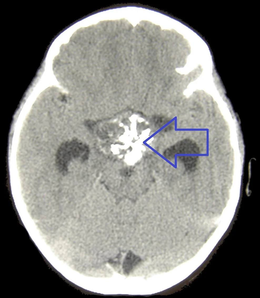
Figure 1Craniopharyngioma1
Long description
Unenhanced CT of a craniopharyngioma showing a calcified cystic structure in the suprasellar region, together with hydrocephalus.

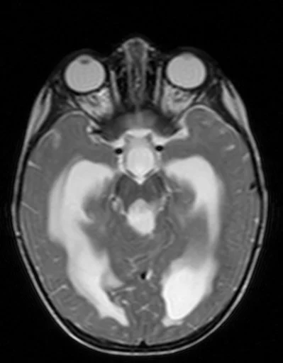
Figure 2Medulloblastom - MRT - T2 axial - 015
Long description
An axial T2-weighted MRI slice from an openly licensed medulloblastoma MRI case, whose file page describes it only as 'Medulloblastoma in the MRI'.

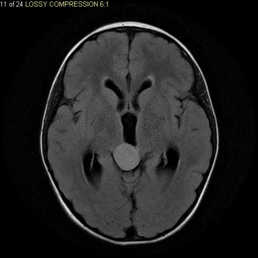
Figure 3Axial FLAIR MRI tectal plate piloctyic astrocytoma
Long description
A single axial CSF-suppressed FLAIR MRI showing a circumscribed hyperintense mass in the tectum of the brainstem presumed to be a pilocytic astrocytoma.

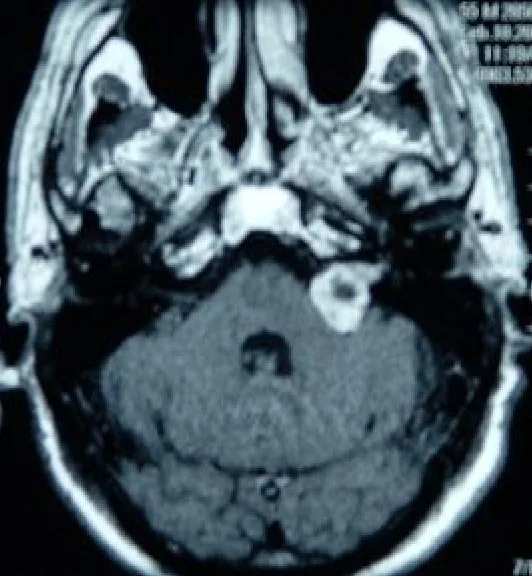
Figure 4Acoustic schwannoma
Long description
An axial MRI of the brain after gadolinium administration demonstrating a left cerebellopontine angle acoustic neuroma in a 51-year-old woman with progressive hearing loss.

Figure 5Tumor Meningioma1
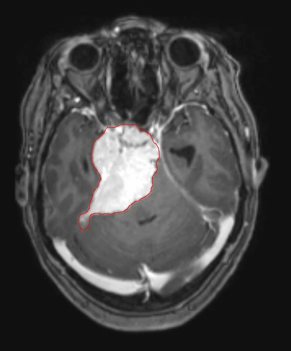
Long description
Axial T1-weighted MRI of a meningioma acquired after contrast administration.

Figure 6Glioblastoma macro
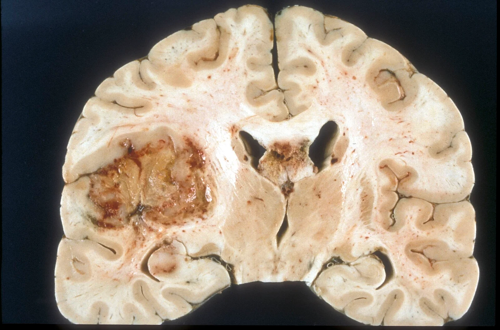
Long description
A macroscopic pathology photograph of a glioblastoma multiforme.

The aim is to define the anatomy, guess the type, and obtain tissue without harming the patient. [1][4]
Standard structural MRI without and with gadolinium contrast (T2-weighted, FLAIR, diffusion-weighted, and 3D T1 pre- and post-contrast sequences) is the first test. [4] It shows the site, size, enhancement pattern, oedema, hydrocephalus and midline shift. If MRI is not possible or is contraindicated, CT before and after contrast is used; plain CT also detects calcification, haemorrhage and bone involvement (important in meningioma and skull-base tumours). [4]
Advanced techniques help when the picture is unclear. MR perfusion, MR spectroscopy and amino acid positron emission tomography (PET) can distinguish tumour from necrosis, define aggressive areas for biopsy, or separate high-grade transformation in a previously low-grade lesion. [1]
NICE recommends CT of the skull when bone involvement is suspected in meningioma, and extracranial imaging (appropriate to the primary) for all suspected brain metastases, before neuro-oncology referral. [4]
Treatment decisions should be based on a tissue diagnosis that includes molecular markers, so upfront surgery has both diagnostic and therapeutic intent. [1] When resection is not safely possible (deep or eloquent location, poor condition), a frame-based or frameless stereotactic biopsy gives a high diagnostic accuracy with low morbidity. [1] Intraoperative fluorescence with 5-aminolevulinic acid helps to ensure adequate sampling. [4]
High-volume specialist centres, not individual surgeons, should manage the surgery. [1] A baseline MRI within 72 hours of resection, and again 3 months after radiotherapy, is considered for all glioma types. [4]
The molecular profile changes both name and treatment. [1][3][4]
| Marker | Why it matters |
|---|---|
| IDH1 R132H immunohistochemistry, then IDH1/2 sequencing if negative | Separates IDH-mutant astrocytoma/oligodendroglioma from glioblastoma; guides prognosis and therapy [1][4] |
| Nuclear ATRX loss or retention | Loss of ATRX in an IDH-mutant tumour indicates astrocytic lineage; retained ATRX triggers 1p/19q testing [1] |
| 1p/19q codeletion | Defines oligodendroglioma; associated with benefit from PCV chemotherapy [1][3] |
| MGMT promoter methylation | Predicts response to temozolomide in IDH-wildtype glioblastoma and helps decide chemo(radio)therapy in the elderly [1][4] |
| TERT promoter mutation, EGFR amplification, +7/-10 | Upgrade IDH-wildtype diffuse astrocytoma to glioblastoma even without necrosis/microvascular proliferation [1] |
| CDKN2A/B homozygous deletion | Upgrades IDH-mutant astrocytoma to grade 4 [1][3] |
| H3 K27M mutation and H3K27me3 loss | Defines diffuse midline glioma (thalamus, brainstem, spinal cord) [1] |
| H3.3 G34R/V mutation | Defines diffuse hemispheric glioma in young patients [1] |
| BRAF V600 mutation / fusion | Identifies pilocytic astrocytoma and other low-grade gliomas; opens targeted therapy [3][4] |
| NTRK fusion | Rare in glioblastoma but targeted inhibitors exist [3] |
Other tests are used as needed: electroencephalography (EEG) for tumour-associated epilepsy or altered consciousness; neuropsychological testing with a standardised battery or at least MMSE/MoCA; and, when metastasis is suspected, staging imaging and sometimes biopsy of the primary or extracranial disease before any brain surgery. [1][4]
There is no single staging system for primary brain tumours comparable with TNM; the WHO grade and molecular type do most of the prognostic work, supported by functional assessment. [3]
- WHO CNS5 (2021) sets the diagnosis for gliomas and many other entities, combining histology with the markers above. [2][3]
- IDH-wildtype grade 2 gliomas with molecular features of glioblastoma (EGFR amplification, TERT promoter mutation, or +7/-10) should be treated like glioblastoma. [1][4]
- Performance status: Karnofsky performance status (KPS) or ECOG performance status, age, and individual risks and benefits guide decisions. [1][4]
- Neurological function: the NANO assessment of nine domains (gait, strength, ataxia, sensation, visual fields, facial movement, language, consciousness and behaviour) is used in neuro-oncology. [1][5]
- Cognition: MoCA or MMSE. [1]
- Brain metastases severity is driven by number and volume of deposits, control of extracranial disease, location, and KPS (NICE uses 70 or more as a cut-off for some local therapies). [4]
Management is organised by a multidisciplinary team that includes neurosurgery, neuroradiology, neuropathology, neuro-oncology and radiation oncology. [1] The NICE guideline also requires a named key worker (often a clinical nurse specialist) to coordinate care. [4]
Raised pressure and oedema. Symptomatic brain oedema is treated with corticosteroids, which remain the drug of choice, but only while they produce clinical benefit because long-term use causes pneumonia, diabetes, hypertension, osteoporosis, myopathy and psychiatric symptoms. [5] EANO supports corticosteroids before surgery unless primary cerebral lymphoma or an inflammatory lesion is suspected. [1] Head-of-bed elevation and, rarely, osmotic therapy are adjuncts. [1]
Seizures. Patients who have had a seizure should receive an antiepileptic drug (secondary prophylaxis). First-choice agents are levetiracetam and lamotrigine; valproic acid is effective and well tolerated, and enzyme-inducing antiepileptic drugs should be avoided. [5] Routine primary prophylaxis in a patient who has never seized does not reduce the risk of a first seizure in glioma, and is not recommended. [1][5] Consent, driving rules and sudden-death advice follow local epilepsy guidance; tumour-associated epilepsy itself is a major determinant of quality of life. [4][5]
When not to give steroids. If a primary CNS lymphoma is suspected, steroids should be withheld until biopsy, because they shrink the tumour and can make the diagnosis impossible. [1] Inflammatory lesions are a similar trap.
Surgery. Maximal safe resection is the standard first step, aiming to remove all enhancing tumour while preserving function. Techniques include intraoperative microscopy, frameless stereotactic guidance, preoperative functional MRI, diffusion tensor imaging overlays, 5-aminolevulinic acid fluorescence-guided resection (offered when an enhancing high-grade glioma looks completely resectable), intraoperative MRI or ultrasound, and awake craniotomy with neuropsychological and speech-and-language support. [4] If resection is not safely possible, biopsy should be performed to obtain a diagnosis. [1][4]
Radiotherapy and chemotherapy for glioblastoma. EANO recommends the Stupp regimen (60 Gy of radiotherapy in 30 fractions with concomitant temozolomide, followed by up to 6 cycles of adjuvant temozolomide) as standard of care for IDH-wildtype glioblastoma in patients under 70 years with a KPS above 70. [1][2] NICE makes the same 60 Gy in 30 fractions offer for people around 70 or under with KPS 70 or more who have had maximal safe resection or biopsy. [4] NICE does not recommend bevacizumab or tumour-treating fields as part of newly diagnosed glioblastoma management, and does not recommend them for recurrent high-grade glioma. [4]
For older adults (around 70 or over with KPS 70 or more), NICE recommends 40 Gy in 15 fractions with concomitant temozolomide and up to 12 cycles of adjuvant temozolomide when the tumour has MGMT promoter methylation, and considers the same schedule when methylation is absent or unknown. [4] Best supportive care alone is considered when KPS is under 70. [4] EANO uses 65-70 years as the elderly cut-off and favours an abbreviated course for poorer performance status, aiming to complete treatment within 2-3 weeks. [1][2]
Radiotherapy and chemotherapy for grade 3 gliomas. NICE offers sequential radiotherapy and 4-6 cycles of PCV (procarbazine, lomustine and vincristine) for anaplastic oligodendroglioma with 1p/19q codeletion, and radiotherapy followed by up to 12 cycles of adjuvant temozolomide for astrocytoma (IDH-wildtype or mutant) without 1p/19q codeletion. [4] EANO recommends radiotherapy of 54-60 Gy in 1.8-2.0 Gy fractions followed by temozolomide or PCV for grade 3 astrocytoma according to IDH status, and radiotherapy followed by PCV (not temozolomide) for anaplastic oligodendroglioma. [1][2] For grade 3 IDH-wildtype astrocytoma, EANO recommends radiotherapy 54-60 Gy in 1.8-2.0 Gy fractions or chemoradiotherapy with temozolomide according to MGMT promoter methylation. [2]
Radiotherapy for grade 2 gliomas. NICE recommends not exceeding 54 Gy at 1.8 Gy per fraction, and adjuvant PCV (up to 6 cycles) after surgery for IDH-mutant low-grade glioma that is 1p/19q codeleted in patients around 40 or over, or with residual tumour. [4] EANO considers radiotherapy of 50-54 Gy in 1.8-2.0 Gy fractions followed by PCV, or concomitant chemoradiotherapy with temozolomide, for astrocytoma, and observation for selected low-risk patients. [1][2] NICE stratifies grade 2 glioma follow-up by molecular subtype (see below) and offers active monitoring for patients under 40 years with no residual tumour. [4]
Systemic (targeted and salvage) therapy. High-dose methotrexate-based regimens are used for primary CNS lymphoma (biopsy is needed first). For glioma, temozolomide and PCV are the backbone chemotherapies. Salvage options for recurrent high-grade glioma include repeat surgery, re-irradiation in selected cases, nitrosoureas, temozolomide rechallenge and, where available, bevacizumab; enrolment in clinical trials is emphasised. [1][2][4] NICE lists PCV or single-agent lomustine for recurrent high-grade glioma and offers best supportive care when unlikely to benefit. [4]
Molecularly targeted options now exist for selected patients. [3]
- IDH inhibitors: vorasidenib (an IDH1/2 inhibitor) prolongs progression-free survival (27.7 versus 11.1 months) in IDH-mutant grade 2 astrocytoma and oligodendroglioma and was approved by the FDA for this indication in August 2024; NCCN lists ivosidenib as an option for recurrent IDH1-mutant grade 2 gliomas, and in selected circumstances for grades 3 and 4. [2][3]
- BRAF V600E: dabrafenib plus trametinib has tumour-agnostic approval and has been studied in gliomas with response rates around 69 percent in low-grade and 33 percent in high-grade cohorts. [3]
- NTRK fusions (1-2 percent of glioblastomas) can be treated with larotrectinib, entrectinib or repotrectinib. [3]
- TMB-high tumours may be treated with pembrolizumab. [3]
- Von Hippel-Lindau disease with CNS haemangioblastomas may be treated with belzutifan; everolimus is used for tuberous sclerosis complex-associated partial-onset seizures; tovorafenib for paediatric low-grade glioma with BRAF alteration. [3]
NICE bases management on grade and extent of excision. [4]
- Grade 1, completely excised: active monitoring.
- Grade 1, incompletely excised: consider further surgery, radiotherapy or active monitoring.
- Grade 1, not excised: consider active monitoring or radiotherapy.
- Grade 2, completely excised: offer a choice between active monitoring and radiotherapy.
- Grade 2, incompletely excised: consider further surgery; offer radiotherapy if surgery is not possible or the tumour is incompletely excised afterwards.
- Grade 3: offer radiotherapy after any surgery, with further surgery where possible.
Choice of radiotherapy technique should take into account tumour grade, size and location (proximity to optic nerves, chiasm and brainstem), and the person's preference. [4]
NICE recommends maximal local therapy (surgery, stereotactic radiosurgery or stereotactic radiotherapy) for people with a single metastasis, taking into account comorbidities, tumour size and location, oedema and preference. [4] Note on single-lesion resection: overall survival does not differ greatly between surgery and stereotactic radiosurgery/radiotherapy; surgery offers faster symptom control and a fresh diagnosis, while radiosurgery offers a higher local control rate as an outpatient. [4]
For multiple metastases, stereotactic radiosurgery or radiotherapy is considered when extracranial disease is controlled or controllable and KPS is 70 or more, taking into account number and total volume. [4] Where that is not suitable, whole-brain radiotherapy (WBRT) is discussed honestly: there is no important overall survival benefit, but it may slow brain disease, at the cost of fatigue, temporary hair loss and accelerated cognitive loss. [4] Memantine and concurrent systemic therapy to enhance WBRT are not offered outside trials. [4] NICE advises against WBRT for people with non-small-cell lung cancer whose metastases are not suitable for surgery or stereotactic treatment and whose KPS is under 70. [4] Do not offer adjuvant whole-brain radiotherapy after surgery or stereotactic treatment of a single metastasis. [4]
Systemic anti-cancer therapy is considered when the metastases are likely to respond (for example germ cell tumours or small-cell lung cancer). [4]
NICE suggests regular clinical review with standard structural MRI. [4] After surgery, a baseline MRI within 72 hours and 3 months after radiotherapy helps detect recurrence. [4] Ongoing intervals vary by tumour grade and molecular subtype. [4] For an outline: grade 1 tumours are often scanned at 12 months with increasing intervals, whereas grades 2-4 have 3-6 monthly imaging initially, lengthening over years. [4] Meningioma follow-up is tailored by grade and residual tumour and ranges from 3-monthly to annual imaging, with discharge considered in selected grade 1 cases after many years. [4] Brain metastases are followed every 3 months in the first year, then 4-6 monthly, then annually. [4]
Brain tumours have unique effects on behaviour, cognition and personality, so health and social care needs should be assessed throughout the pathway, with a named coordinator. [4] Late side effects of radiotherapy and chemotherapy, including stroke risk and cognitive change, should be surveilled. [4][5]
- Oedema is common in both malignant and benign tumours: 40 to 92 percent of meningiomas have peritumoural oedema. [5] Corticosteroids help symptoms but should be used for the shortest time possible because of their systemic side effects. [5]
- Seizures (see above) and recurrence of seizures despite medication. [5]
- Stroke: primary brain tumour patients have one of the highest fatal stroke risks among cancer patients, highest in the first 6 months after diagnosis; radiotherapy contributes about a quarter of strokes in this group. Thrombolysis is generally avoided because of haemorrhage risk. [5]
- Pseudoprogression: enlargement of enhancement in the first months after radiochemotherapy can be treatment effect rather than tumour; MGMT-methylated glioblastomas are especially prone. [1][3]
- Hydrocephalus from obstructed cerebrospinal fluid, especially in posterior fossa tumours and medulloblastoma. [2]
- Treatment complications: surgical morbidity, infection, wound issues, and radiotherapy effects on cognition and endocrine function. [4][5]
- Recurrence: glioblastoma almost always recurs; recurrent disease may be managed with surgery, re-irradiation, nitrosoureas or best supportive care. [1][4]
Prognosis is dominated by tumour type and molecular markers. [3]
- Glioblastoma remains a lethal disease. In one meta-analytic summary, median overall survival for IDH-wildtype glioblastoma is 14.11 months with unmethylated MGMT promoter methylation and 24.59 months with methylated MGMT promoter methylation. [3]
- IDH-mutant astrocytomas behave less aggressively and grade 4 IDH-mutant astrocytoma usually outlives glioblastoma. [1][3]
- Oligodendrogliomas have the best prognosis among diffuse gliomas in adults and benefit particularly from PCV chemotherapy added to radiotherapy. [3]
- Meningiomas of WHO grade 1 are usually controlled after complete resection; grades 2 and 3 recur more often and may need radiotherapy. [4]
- Medulloblastoma outcome varies by age, stage and molecular subgroup; modern combined therapy cures a substantial proportion of standard-risk disease in children. [2]
- Pilocytic astrocytomas are usually cured surgically when accessible. [3]
- Brain metastases prognosis depends on the primary cancer, number and volume of deposits, and performance status. [4]
- Primary CNS lymphoma is chemosensitive, but relapses are common; immunosuppression worsens outcome.
There is no known way to prevent most primary brain tumours. [1] Avoidable or therapeutic radiation exposure should be minimised, especially in children. [1] Screening imaging is appropriate for familial syndromes at the initial work-up, but repeated imaging of asymptomatic relatives is not recommended unless symptoms develop. [1]
For metastases, screening brain imaging is used selectively in cancers that commonly spread to brain (such as small-cell lung cancer, melanoma, and testicular germ cell tumours) according to oncology protocols; routine screening is not recommended for all cancers.
Children. Common tumours differ: pilocytic astrocytoma, medulloblastoma, ependymoma and diffuse midline glioma are much more likely than glioblastoma. [2][3] EANO's adult diffuse glioma guidelines do not cover children; treatment is delivered by paediatric neuro-oncology teams using combined surgery, craniospinal radiotherapy and chemotherapy. Medulloblastoma can seed through the cerebrospinal fluid. [2]
Pregnancy. Management is individualised by a multidisciplinary team. Surgery is usually possible in the second trimester; radiotherapy and chemotherapy are generally deferred where possible because of fetal risk, and early delivery may be planned. Pharmacological choices must consider placental and fetal effects, and seizure control should be maintained with the safest effective agent.
Older adults. The temozolomide and radiotherapy schedules above are adjusted for age and KPS. MGMT promoter methylation status is used to choose whether temozolomide adds benefit in the elderly. [2][4] Best supportive care alone is appropriate when performance status is poor. [4]
Immunocompromised patients. Primary CNS lymphoma and opportunistic infections are more common; biopsy should be obtained before steroid therapy, and testing for HIV and other immunodeficiency is important. [1]
- Standard structural MRI with and without contrast may be unavailable; contrast-enhanced CT can be the diagnostic test, and a clear surgical and pathology pathway matters more than advanced imaging. [4]
- Maximal safe resection and histopathology (with at least basic markers such as IDH immunohistochemistry) should be prioritised; molecular testing can be limited, so treatment follows grade, age and performance status. [1][4]
- Corticosteroids for symptomatic oedema, antiepileptics after a seizure, and focal radiotherapy may be accessible where full neuro-oncology resources are not.
- Stereotactic radiosurgery, PCV chemotherapy components and advanced systemic agents may be unavailable; escalation to regional cancer centres should be planned early. [4]
- Palliative care and rehabilitation (motor, speech, cognitive) are an essential part of management and should be established early. [4]
This table revisits the main diagnostic traps from above and adds a clinical one.
| Feature | Primary brain tumour / metastasis | Closest mimic |
|---|---|---|
| First seizure in an adult | Structural lesion with focal onset, may be slow-growing | Idiopathic epilepsy: no lesion on MRI; normal examination |
| Subacute focal deficit | Progressive over weeks; imaging mass with oedema | Stroke: sudden; restricted diffusion early |
| Raised intracranial pressure | Morning headache, vomiting, papilloedema, mass on imaging | Idiopathic intracranial hypertension: no mass; normal imaging; papilloedema; obese young woman |
| Ring-enhancing lesion | Irregular rim, central necrosis, mass effect | Abscess: thin regular rim, restricted diffusion, fever/source |
| Periventricular enhancement | Primary CNS lymphoma: homogeneous, deep, often older adult | Toxoplasmosis: ring-enhancing, multiple, HIV positive |
| Dural mass | Meningioma: extra-axial, dural tail, homogeneous | Metastatic dural disease: known cancer; often multiple; destructive bone |
| Cerebellopontine angle mass | Vestibular schwannoma: intracanalicular extension | Meningioma: broad dural base, no canal extension |
| Oedema after treatment | Recurrent tumour | Radiation necrosis/pseudoprogression: time course and perfusion findings differ |
Approach to a suspected brain tumour in adults
- Clinical presentationHeadache, seizure, focal deficit, cognitive or personality change, cranial-nerve palsy
- Red flagsProgressive focal deficit, first seizure in an adult, papilloedema, altered consciousness
- Contrast-enhanced MRI of brain and (if indicated) spine
- Question: Is there a mass lesion?
No
- Reconsider differential diagnosis (vascular, inflammatory, functional) and investigate accordingly
Yes
- MDT review and classification: primary vs metastatic; intra-axial vs extra-axial
- Question: Immediate risks?
Raised pressure, hydrocephalus or unsafe herniation risk
- Corticosteroids if not suspected PCNSL; neurosurgical review; treat hydrocephalus
Stable
- Plan tissue diagnosis
- Tissue diagnosis: resection if feasible, otherwise stereotactic biopsy; integrated histology and molecular markers
- Question: Tumour type and grade
Glioblastoma, IDH-wildtype
- Maximal safe resection then Stupp: 60 Gy in 30 fractions with temozolomide
IDH-mutant astrocytoma/oligodendroglioma
- Resection; grade, MGMT and 1p/19q guide radiotherapy and PCV or temozolomide
Meningioma
- Grade and Simpson grade direct surgery/radiotherapy/monitoring
Metastasis
- Staging; then surgery/SRS/SRT or WBRT by clinical factors
Suspected PCNSL
- Biopsy before steroids; high-dose methotrexate-based regimen
- Aftercare: MRI surveillance, supportive care, rehabilitation, follow-up by grade and protocol
- Brain tumours are classified by WHO CNS5 (2021), combining histology with molecular markers. [2][3]
- Gliomas occur at roughly 6 per 100,000 per year worldwide and are diagnosed more often in men. [1]
- Glioblastoma is IDH-wildtype grade 4; IDH-mutant grade 4 astrocytoma is a separate entity. [1]
- IDH-wildtype diffuse astrocytomas with EGFR amplification, TERT promoter mutation or +7/-10 behave like glioblastomas. [1]
- Enhanced MRI is the first investigation; tissue with molecular testing is needed before definitive treatment. [1][4]
- Corticosteroids relieve symptomatic oedema but should be used for the shortest possible time; avoid them if primary CNS lymphoma is suspected. [1][5]
- Seizures receive secondary prophylaxis; primary prophylaxis does not prevent a first seizure in glioma. [1][5]
- Standard glioblastoma treatment is the Stupp regimen (60 Gy in 30 fractions with concomitant and adjuvant temozolomide). [1][4]
- Older patients are treated with 40 Gy in 15 fractions and temozolomide, guided by MGMT promoter methylation. [4]
- Brain metastases are managed with surgery, stereotactic radiosurgery/radiotherapy or WBRT according to number, volume and performance status. [4]
- WHO grade, molecular markers (especially MGMT) and performance status determine prognosis more than site alone. [3][4]
- There is no known way to prevent most primary brain tumours; screening is limited to familial syndromes. [1]
Answer each one in your head first, then open the answer.
A 68-year-old man has a right temporal ring-enhancing mass with central necrosis. Molecular testing shows IDH-wildtype, TERT promoter mutation, MGMT promoter methylated. What is the diagnosis and the standard initial treatment?
Show answerHide answer
Glioblastoma, IDH-wildtype (grade 4). After maximal safe resection, the standard treatment is 60 Gy of radiotherapy in 30 fractions with concomitant temozolomide, followed by up to 6 cycles of adjuvant temozolomide.
Why should steroids not be given, or only briefly, before biopsy when a periventricular tumour in an older patient looks like primary CNS lymphoma?
Show answerHide answer
Steroids can induce rapid shrinkage of lymphoma, obliterating the diagnosis; biopsy should be obtained first whenever it is safe.
A 42-year-old has had three generalised tonic-clonic seizures in a year and MRI shows a non-enhancing frontal lesion. Molecular testing shows IDH mutation and no 1p/19q codeletion. How does this change management compared with glioblastoma?
Show answerHide answer
The tumour is an IDH-mutant astrocytoma, which has a slower course and better prognosis than glioblastoma. Treatment uses radiotherapy (50-54 or 54-60 Gy according to protocol) followed by temozolomide or PCV according to grade and risk, rather than the Stupp 60 Gy regimen alone, and study of targeted IDH inhibition may be relevant.
For a 75-year-old with newly diagnosed glioblastoma and KPS 75, how does NICE recommend adjusting radiotherapy compared with a younger, fitter patient?
Show answerHide answer
NICE recommends 40 Gy in 15 fractions with concomitant temozolomide and up to 12 cycles of adjuvant temozolomide if MGMT is methylated, or considers the same schedule when methylation is absent or unknown, instead of 60 Gy in 30 fractions.
A patient has a meningioma completely removed (Simpson 1 to 3) and pathology shows WHO grade 1. What does NICE recommend?
Show answerHide answer
[4]Active monitoring, with imaging intervals that lengthen over the years and possible discharge after a long interval without recurrence.
What are the NICE criteria for considering stereotactic radiosurgery or radiotherapy in a person with multiple brain metastases?
Show answerHide answer
Controlled or controllable extracranial disease and Karnofsky performance status of 70 or more, taking into account the number and total volume of metastases.
- Weller M, van den Bent M, Preusser M, Le Rhun E, Tonn JC, et al. EANO guidelines on the diagnosis and treatment of diffuse gliomas of adulthood. Nature Reviews Clinical Oncology 2021;18(3):170-186. doi:10.1038/s41571-020-00447-z. Accessed .
- Kim YZ, Kim CY, Lim DH. The Overview of Practical Guidelines for Gliomas by KSNO, NCCN, and EANO. Brain Tumor Research and Treatment 2022;10(2):83-93. doi:10.14791/btrt.2022.0001. Accessed .
- Alnahhas I. Molecular Testing in Gliomas: What is Necessary in Routine Clinical Practice?. Current Oncology Reports 2024;26(11):1277-1282. doi:10.1007/s11912-024-01602-w. Accessed .
- National Institute for Health and Care Excellence (NICE). Brain tumours (primary) and brain metastases in over 16s (NG99). NICE guideline, published 2018, last updated 2021-01-29. Accessed .
- Popescu SO, Popa DC, Alexandru O, Dricu A. Obstacles and Trials in Treating Primary Brain Tumors. International Journal of Molecular Sciences 2026;27(16):7390. doi:10.3390/ijms27167390. Accessed .
Figure credits
- Figure 1: see above, via Wikimedia Commons, CC BY 2.0, CC BY 2.0.
- Figure 2: Hellerhoff, via Wikimedia Commons, CC BY-SA 3.0, CC BY-SA 3.0.
- Figure 3: Jto410, via Wikimedia Commons, CC BY-SA 3.0, CC BY-SA 3.0.
- Figure 4: see above, via Wikimedia Commons, CC BY 2.0, CC BY 2.0.
- Figure 5: Tdvorak, via Wikimedia Commons, CC BY-SA 3.0, CC BY-SA 3.0.
- Figure 6: Sbrandner, via Wikimedia Commons, CC BY-SA 4.0, CC BY-SA 4.0. resized.
Educational content for medical students and doctors. It is not a substitute for clinical judgement or local guidelines.