Antepartum haemorrhage: placenta praevia and abruption
Painful and painless third-trimester bleeding from placenta praevia, abruption, vasa praevia and uterine rupture, with ultrasound and emergency management.
- Antepartum haemorrhage (APH) is bleeding from the genital tract after 20 to 24 weeks of pregnancy and before delivery. It is a major cause of perinatal death and maternal morbidity in the developed world, and in about half of cases placental abruption or placenta praevia is found. [2]
- Placenta praevia is a placenta that reaches or covers the internal cervical os. Placental abruption is premature separation of a normally situated placenta. Both can cause major bleeding; abruption is often painful with a hard uterus, while a typical previa bleed is painless.
- Vasa praevia is fetal vessels crossing the membranes over the cervix unprotected; if they tear, the fetus can exsanguinate within minutes. Undiagnosed, perinatal mortality is about 37 to 44%; diagnosed antenatally it is close to nil. [4]
- Uterine rupture, most often of a scarred uterus, is a catastrophic event in late pregnancy or labour. Prior classical caesarean leaves a 1 to 12% risk of rupture in the next pregnancy. [3]
- Ultrasound locates a low or overlying placenta and diagnoses praevia; it cannot exclude abruption. Management is resuscitation first, then delivery when maternal or fetal status, bleeding or gestation demands it, with corticosteroids up to 34+6 weeks and planned delivery by 37+6 weeks for uncomplicated praevia. [1][3] [4]
By the end of this page you should be able to:
- Define APH and classify the four main placental causes of late-pregnancy bleeding.
- Explain why a praevia bleed is usually painless and an abruption bleed is usually painful.
- List the risk factors and ultrasound findings that distinguish placenta praevia, abruption, vasa praevia and uterine rupture.
- Order the investigations and interpret the findings, including why ultrasound is reliable for praevia but not for abruption.
- Manage acute bleeding in late pregnancy step by step, including resuscitation, investigations and decision to deliver.
- Apply the gestational-age cut-offs for corticosteroids, magnesium sulphate, hospitalisation, screening and planned birth.
- Recognise vasa praevia and uterine rupture as causes of fetal distress or shock and act without delay.
- State what the RCOG 2026, ACOG/SMFM 2011, NICE and WHO guidance each advise, and where they differ.
Figure 1Placental previa
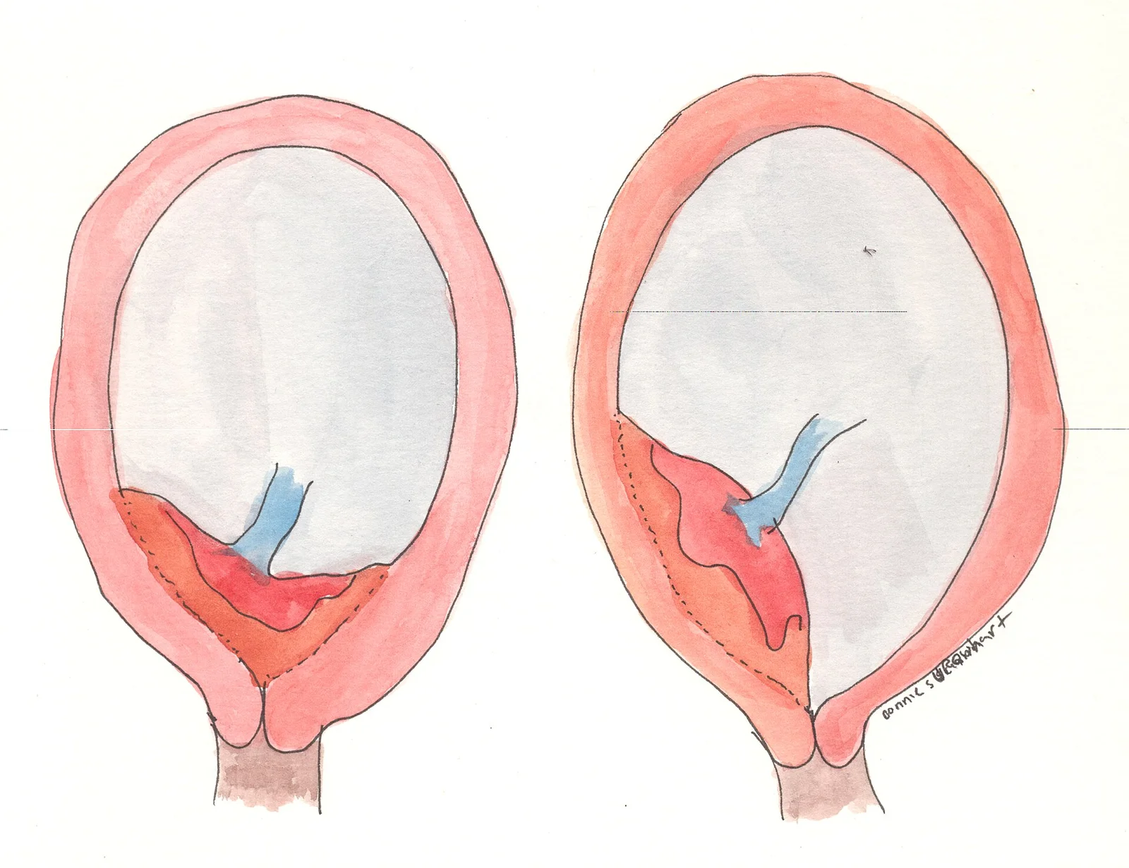
Long description
A watercolor drawing showing marginal and complete placenta previa.

DefinitionAntepartum haemorrhage
Bleeding from the genital tract after the age of viability (about 20 to 24 weeks of gestation) and before the delivery of the baby. It complicates an estimated 3 to 5% of pregnancies and is a leading cause of perinatal death and maternal morbidity. [2]
APH is a presentation, not a diagnosis. The underlying cause must be sought, but the first task is always maternal stabilisation. The main placental causes are:
- Placenta praevia: the placenta lies wholly or partly within the lower uterine segment and reaches or covers the internal cervical os. [2]
- Placental abruption: premature separation of a normally situated placenta from the uterine wall, with haemorrhage. [2]
- Vasa praevia: fetal vessels run through the membranes over the internal os, unprotected by placenta or cord.
- Uterine rupture: tearing of the uterine wall, usually through a scar or previously damaged area, during pregnancy or labour.
Lesser causes of late bleeding (cervical change or "show", cervicitis, trauma, infection, vulval varicosities, genital tract tumours, haematuria, vasa praevia) are often evident on speculum examination. [2]
Placenta praevia. The prevalence of clinically evident placenta praevia at term is roughly 4 to 5 per 1000 pregnancies. [2] In the second half of pregnancy the reported prevalence of low-lying or praevia placentas ranges from 1 in 50 to 1 in 500. [1] In two million pregnancies, the incidence of praevia rises from 10 per 1000 births after one previous caesarean birth to 28 per 1000 after more than three. [1] In singleton IVF pregnancies the risk is markedly increased (odds ratio 3.76, 95% CI 3.09 to 4.59). [1]
Placental abruption. This complicates about one in 80 deliveries, or 5.9 to 6.5 per 1000 singleton births and 12.2 per 1000 twin births. [2] Between 0.65 and 1.0% of pregnancies are complicated by abruption overall. [3] Perinatal mortality is reported as 119 per 1000 abruption births, and the risk of recurrence in a later pregnancy is increased up to 10-fold. [2]
Vasa praevia. A systematic review estimates a mean incidence of about 7.9 per 10000 pregnancies (95% CI 5.9 to 10.1), but national population studies find lower rates: a population-based UK study found 6.64 per 100000 maternities (about one in 15062). [4] [2]
Uterine rupture. A prior classical (upper-segment) caesarean occurs in 0.3 to 0.4% of pregnancies and carries a 1 to 12% risk of uterine rupture in the next pregnancy. [3] The true incidence of rupture depends on how many women with previous uterine surgery become pregnant again.
The condition results from implantation of the blastocyst low in the uterine cavity. Risk factors include increasing maternal parity and age, multiple pregnancy, endometrial damage (previous dilatation and curettage), previous caesarean birth, uterine scars and pathology such as previous myomectomy or endometritis, placental pathology (marginal cord insertions, succenturiate lobes), a previous praevia and cigarette smoking. [2][1] Endometriosis roughly triples the risk, and a short inter-pregnancy interval of less than one year after a caesarean birth also increases it. [1]
Placenta accreta spectrum (PAS, abnormal placental attachment into a scar) complicates 9.3% of praevias. [2]
The precise cause is unknown. Haemorrhage into the decidua basalis forms a haematoma that pushes the placenta off the wall. Absence of the normal transformation of spiral arterioles into low-resistance vessels is implicated, leading to underperfusion and vessel rupture. Risk factors include gestational hypertensive disease, advanced maternal age, increasing parity, multiple pregnancy, polyhydramnios, chorioamnionitis, prolonged rupture of membranes, trauma and possibly thrombophilia. Preventable contributors include maternal cocaine and tobacco use. [2] Raised maternal serum alpha-fetoprotein in the second trimester is also associated. [2]
Type 1: velamentous cord insertion, so fetal vessels run unprotected in the membranes. Type 2: vessels run between lobes of a bilobed or succenturiate placenta. Type 3 (described more recently): vessels leave and re-enter the placenta in a boomerang curve. [4] Risk factors include low-lying placenta, velamentous or marginal cord insertion, bilobed or succenturiate placentation, IVF conception and multiple pregnancy. [4]
The highest-risk scars are vertical incisions through the upper muscular part of the uterus (classical caesarean) and extensive myomectomy. [3] Other risks include previous uterine rupture or dehiscence, shoulder dystocia injury, forceps-related trauma, prolonged or obstructed labour and labour after previous caesarean. Uterine rupture after previous muscular-level surgery can occur before labour begins. [3]
Placenta praevia. As the lower segment forms in the third trimester, a placenta attached low stretches, tears and bleeds. The first bleed is classically a small "warning" haemorrhage that stops spontaneously, with recurrent bleeds as gestation advances. [2] Bleeding is usually painless because the separation is in the lower segment, away from the pain-sensitive upper myometrium.
Placental abruption. Haematoma growth raises hydrostatic pressure, separating more placenta. The haemorrhage may be revealed (visible vaginal bleeding, 65 to 80% of cases), concealed (retroplacental, with the blood trapped behind the placenta, 20 to 35% of cases) or mixed. [2] Bleeding can track into the myometrium, giving a Couvelaire uterus. The amount of visible blood does not reflect total blood loss, which is why shock can appear out of proportion. [2] Small separations produce mild, self-limited bleeding; separation of more than half of the placental area sharply raises the stillbirth rate. [2]
Vasa praevia. Fetal vessels overlying the internal os are unprotected. When membranes rupture, or the cervix dilates, they shear and the fetus (not the mother) bleeds. Because the fetal blood volume is small, exsanguination is rapid. [4]
Uterine rupture. A thinned or scarred wall tears under load, especially during contractions or as the lower segment distends late in pregnancy. Blood loss can be sudden and massive; the fetus is compromised because the placenta detaches from its supply. [3]
- Sudden vaginal bleeding, bright red. Ask about pain, contractions, trauma, fluid loss, fetal movements and syncope.
- A painless bleed late in pregnancy is classically placenta praevia until excluded; bleeding with abdominal or back pain and uterine tenderness is abruption.
- Recurrent minor bleeds ("warning haemorrhage") suggest praevia. [2]
- Risk factors are part of the history: previous caesarean, uterine surgery, IVF, smoking, cocaine, hypertension.
- Vasa praevia (and ruptured vessels at membrane rupture) classically presents as a small vaginal bleed followed by fetal bradycardia after amniotomy; the mother is usually well. [2]
- Assess airway, breathing and circulation first: pallor, cool clammy skin, tachycardia, hypotension. A large abruption can cause shock with little visible blood.
- Abdomen: uterine size and tone, fundal height, fetal lie and presentation (one-third of praevias show malpresentation). Tenderness, irritability or a firm "woody" uterus points to abruption. [2]
- Fetal heart: rate, variability, decelerations. Bradycardia after a small bleed is a red flag for vasa praevia.
- Do not perform a digital vaginal examination when praevia or abruption is suspected; it can provoke brisk haemorrhage. [2] A speculum examination is safe and may reveal cervicitis, polyps or visible bleeding.
- Ultrasound is the definitive first imaging step; maternal resuscitation must not be delayed for it.
| Cause | Typical features | What distinguishes it |
|---|---|---|
| Placenta praevia | Painless bright-red bleeding, often recurrent; uterine relaxed; fetal heart usually normal. [2] | Placenta reaches or covers the internal os on transvaginal ultrasound. [1] |
| Placental abruption | Painful bleeding with a hard, tender uterus; uterine hypertonicity; fetal distress or demise. May be concealed. [2] | Clinical diagnosis; ultrasound is insensitive. Look for Couvelaire uterus, DIC. [2] |
| Vasa praevia | Small bleed then sudden fetal bradycardia, often after membrane rupture; mother stable. [2] | Fetal vessels over the internal os on colour Doppler; Apt test shows fetal blood. [2] |
| Uterine rupture | Sudden severe pain, shock, loss of fetal station, "shoulder tip" signs; fetal bradycardia or absent heart. [3] | Usually occurs in labour in a scarred uterus; requires emergency laparotomy. [3] |
| Cervical causes (ectropion, cervicitis, polyps, trauma) | Small, post-coital or post-examination bleeding; cervix visibly abnormal. | No placental abnormality on ultrasound. |
| "Show" / preterm labour | Mucus blood-tinged, mild contractions, cervical shortening. | Different pattern from frank haemorrhage. |
| Vulval or urinary bleeding | Local lesions, varicosities, haematuria. | Source seen locally. |
Ultrasound is the investigation of choice for placental location and should be available for every APH. [2][1]
- Placenta praevia. Diagnosed when the placental edge reaches or covers the internal os after 16 weeks. [1] Report as low-lying if the leading edge is less than 20 mm from (but not covering) the internal os. [1]
- Transvaginal scanning at 32 to 36 weeks is more accurate than transabdominal, especially for posterior placentas and high BMI, and is safe even with bleeding. [1] It reclassifies up to 60% of cases thought to be low-lying on the transabdominal scan. [1]
- Cervical length under 30 mm at 28 to 34 weeks predicts antenatal bleeding and preterm birth in praevia. [1]
- Placental migration (trophotropism). Over 90% of placentas called low-lying at mid-pregnancy are no longer low-lying by 37 weeks; about half of those still low at 32 weeks resolve by 36 weeks, with no further change after 36 weeks. [1]
- Placental abruption. Ultrasound is insensitive and unreliable for detecting or excluding abruption; a negative scan is common with clinically significant separation. [2] The diagnosis is clinical and confirmed postpartum by a retroplacental clot or depression on the maternal placental surface. [2]
- Vasa praevia. Suggested by parallel echogenic lines near the cervix; confirmed by colour Doppler and transvaginal sonography for vessels over the internal os. [2]
- PAS. Irregular vascular lacunae, loss of the retroplacental clear space, myometrial thinning under 1mm and bladder-wall abnormalities at 18 to 20 weeks in a woman with risk factors. [1]
- Full blood count, group and save or crossmatch, coagulation profile (prothrombin time, activated partial thromboplastin time, fibrinogen), renal function.
- In abruption with intrauterine death, begin replacement of at least 2 units of blood, because average loss is about 1 litre. [2]
- Kleihauer-Betke test to quantify fetomaternal haemorrhage in vasa praevia or sensitisation events, and anti-D immunoglobulin for rhesus-negative women.
- "Apt test" (or alkali denaturation) to distinguish fetal from maternal blood when vasa praevia is suspected. [2]
Continuous fetal heart monitoring is mandatory in any significant bleed. Abnormal traces, including reduced variability, late decelerations or bradycardia, guide timing of delivery.
There is no single numerical score, but three practical categories guide management: [2][3]
- Placenta praevia classified by relation to the internal os (modern ultrasound definition used by RCOG 2026): praevia (edge reaches or covers the os); low-lying (edge less than 20 mm from os). [1]
- Placental abruption, by clinical severity (Sher/Statland): grade 1 (mild, often diagnosed only after delivery by a retroplacental clot); grade 2 (moderate, with clinical signs and a live fetus); grade 3 (severe, fetal death, coagulopathy may be present). [2]
- Vasa praevia, by prenatal diagnosis: detected (diagnosed before labour) versus peripartum (suspected during labour or after birth). [4]
- Concealed versus revealed haemorrhage in abruption: 20 to 35% concealed, 65 to 80% revealed. [2]
- Uterine scar type that predicts rupture (classical upper-segment versus lower-segment transverse). [3]
- Resuscitate: secure airway, high-flow oxygen, large-bore intravenous access, crystalloid and blood.
- Keep the woman nil by mouth, have cross-matched blood ready, inform senior obstetrician, anaesthetist and haematologist.
- Lie her in a left-lateral position; avoid digital vaginal examination.
- Continuous maternal and fetal monitoring; check blood pressure, pulse, oxygen saturation and urine output.
- Confirm gestation, placental site on (transvaginal) ultrasound and check rhesus status.
- Give anti-D if rhesus negative.
The choice between expectant, vaginal, or operative delivery depends on the cause, gestation and maternal-fetal condition.
Asymptomatic or minor bleeding. Most asymptomatic women can be managed as outpatients with safety measures and ready access to hospital. [1] Hospitalise women who are symptomatic (bleeding, pain, contractions) with care tailored to clinical need and social circumstances such as distance from hospital. [1]
Medically indicated delivery timing. Planned birth for an uncomplicated low-lying or praevia placenta should be considered no later than 37+6 weeks. [1] (RCOG Green-top Guideline No. 27a, 2026). For an uncomplicated praevia, ACOG/SMFM guidance is 36 to 37 weeks gestation. [3] An ACOG/SMFM decision model on 230 praevia cases puts the risk of an emergent bleed at 4.7% at 35 weeks, 15% at 36 weeks, 30% at 37 weeks and 59% at 38 weeks. [3] Corticosteroid administration at 35+5 weeks followed by planned birth at 36 weeks has been modelled to optimise maternal and neonatal outcomes. [1]
To stop bleeding. Tocolysis may be used for 48 hours to allow antenatal corticosteroids when birth is imminent, but cervical cerclage is not recommended. [1] Outpatient surveillance and repeated transvaginal ultrasound guide ongoing care. Magnesium sulphate is used up to 30+0 weeks for neuroprotection if preterm birth is planned. [1]
Surgical care. Birth should take place in a maternity unit with on-site blood transfusion and access to critical care. [1] Have at least 4 units of cross-matched blood ready even if the woman has never bled. [2] Intraoperative cell salvage is beneficial and safe, especially for women who decline blood products. [1] If uterotonics and tranexamic acid fail to control haemorrhage, use intrauterine balloon tamponade and consider uterine compression sutures. [1] Deliveries should be led by the most experienced obstetrician and anaesthetist available. [2]
Antenatal corticosteroids (to reduce neonatal respiratory morbidity) are recommended between 24+0 and 34+6 weeks when imminent preterm birth is anticipated, and magnesium sulphate up to 30+0 weeks for neuroprotection (RCOG Green-top No. 74). [1] NICE NG25 allows magnesium sulphate to be considered up to 33+6 weeks. [7] NICE NG25 also guides repeat courses of steroids in recurrent bleeding.
Management depends on severity, gestation and maternal-fetal condition. [2]
- Mild (grade 1) abruption. Bleeding stops, monitoring is reassuring, and the pregnancy can often continue; outpatient care is reasonable if observation (24 to 48 hours) is reassuring. [2]
- Moderate (grade 2). Resuscitate, then deliver the baby: a trial of vaginal delivery is advocated whenever tolerated by the mother and fetus, with continuous fetal heart monitoring. Labour is often rapid. Proceed to caesarean if there is fetal distress or failure to progress. [2]
- Severe (grade 3) or fulminant abruption. regarded as an emergency requiring multidisciplinary input. Deliver without delay, as delivery is the only way to stop consumptive coagulopathy. Replace blood and components before and during surgery. Arterial and central venous monitoring, high-dependency care and urine output surveillance are appropriate, because renal failure is a complication. [2]
- Preterm abruption. Expectant management with or without tocolysis has been reported to be safe in selected preterm cases, prolonging gestation by several days, but the evidence is observational and tocolysis remains uncertain outside research. [2]
- If intrauterine death has occurred, start blood replacement of at least 2 units. [2]
RCOG Green-top Guideline No. 27b (2019) covers diagnosis and management. [5]
- Prenatal detection is critical: 97% infant survival when diagnosed antenatally versus 44% when not. [2]
- Antenatal steroids are considered from 28 to 32 weeks, prophylactic hospitalisation from 30 to 32 weeks, and birth by planned caesarean at 34 to 37 weeks. [4][5]
- UK population data show antenatally diagnosed cases with no perinatal deaths, hospitalisation at a median of 32 weeks and caesarean at a median of 36 weeks (range 34 to 39), with 89% regional anaesthesia. [4]
- If vasa praevia is diagnosed during labour or after membrane rupture with bleeding and fetal bradycardia, perform immediate (category 1) caesarean. [4]
- Screening policy differs by country. The UK does not recommend universal screening; ultrasound with colour Doppler is advised for women with risk factors. [4][5] WHO guidance in this area is general (maternal and fetal assessment within antenatal care), so individual practice should follow local guidelines. [8]
- This is an obstetric emergency: maternal shock and sudden fetal compromise. [3]
- Resuscitate and deliver immediately, usually by laparotomy and caesarean birth; be prepared for transfusion and hysterectomy.
- For women with a previous classical (upper-segment) caesarean or extensive myomectomy where caesarean delivery is planned, ACOG/SMFM timing guidance is delivery at 36 to 37 weeks. [3] Where prior surgery is less extensive, timing can be individualised up to 38 weeks. [3]
- Rupture can occur before labour, so women with a high-risk scar need counselling and a clear delivery plan from 34 weeks. [3]
- Attempted vaginal birth after caesarean (VBAC) carries its own uterine rupture risk, which is outside this topic; it needs its own shared decision.
- Maternal. Haemorrhagic shock, blood transfusion, coagulopathy and disseminated intravascular coagulation (DIC, especially in abruption), renal failure, need for hysterectomy. [2][1]
- Fetal. Stillbirth, fetal hypoxia/acidosis, prematurity, fetal anaemia (vasa praevia) and low birth weight. Abortion of more than half the placenta sharply raises stillbirth. [2]
- Abruption complications include Couvelaire uterus, infection and, rarely, maternal death. [2]
- Praevia complications include emergency caesarean birth, intraoperative haemorrhage, postpartum haemorrhage and hysterectomy. [1]
- PAS can turn a praevia into an operation requiring a specialised team and possible hysterectomy; routine ureteric stents and interventional radiology are not supported by evidence. [1]
- Uterine rupture carries maternal shock and emergency laparotomy and fetal asphyxia or stillbirth. [3]
- Most mild praevia and unexplained APH bleeds settle spontaneously. About 15% of women with unexplained APH go into spontaneous labour within 2 weeks. [2]
- Even a single episode in praevia raises the risks of transfusion, emergency caesarean and preterm birth. [1]
- With severe abruption the prognosis deteriorates with delay and with the size of separation.
- Vasa praevia prognosis depends entirely on antenatal diagnosis: 97% survival if diagnosed antenatally versus 44% if not; 47% of peripartum-diagnosed survivors develop hypoxic ischaemic encephalopathy. [2][4]
- Praevia recurs, and the risk rises with each caesarean birth. [1][3]
- National screening programmes for placenta praevia itself are not standard, but the routine 18+0 to 20+6 week anomaly scan (including placental location) detects almost all cases before symptoms. [1][6]
- Low-lying or praevia at the mid-trimester scan is followed up by transvaginal ultrasound at 32 to 36 weeks. [1]
- Vasa praevia is not universally screened for in the UK, although risk-factor-targeted colour Doppler screening is advocated in several countries, and evidence of benefit is growing. [4][5]
- Modifiable risk factors are smoking and cocaine use; prevention of unnecessary first caesarean births indirectly reduces future praevia. [2][3]
- Multiple pregnancy. Low-lying placentas in twins may need follow-up beyond 32 weeks and sometimes to 36 weeks. Antenatal care should be consultant-led. [1]
- IVF pregnancies. Higher praevia risk, partly independent of multiple pregnancy; plan delivery at a centre able to manage haemorrhage. [1]
- Women who decline blood products. Cell salvage is beneficial and safe. [1]
- Children and adolescents. APH from vasa praevia after membrane rupture can mimic abruption; a fetal bradycardia after a small bleed demands immediate caesarean. [2]
- Older women. Advanced maternal age carries a small increased risk of praevia, largely confounded by parity. [1]
Where ultrasound, cross-matched blood or critical care are not available, three priorities from the WHO guidance are: screen the pregnancy by assessing risk, recognise bleeding early, and refer before shock. [8]
- Recognition. Know the difference between a painless, non-tender bleed (suspect praevia) and a painful tender uterus with shock out of proportion to visible loss (suspect abruption). [2]
- Avoid harm. Never do a digital vaginal examination with an undiagnosed APH; a speculum examination is safe and often reveals the source. [2]
- Stabilise and transfer. Give crystalloid, keep the woman left lateral, nil by mouth, and refer to a unit with blood and surgical capability; aim to have blood cross-matched at the receiving unit.
- Kleihauer and anti-D issues. Give anti-D when the woman is rhesus negative, even if transfusion seems unlikely.
- WHO estimate. Around 15% of all pregnant women develop a potentially life-threatening complication needing skilled care, and some need a major obstetric intervention to survive. [8] This is why every unit should have a massive haemorrhage protocol and rehearsed drills.
- Where possible, get ultrasound to locate the placenta at the 18 to 20 week visit and arrange a transvaginal follow-up if it is low.
Approach to bleeding in the second half of pregnancy
- Bleeding after 20–24 weeksCall for help; assess airway, breathing, circulation; left lateral, nil by mouth
- Maternal stabilisationLarge-bore IV access, fluids and blood as needed, oxygen, continuous maternal monitoring, cross-match
- Question: Is the mother or fetus unstable?
Yes
- Deliver nowCaesarean unless vaginal delivery is immediately achievable; multidisciplinary care
No
- Transvaginal ultrasound for placental locationPraevia if edge reaches or covers internal os; low-lying if edge less than 20 mm
- Question: Placental edge covers or reaches the os?
Yes
- Placenta praeviaObserve inpatient if symptomatic; planned birth no later than 37+6 weeks; 36–37 weeks for ACOG/SMFM uncomplicated; 4 units cross-matched
No
- Consider abruption, uterine rupture, vasa praevia, cervical causesAbruption is clinical; concealed bleeding possible; vasa praevia shows fetal vessels over the os on Doppler
- Question: Fetal bradycardia after a small bleed?
Yes
- Suspect vasa praeviaImmediate category 1 caesarean; Apt test for fetal blood
No
- Manage by severity and gestationMild observe; moderate deliver vaginally if tolerated; severe deliver now with blood and component support
- Corticosteroids and magnesiumCorticosteroids 24+0 to 34+6 weeks; magnesium sulphate up to 30+0 weeks (RCOG) or consider to 33+6 (NICE)
- Documentation and handoverDiagnosis working list, gestation, blood loss, CTG, plan, owner and escalation triggers
- APH is bleeding after 20 to 24 weeks. Placental abruption or placenta praevia accounts for roughly half of cases; a cause remains unknown in the other half. [2]
- A praevia bleed is classically painless; an abruption bleed is painful, with a tender, hypertonic uterus. [2]
- Ultrasound reliably diagnoses praevia but cannot exclude abruption, which is a clinical diagnosis. [2]
- Concealed abruption can cause shock with little visible blood; blood loss is out of proportion. [2]
- Vasa praevia presents as a small bleed followed by fetal bradycardia; antenatal diagnosis converts a 37 to 44% perinatal mortality to near zero. [2][4]
- Uterine rupture is usually of a scarred (classical or myomectomy) uterus, can occur before labour, and is an immediate-delivery emergency. [3]
- Planned birth for uncomplicated praevia should be no later than 37+6 weeks; RCOG (2026) supports this and ACOG/SMFM advises 36 to 37 weeks. [1][3]
- Place 4 units of cross-matched blood on standby for any placenta praevia delivery, even with no prior bleeding. [2]
- Corticosteroids 24+0 to 34+6 weeks and magnesium sulphate up to 30+0 weeks if preterm birth is imminent. [1]
- Previa risk rises steeply with previous caesareans: 10 per 1000 after one and 28 per 1000 after more than three. [1]
- Do not do a digital vaginal examination in undiagnosed APH. [2]
- Vasa praevia screening is targeted, not universal, in the UK, though about 7.9 per 10000 pregnancies are affected. [4][5]
- WHO reminds us that about 15% of pregnant women develop a life-threatening complication needing skilled care. [8]
- RCOG 2026: low-lying or praevia reported when leading edge is less than 20 mm from the internal os; praevia when it reaches or covers the os. [1]
Answer each one in your head first, then open the answer.
A 38-year-old woman at 34 weeks with two previous caesareans presents with a sudden, painless heavy bleed. Her uterus is soft and non-tender and the fetus is not compromised. What is the most likely diagnosis, what test confirms it, and what is your plan?
Show answerHide answer
Placenta praevia. A transvaginal ultrasound showing the placental edge reaching or covering the internal os confirms it. She needs admission, blood cross-matched (4 units), senior obstetric and anaesthetic care, and planned delivery no later than 37+6 weeks, or 36 to 37 weeks if uncomplicated per ACOG/SMFM, after discussion. [1][2]
A nulliparous woman at 30 weeks reports dark vaginal spotting. Her abdomen is tender and the uterus is hard between contractions; the fetal heart rate shows late decelerations. Ultrasound is reported as normal. What is your diagnosis and why is the ultrasound normal?
Show answerHide answer
Placental abruption. Ultrasound is insensitive for abruption: a negative scan is common with clinically significant separation. The tender, hypertonic uterus and fetal distress fit. Manage as moderate-to-severe: resuscitate, cross-match, and deliver by the safest quickest route, with coagulation monitoring. [2]
Five minutes after artificial rupture of membranes at 36 weeks, a woman has a small vaginal bleed and the fetal heart rate falls to 70 beats per minute. The mother feels well. What must you do immediately, and why?
A woman at 18 weeks in her second pregnancy has a placenta reaching the internal os on the anomaly scan. What proportion of such placentas will no longer be low at term, what is the safe follow-up, and when should she be told to attend urgently?
Show answerHide answer
Over 90% resolve by 37 weeks. Offer transvaginal ultrasound at 32 to 36 weeks and discuss mode of birth based on the final distance from the internal os. She should attend urgently for any bleeding, pain or reduced fetal movements. [1]
A woman with a previous classical caesarean birth asks when she should be delivered next time. What does current guidance advise and for what reason?
Show answerHide answer
Plan delivery at 36 to 37 weeks because of a 1 to 12% uterine rupture risk with a classical scar, with earlier delivery if labour or any complications begin. [3]
Which drug timings does the RCOG 2026 guideline recommend in a woman with placenta praevia in whom preterm birth is likely within days?
You are working in a district hospital with ultrasound unavailable. A woman arrives at 33 weeks with a painful tender uterus and fresh bleeding, and her pulse is 120 per minute, but only a small stain is visible. What is the likely diagnosis and the first three actions?
How does placenta praevia differ from placenta accreta spectrum, and when should the patient be referred to a specialist centre?
Show answerHide answer
Praevia is a placenta overlying the internal os; PAS is invasion into the scarred wall after delivery of the fetus is impossible to separate. Refer as soon as a high-risk woman (previous caesarean with an anterior praevia) has screening at 18 to 20 weeks; deliver at 36+1 to 37+0 weeks at a specialist centre. [1]
Antepartum fetal surveillance; diabetes in pregnancy; fetal growth restriction and small-for-gestational-age fetus; gestational and chronic hypertension in pregnancy; pre-eclampsia, HELLP and eclampsia; induction of labour; multiple pregnancy; placenta accreta spectrum (planned topic); preterm labour and birth; previous stillbirth; reduced fetal movements; rupture of membranes; third-trimester bleeding (MedGrasp obstetrics topics linked as plain text until verified).
- Jauniaux E, Bhide A, Hussein AM, Lees C, D'Antonio F, Robson SC, Kaelin Agten A, Jurkovic D; Royal College of Obstetricians and Gynaecologists. Placenta Praevia and Placenta Accreta Spectrum: Diagnosis and Management (Green-top Guideline No. 27a). BJOG 2026;133(10):e93-e128.. Accessed .
- Giordano R, Cacciatore A, Cignini P, Vigna R, Romano M. Antepartum haemorrhage. J Prenat Med 2010;4(1):12-16.. Accessed .
- Spong CY, Mercer BM, D'Alton M, Kilpatrick S, Blackwell S, Saade G. Timing of Indicated Late-Preterm and Early-Term Birth. Obstet Gynecol 2011;118(2 Pt 1):323-333.. Accessed .
- Attilakos G, David AL, Tunn R, Knight M, Brocklehurst P. Incidence and outcomes of vasa praevia in the United Kingdom. NIHR Open Res 2024;4:49.. Accessed .
- Jauniaux E, Alfirevic Z, Bhide A, et al.; Royal College of Obstetricians and Gynaecologists. Vasa praevia: diagnosis and management (Green-top Guideline No. 27b). BJOG 2019;126(1):e49-e61.. Accessed .
- National Institute for Health and Care Excellence. Antenatal care (NICE guideline NG201). NICE guideline NG201; published 2021, last reviewed 27 December 2024.. Accessed .
- National Institute for Health and Care Excellence. Preterm labour and birth (NICE guideline NG25). NICE guideline NG25; published 2015, updated 2022.. Accessed .
- World Health Organization, UNFPA, UNICEF. Managing complications in pregnancy and childbirth: a guide for midwives and doctors (2nd edition). Geneva: WHO; 2017.. Accessed .
Figure credits
Educational content for medical students and doctors. It is not a substitute for clinical judgement or local guidelines.