Fetal malpresentation
How breech, transverse, face and brow presentations are classified, found and managed, including external cephalic version and current RCOG, ACOG, NICE and.
- A malpresentation is any fetal presentation other than a well-flexed vertex: the common ones are breech (buttocks or feet first), transverse (shoulder or side first), face (chin first) and brow (forehead first). [1][2][8]
- Breech presentation complicates 3 to 4% of term deliveries, is more common with prematurity and with nulliparity, and is the malpresentation a student will meet most often. [1][2][6]
- Transverse lie, face and brow presentations are much rarer (non-vertex malpresentations in total under 4% at term); a transverse lie or a persistent brow presentation is an absolute fetopelvic disproportion and cannot be delivered vaginally. [7][8]
- The first management question for a breech is almost always whether external cephalic version (ECV) can turn the baby head-down: it succeeds in about half of attempts and, when successful, roughly 84% of women deliver vaginally. [1][6]
- If ECV fails or is not suitable, planned caesarean section is the usual mode for a singleton term breech; a carefully selected planned vaginal breech birth remains an option where skill and a protocol exist. [3][4]
- Face presentation can be delivered vaginally when the chin is anterior; brow presentation almost never is. [8]
By the end of this page you should be able to:
- Define malpresentation, and classify fetal lie and the types of breech, transverse, face and brow presentation.
- Explain why some babies present by the breech, and list the main risk factors.
- Recognise malpresentation on history, abdominal palpation, vaginal examination and ultrasound, and state when each test is decisive.
- Offer and conduct external cephalic version safely, including timing, tocolysis, contraindications and aftercare.
- Choose the mode of birth for breech, transverse, face and brow presentation, and follow the RCOG, ACOG, NICE and WHO guidance.
- Manage a transverse or brow presentation as an obstetric emergency, including low-resource options.
Figure 1Haultain and Ferguson - foetal positions
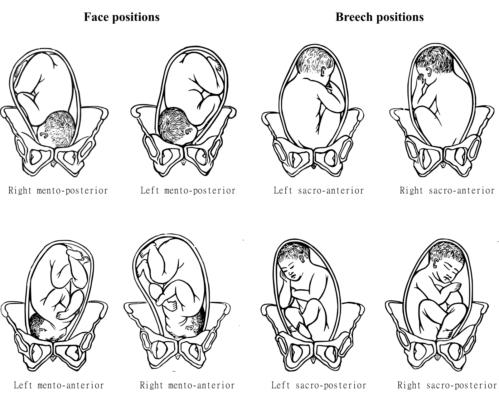
Long description
A public-domain engraving titled "Breech and face positions of the foetus before birth" from a 1902 obstetric nursing handbook.

A malpresentation is any fetal presentation that is not a well-flexed vertex: the fetal head is not dipped and flexed so that the occiput presents first. Presentation differs from position (where the presenting part faces) and from lie (the angle between the fetal spine and the maternal spine). A longitudinal lie may be cephalic or breech; a transverse lie is perpendicular to the maternal long axis. [1][2][5]
DefinitionFetal malpresentation
Any fetal presentation other than a well-flexed vertex, whether breech (buttocks or lower limb first), transverse or oblique (shoulder or trunk first), face (chin first) or brow (forehead first). Breech is the commonest; transverse, face and brow are rare but important.
In a breech the fetal buttocks or a lower limb enters the pelvis first. The type depends on hip and knee position: [8]
- Frank breech: hips flexed, knees straight, so the feet lie near the face. This is the commonest type.
- Complete breech: hips and knees both flexed, so the fetus sits cross-legged.
- Footling (incomplete) breech: one hip or knee extended, so a single foot (footling) or both feet (double footling) present.
- Knee presentation: one or both knees present (uncommon; behaves like a footling breech).
The fetal spine lies across the maternal abdomen; the shoulder or trunk is the presenting part. It may be dorso-anterior or dorso-posterior, back up or back down. A transverse lie is an absolute fetopelvic disproportion: the shoulder cannot engage in the pelvis and vaginal delivery is impossible. [7]
Both are forms of cephalic presentation in which the fetal neck is extended instead of flexed:
- Face presentation: the neck is fully extended and the mentum (chin) leads. It occurs in about 1 in 600 to 800 live births. [8]
- Brow presentation: the neck is partly extended and the area between the anterior fontanelle and the orbital ridges leads. It is the rarest malpresentation, occurring in 1 in 500 to 1 in 1400 deliveries. [8]
On descent both may convert spontaneously: more than half of brow presentations convert to vertex or face, while face presentation can deliver vaginally once the chin rotates anteriorly. [8]
Breech presentation complicates 3 to 4% of all term deliveries. [1][2][6] It is more common in preterm births: about 7% of fetuses are breech at 32 weeks and 25% at 28 weeks or less. [8] Breech is more common in nulliparous women and becomes slightly more common again very close to term as some vertex fetuses rotate late. [2]
Other malpresentations are rare. Non-vertex malpresentations (breech, transverse, face, brow and compound) in total occur in less than 4% of fetuses at term, with face presentation in 1 in 600 to 800 live births and brow presentation in 1 in 500 to 1 in 1400 deliveries. [8]
Recurrence is substantial: after one breech delivery the recurrence rate is about 10% in the next pregnancy and 27% in a third. A previous caesarean delivery also doubles the incidence of breech presentation. [8]
Masses of data come from high-income systems. Where caesarean access is limited, malpresentation is a major contributor to obstructed labour and stillbirth; transverse lie in particular is an emergency. [7]
Breech presentation usually arises from factors that change fetal motility or the vertical polarity of the uterine cavity, leaving the fetus unable to turn vertex in the third trimester: [1][2][8]
- Prematurity and low birth weight
- Multiple pregnancy (the second twin may be non-vertex)
- Placenta praevia or an unusually low implantation, so the presenting part cannot engage
- Uterine abnormalities (septate or bicornuate uterus) and large uterine fibroids
- Fetal abnormalities (aneuploidy, congenital anomalies, hydrocephalus, fetal neck masses, anencephaly); fetal factors may account for up to 60% of face presentations [8]
- Polyhydramnios or oligohydramnios
- Pelvic contraction or cephalopelvic disproportion
- Fetal macrosomia (brow presentation is more likely with a large fetus)
- Previous breech delivery or previous caesarean delivery (the latter doubles the incidence)
- Multiparity with lax abdominal wall
- Grand multiparity (five or more deliveries) for transverse lie [7]
Face and brow presentations share these factors; anything that prevents flexion of the fetal head favours extension. Contracted pelvis and cephalopelvic disproportion occur in 10 to 40% of face presentations, and prematurity or grand multiparity are common for brow presentation. [8]
A baby in a breech presentation is folded differently from a vertex baby, and this shape is what produces the clinical picture:
- The head is the largest and least compressible part, so when it presents last it can become stuck. A large or poorly flexed (extended) head is at particular risk.
- In a frank breech the thighs are straight and together, forming a cylinder that engages the pelvis like a plug. This is the most stable breech and the one most suitable for a careful vaginal birth.
- In a complete breech the legs are flexed and can fall out or fall back, so the breech is less engaged and the cord can slip past.
- In a footling breech a single small foot, or two feet, engages poorly; the membranes are more likely to break with the head still high, risking cord prolapse.
- A transverse lie cannot engage at all: the wide shoulder and trunk neither dilate the cervix nor deliver, so the uterus contracts against an obstruction, risking rupture and fetal asphyxia.
- In face and brow presentation the neck is extended, so the diameter negotiating the pelvis is 10.2 cm (submentobregmatic) or the even larger occipitomental diameter, instead of the 9.5 cm of a flexed vertex. Hence engagement is late and slow, and labour arrests. [8]
Most malpresentations are asymptomatic and are discovered before labour: the mother may notice less movement in the upper abdomen, or a lump (the breech or head) in the fundus. The fetal lie should be recorded at every visit from 36 weeks, and always if membranes rupture early. [5][7]
Take particular note of:
- Gestational age: malpresentation at term differs from one at 28 to 32 weeks, when many will still turn.
- Previous breech presentation (high recurrence) and previous caesarean section.
- Multiple pregnancy, bleeding, pain, reduced movements, which suggest a cause (placenta praevia, transverse lie with risk of cord prolapse).
- Fever or infection signs if the membranes have ruptured with a high head (risk of ascending infection).
Abdominal palpation with the Leopold manoeuvres is the core skill. In a breech the hard, round fetal head is felt in the fundus and a softer, less regular presenting part at or above the symphysis; in a transverse lie the head is felt on one side and the breech on the other, with a fundal height under 30 cm near term. [8][7]
Vaginal examination is confirmatory:
- Breech: soft buttocks, possible fetal anus and genitalia, feet in a footling breech; the hard head may be felt as caput and mistaken for occiput if the labour is advanced. Ultrasound removes the doubt. [8]
- Transverse: an empty pelvis or a presenting shoulder with sometimes an arm prolapsing. [7]
- Face: firm orbital ridges, nose, mouth and chin, often described as a triangular arrangement; the mouth may be confused with the anus.
- Brow: orbital ridges, anterior fontanelle and forehead, but neither the mouth and chin (which would indicate face) nor the posterior fontanelle (which would indicate vertex).
Breech may still be missed on palpation; if examination and ultrasound disagree, trust ultrasound.
The main challenge is to tell malpresentation from a malposition (vertex but occiput posterior or transverse), and to tell one malpresentation from another. Premature rupture of membranes with a high head, placenta praevia or a compound presentation (a hand or foot beside the head) can mimic them. [7][8]
| Presentation | What leads | Palpates as | Vaginal delivery possible? |
|---|---|---|---|
| Vertex (normal) | Occiput, well flexed | Hard round head, engaged | Yes |
| Frank breech | Buttocks, with extended legs | Fundal head, soft buttocks | Selected cases, only with skill |
| Complete breech | Buttocks and flexed legs | Fundal head, compact breech | Selected cases, only with skill |
| Footling breech | One or both feet | A foot at the cervix | Usually no (cord prolapse risk) |
| Transverse lie | Shoulder or trunk | Head and breech on opposite sides, high wide uterus | No |
| Face (mentum anterior) | Chin, neck hyperextended | Orbital ridges, nose, mouth, chin | Yes, with progress |
| Face (mentum posterior/transverse) | Chin, neck hyperextended | Orbital ridges, nose, mouth, chin | No |
| Brow | Forehead | Orbital ridges, fontanelle, no mouth or chin | Rarely, only small fetus or large pelvis |
Malpresentation is diagnosed clinically and confirmed by ultrasound; nothing else is needed for the diagnosis. The following table lists the findings and thresholds from the guidelines used on this page. [1][2][5][7][8]
| Test or finding | What it shows | Threshold or note |
|---|---|---|
| Abdominal palpation (Leopold manoeuvres) | Lie and presenting part | Fundal head suggests breech; head and breech on opposite sides with fundal height under 30 cm near term suggests transverse lie |
| Vaginal examination | Confirm presenting part and engagement | Breech: soft buttocks; face: chin and mouth; brow: fontanelle without mouth or chin |
| Ultrasound | Definitive diagnosis and ECV planning | Confirm lie, presentation, breech type, neck attitude, estimated fetal weight, amniotic fluid volume and placental site |
| Estimated fetal weight (breech eligibility) | Whether vaginal breech birth is reasonable | Commonly accepted band 2500 to 4000 g; abnormal lie, extended head or footling breech fall outside usual criteria |
| Amniotic fluid (ECV planning) | Chance of successful version | A vertical pool at least 4.6 cm (about 2 in) favours success; oligohydramnios and polyhydramnios are relative problems [6] |
| Fetal heart rate before and after ECV | Fetal wellbeing | Non-stress test or auscultation should be reassuring before ECV; monitoring for at least 30 minutes afterwards is common practice |
| Rhesus status and anti-D | Fetomaternal haemorrhage | Give anti-D within 72 hours to Rh-negative women after ECV or external version |
Once a breech is confirmed at 36 to 37 weeks, discuss with the woman:
- the option of external cephalic version;
- if version is declined or fails, the choice between planned caesarean section and, where skill exists, planned vaginal breech birth;
- the fact that planned caesarean section gives a small reduction in perinatal mortality compared with planned vaginal breech birth, while caesarean section carries its own serious long-term consequences. [2][4]
Do not make this an emergency conversation: decision making should occur at 36 to 37 weeks where possible. [1][2]
ECV is the manual turning of the fetus through the maternal abdomen from breech (or transverse) into a vertex presentation. It is the single most effective way to avoid a caesarean section for malpresentation. [1][3][4]
Success and effect. A systematic review embedded in the RCOG guideline found that ECV at term reduces non-cephalic presentation at delivery (relative risk 0.42) and caesarean section (relative risk 0.57). Overall, the success rate is approximately 47 to 54% in large series, higher in multiparous women (about 60 to 76%) than in nulliparous women (about 40%). Only 3 to 7% of babies remain breech after an unsuccessful attempt, and only about 3% revert to breech after a successful version. [1][6]
When to offer. Offer ECV at term, from 37 weeks 0 days (nulliparous women may be offered it from 36 weeks 0 days, because spontaneous version after that is rare). There is no upper gestational-age limit provided there are no contraindications. [1][3][6]
Preparation. Perform ECV where facilities for monitoring and surgical delivery are available, by a trained practitioner or a supervised trainee. [1][3] Before the procedure confirm presentation and placental site by ultrasound, assess fetal wellbeing with a non-stress test, obtain consent (including the possibility of discomfort: the procedure is painful for some women), and advise Rh-negative women about anti-D. [1][3][6]
Tocolysis. Use of tocolysis with betamimetics improves success. A commonly used regimen is terbutaline (beta-agonist) 250 micrograms subcutaneously, or a ritodrine infusion, before the attempt. If a beta-agonist is contraindicated (significant cardiac disease, hypertension or beta-blocker therapy), oral nifedipine 20 to 30 mg non-sustained release may be used as an alternative. Neuraxial analgesia is not recommended routinely but may be considered for a repeat attempt or for women who cannot tolerate the procedure. [1] [6]
Technique. Disengage the breech from the pelvis, then rotate the fetus by the shortest route (forward roll is attempted first, then backward roll if needed), with the fetal heart rate checked during the procedure. The attempt should be abandoned for prolonged bradycardia, significant maternal pain, or difficult progress. [1][7]
Aftercare. Continue fetal heart-rate monitoring for about 30 minutes afterwards; ultrasound confirms the presentation. Women who are Rh-negative need testing for fetomaternal haemorrhage and anti-D within 72 hours if delivery is not imminent. [1][6]
Complications. ECV has a very low complication rate. About 1 in 200 attempts (0.5%) requires an emergency caesarean section within 24 hours, usually for placental abruption or fetal compromise; transient fetal bradycardia (usually under one minute) is the commonest minor event. Emergency caesarean section for a serious complication is far less common than for women in spontaneous labour. [1][6]
Predictors of failure. Primiparity, an anterior placenta and low amniotic fluid (maximum pool under 4.6 cm) independently predict failure, while maternal age, fetal weight and tocolysis used as a sole add-on are weaker predictors in smaller studies. [6]
Contraindications. These divide into absolute and relative: [1][3][6][7]
- Absolute: placenta praevia (or any other obstacle to vaginal birth), placental abruption, abnormal fetal heart rate, ruptured membranes, rhesus isoimmunisation, multiple pregnancy (except after delivery of the first twin), any other reason a caesarean section is already indicated, and inability to obtain informed consent.
- Relative: severe pre-eclampsia; severe fetal growth restriction with abnormal Doppler; oligohydramnios or polyhydramnios; major fetal anomaly; uterine anomaly (lower chance of success); maternal obesity; untreated HIV infection.
ECV is not contraindicated after one caesarean delivery: the largest analyses show no greater risk than with an unscarred uterus, although numbers are too small to exclude a low risk of uterine rupture. [1][3] ECV can also be performed during early labour if the membranes are intact and the woman consents. [3][6]
Women's experience. Most women tolerate ECV, but they should be warned that it can be painful, and analgesia or a repeat attempt may be needed. Overall, only a few babies spontaneously turn after 36 weeks, so offering ECV increases vaginal cephalic births rather than watching and waiting. [1]
RCOG and ACOG agree on the broad message, with nuance: [1][2][3][4]
- Offer ECV at term unless there is an absolute contraindication, and advise on its risks, benefits and implications for mode of birth.
- If ECV fails or is declined, counsel the woman on planned vaginal breech birth versus planned caesarean section.
- Planned caesarean section leads to a small reduction in perinatal mortality compared with planned vaginal breech birth, but caesarean section has serious long-term consequences; the decision should weigh maternal preferences, risk, and local skill.
- External cephalic version should be attempted only in settings where caesarean delivery is readily available.
Planned vaginal breech birth remains reasonable for selected women when hospital-specific protocol criteria and a skilled accoucheur are in place. Typical eligibility criteria from the largest reports include: gestation over 37 weeks; frank or complete breech; no fetal anomalies; estimated fetal weight between 2500 and 4000 g; a flexed (not extended) fetal head; adequate liquor (at least a 3 cm vertical pocket); and no labour abnormality once labour begins (augmentation with oxytocin is not offered, and caesarean section is chosen for poor progress). [8]
During an unplanned vaginal breech labour: management depends on stage, the presence of complicating factors, availability of expertise and consent. Women who are near or in active second stage should not be routinely offered caesarean section; if time and circumstances permit, ultrasound is used to check neck and leg position and estimated weight, and the woman is counselled as for a planned breech birth. Rapid progress, footling breech, an extended head or an inadequate pelvis are reasons to move to caesarean section. [2]
Low-resource settings need simulation-based training in vaginal breech delivery, because ECV and caesarean access may be limited and undiagnosed breech in labour is common. [6][8]
A transverse lie is an absolute fetopelvic disproportion: vaginal delivery is impossible and the condition is an obstetric emergency because labour is obstructed and the uterus may rupture. [7]
- Antenatal: offer external version 4 to 6 weeks before delivery in a comprehensive facility (EMOC) if suitable. If version fails or is unsuitable, plan a caesarean section, either planned or at the onset of labour. [7]
- External version is contraindicated in a twin pregnancy; if the first twin is transverse, schedule caesarean section. For a second twin in transverse lie, deliver the first twin and wait briefly for the second to turn longitudinal; only then consider external or internal version with an experienced operator. [7]
- If diagnosed in labour with intact membranes and a live fetus: attempt gentle external version between contractions as early as possible, then proceed as for a normal delivery; if this fails, perform caesarean section. [7]
- If the membranes have ruptured and the fetus is alive: at full dilation a multiparous woman with a relaxed uterus, a mobile fetus and an experienced operator may have internal version and total breech extraction; a primipara, a tight uterus or scarred uterus, an immobile fetus or an engaged arm requires caesarean section. With incomplete dilation, perform caesarean section. [7]
- If the fetus is dead: embryotomy is appropriate, with caesarean section if the cervix will not admit. [7]
- Caesarean section can be difficult because of uterine retraction; a vertical hysterotomy is preferred, with extraction by grasping a foot in the fundus. [7]
In remote settings without surgery, try referral first; if that fails, use combined external and internal version with Trendelenburg or knee-chest positioning after full dilation, and embryotomy for a dead fetus. [7]
Face and brow presentation are the rarer end of the spectrum. [8]
- Face presentation: vaginal delivery is possible when the mentum (chin) is anterior, because the neck can flex under the pubic arch; a persistent mentum posterior or transverse position requires caesarean section, because the neck is already maximally extended. The mentum is anterior in over 60% of cases, transverse in 10 to 12% and posterior in 20 to 25%, and posterior positions rotate anteriorly in 25 to 33% of cases. Manual rotation or conversion to vertex are contraindicated in modern practice; breech extraction and operative vaginal delivery are discouraged and reserved for places without caesarean access. [8]
- Brow presentation: usually a transitional state. More than 50% convert spontaneously to vertex or face. If a brow persists, caesarean section is required because the occipitomental diameter is the widest diameter of the fetal head and cannot engage; vaginal delivery is only possible in a very small baby with a very large pelvis. Oxytocin is used cautiously, if at all, for brow. [8]
- Both: continuous electronic fetal monitoring is recommended, because abnormal traces are common; apply a scalp electrode carefully to the face (mentum) to avoid ocular injury. Labour may be augmented with oxytocin for face presentation when progress is normal; an internal scalp electrode and intrauterine pressure catheter need care. [8]
- Low-resource management: manage face and brow conservatively with expectant observation and close monitoring if progress is good; move to caesarean section for arrest, fetal distress or a persistent posterior chin. Forceps and manual conversion are not the modern standard. [7][8]
- Breech: perinatal morbidity and mortality are higher than for vertex, principally from prematurity, congenital anomalies, birth asphyxia and birth trauma, independent of mode of delivery. Planned caesarean section lowers short-term perinatal mortality; vaginal breech birth carries risks of cord prolapse, head entrapment and trauma where skill is lacking. [2][8]
- Transverse lie: obstructed labour, uterine rupture and fetal asphyxia if undiagnosed; caesarean section itself is technically demanding with uterine retraction. [7]
- Face and brow: prolonged and dysfunctional labour, fetal distress, facial and skull oedema lasting 24 to 48 hours, and laryngeal oedema requiring intubation in some neonates. Caesarean rates and neonatal unit admissions are higher than for vertex. [8]
- ECV: transient fetal bradycardia; fetomaternal haemorrhage needing anti-D; placental abruption or fetal compromise needing emergency caesarean section in about 1 in 200 attempts. [1][6]
- Vaginal examination pitfalls: a frank breech can be mistaken for a vertex, and face presentation for a breech, especially in advanced labour. [8]
With planned management, breech at term does well. Most women who have a successful ECV deliver vaginally (about 84%) and hospital stay is shorter than after caesarean section. [6] After planned caesarean section for breech, recovery is surgical and future pregnancies carry the risks of a scarred uterus. A woman with one breech has about a 10% chance of breech in the next pregnancy and 27% in a third, so recurrence counselling matters. [8]
Mortality for face and brow presentation has fallen with better ultrasound diagnosis and wider caesarean access. [8]
There is no primary prevention of anatomic malpresentation, but recognition removes most of the harm:
- Routine third-trimester assessment of fetal lie and presentation from 36 weeks, at the latest pre-labour visit. [2][5]
- Third-trimester ultrasound can prevent most "surprise" term breech presentations. [2]
- Offer ECV at 36 to 37 weeks instead of watching a breech persist. [1][3]
- External version for transverse lie offered 4 to 6 weeks before delivery in a comprehensive facility. [7]
- Calcium and micronutrient status, pelvic structure and multiple pregnancy are not modifiable here; the benefit comes from timely detection and a documented plan.
- Training and simulation for clinicians in vaginal breech delivery, so skills are not lost even where caesarean is the default. [2][8]
- Preterm pregnancy: malpresentation is more common (7% at 32 weeks, 25% at 28 weeks or less). Management is individualised; preterm breech is usually delivered by caesarean section, and ECV is offered less often than at term. [2][8]
- Multiple pregnancy: the non-presenting twin may be breech or transverse after the first is born; if the first twin is transverse, caesarean section is scheduled. [7]
- Uterine anomaly (bicornuate or septate uterus), fibroids: these cause malpresentation and recurrence. [1][8]
- Fetal anomalies (anencephaly, goitre, neck masses): common in face presentation; arrange delivery in a unit with neonatal and surgical support. [8]
- Women with a previous caesarean section: ECV is allowed; plan scarred-uterus precautions and discuss the small risk of rupture. [1][3][6]
- Low birth weight and very preterm babies: a small fetus may tolerate a breech or even a brow vaginal delivery, but evidence is limited and management should be consultant-led. [8]
- Rh-negative women: anti-D after ECV or external version. [1][6]
- Diagnose before labour: palpate lie and presentation at every late-pregnancy contact, and confirm with ultrasound where it is available. [2][7]
- Offer ECV only where monitoring and surgical delivery are available; do not attempt it in a remote setting without those. [1][7]
- Transverse lie or persistent brow in labour are emergencies: attempt gentle external version if the fetus is alive and membranes intact; use internal version and total breech extraction only with the criteria above; otherwise arrange referral and, if that fails, combined external and internal version with positioning. [7]
- Caesarean section for transverse lie is difficult: a vertical hysterotomy is preferred. [7]
- Face presentation usually tolerates expectant management and normal labour; move to caesarean section for arrest or fetal distress. [8]
- Maintain skills by simulation; do not let familiarity with caesarean birth eliminate vaginal breech training. [2][8]
| Source | Edition used | Main positions reflected on this page |
|---|---|---|
| RCOG | Green-top Guideline No. 20a (2017, reviewed 2024 and 2025, no update; next review 2027) and No. 20b (2017, update in development) | ECV from 37 weeks (36 weeks in nulliparous women), tocolysis improves success, about 50% success, very low complication rate; counselling on ECV versus planned caesarean or planned vaginal breech birth; women near or in second stage not routinely offered caesarean. [1][2] |
| ACOG | Practice Bulletin No. 221 (2020, reaffirmed 2026) and Committee Opinion No. 745 (2018, reaffirmed 2026) | ECV offered for term singleton breech desiring vertex birth, only where caesarean is readily available; neuraxial analgesia plus tocolysis may increase success; planned vaginal breech birth may be reasonable under hospital protocol with detailed consent. [3][4] |
| NICE | NG235 (2023, last updated 9 June 2026) | Malpresentation (breech or transverse lie) is a factor indicating increased risk, suggesting planned birth in an obstetric unit; intrapartum diagnosis and care in the hospital setting. [5] |
| WHO | EMRO report of ECV at term (Eastern Mediterranean Health Journal 2013;19(2)) and platform guidance for national services | Breech complicates 3 to 4% of term deliveries; ECV for uncomplicated breech at term 37 to 42 weeks is recommended; success about 54%; there are no fetal complications reported in the cohort. [6] |
Approach to fetal malpresentation
- Confirm gestational age and presentationPalpation, vaginal examination and ultrasound: identify lie, presentation, breech type, head attitude, fluid and placenta.
- Question: What is the presentation?
Breech
- Offer ECV at 36 to 37 weeks if suitableTocolysis, consent, monitoring and anti-D; abandon for bradycardia or pain.
- Question: ECV successful?
Yes
- Continue pregnancy; plan vertex birth
No or unsuitable
- Counsel on planned caesarean versus planned vaginal breech birthCaesarean gives small reduction in perinatal mortality; skilled accoucheur and protocol required for vaginal birth.
- Birth under hospital protocolCaesarean if criteria not met; unplanned breech in labour managed by stage, expertise and consent.
Transverse or oblique
- Antenatal: external version 4 to 6 weeks before delivery in a comprehensive facilityIf unsuccessful or unsuitable, plan caesarean section.
- In labourAlive and membranes intact: gentle external version, then normal delivery; failed version to caesarean. Ruptured membranes: version and extraction only in a multipara with criteria, otherwise caesarean.
- Dead fetusEmbryotomy if feasible; otherwise caesarean section.
Face
- Question: Mentum anterior?
Yes
- Expectant labour with monitoring; caesarean if arrest or distress
No (posterior or transverse)
- Caesarean section
Brow
- Observe: most convert to vertex or facePersistent brow: caesarean section; vaginal delivery only for a small fetus and a large pelvis.
- Postnatal reviewRecord presentation, management, perinatal outcome and recurrence counselling; Rh-negative mothers receive anti-D after ECV or version.
- Malpresentation is any presentation other than well-flexed vertex: breech, transverse, face and brow. [1][2]
- Breech complicates 3 to 4% of term deliveries and is more common in preterm and nulliparous women; recurrence after one breech is about 10%. [1][2][8]
- Transverse lie, face and brow presentations are rare but important; a transverse lie and a persistent brow are absolute fetopelvic disproportion. [7][8]
- Diagnosis is clinical (Leopold manoeuvres, vaginal examination) and confirmed by ultrasound, which also guides ECV and birth planning. [5][8]
- ECV is offered at 37 weeks (36 weeks in nulliparous women), succeeds in roughly half of attempts, reduces caesarean section, and has a very low complication rate. [1][6]
- Tocolysis with a beta-agonist (for example terbutaline 250 micrograms subcutaneously) improves ECV success; oral nifedipine 20 to 30 mg is an alternative when beta-agonists are contraindicated. [1][6]
- ECV is contraindicated by placenta praevia, abruption, ruptured membranes, abnormal tracing, rhesus isoimmunisation (except after first twin), multiple pregnancy and inability to consent; it is allowed after one caesarean section. [1][3]
- Planned vaginal breech birth is reasonable under protocol with a skilled accoucheur; planned caesarean section gives a small reduction in perinatal mortality. [2][4][8]
- Transverse lie in labour is an emergency: gentle external version if membranes are intact, version and extraction only in selected multiparae, otherwise caesarean section; vertical hysterotomy is preferred. [7]
- Face presentation can be delivered vaginally when the mentum is anterior; brow presentation almost never can. [8]
- In low-resource settings the gain comes from diagnosis before labour, timely referral and simulation training. [7][8]
Answer each one in your head first, then open the answer.
A 36-year-old nulliparous woman at 36 weeks has a breech presentation confirmed by ultrasound, with an estimated fetal weight of 3200 g and a 5 cm vertical pocket of fluid. She asks what her options are. What do you offer and when?
Show answerHide answer
Offer external cephalic version from 36 to 37 weeks, because she is nulliparous and spontaneous version is unlikely at term. Counsel on risks (transient bradycardia, about 1 in 200 emergency caesarean section), benefits (about 54% success, reducing caesarean section) and implications for mode of birth. Use terbutaline 250 micrograms subcutaneously unless contraindicated, confirm wellbeing before and after with a non-stress test, and monitor for 30 minutes. If successful she usually delivers vaginally; if not, counsel on planned caesarean section versus planned vaginal breech birth under a hospital protocol. [1][6]
Why is a transverse lie impossible to deliver vaginally, and what must be done if it is found before labour?
Show answerHide answer
The shoulder and trunk cannot engage the pelvis, so vaginal birth is impossible and the condition is an obstetric emergency with risk of uterine rupture and fetal asphyxia. If found before labour in a singleton, offer external version 4 to 6 weeks before delivery in a comprehensive facility; if this fails or version is unsuitable, plan caesarean section. [7]
A first-labour woman is found at 6 cm to have a face presentation. The chin is mentum posterior. What is the safest management?
Show answerHide answer
Persistent mentum posterior face presentation cannot deliver vaginally because the neck is maximally extended. Caesarean section is the safest mode. Manual rotation or breech extraction are not modern practice and should be avoided where caesarean delivery is available. [8]
A brow presentation is diagnosed early in labour and the cervix is 4 cm dilated. What happens next?
Show answerHide answer
Most brow presentations convert spontaneously to vertex or face as labour proceeds; expectant management with careful monitoring is appropriate. If the brow persists, caesarean section is required because the occipitomental diameter is the widest and cannot engage. Oxytocin should be used cautiously if the brow is not resolved. [8]
Which of the following is an absolute contraindication to external cephalic version: placenta praevia, primiparity, maternal age 40, oligohydramnios, previous caesarean section?
Show answerHide answer
A woman with a transverse lie is in labour at a remote clinic without caesarean facilities; membranes have ruptured and the cervix is fully dilated. What options remain?
Show answerHide answer
Refer urgently first. If referral is impossible and criteria are met (multipara, relaxed uterus, mobile fetus, experienced operator), consider internal version and total breech extraction. Otherwise, place the woman in knee-chest position, try to push the fetus back and engage the head, with vacuum extraction and symphysiotomy at the slightest difficulty. Embryotomy is appropriate only for a dead fetus. [7]
Related MedGrasp topics (plain text): fetopelvic disproportion and obstructed labour; labour stages and failure to progress; caesarean birth; obstetric exam and fetal heart rate monitoring; multiple gestation; pregnancy diagnosis and physiology; preterm labour and premature rupture of membranes; invasive prenatal testing and prenatal care; maternal resuscitation and collapse; placenta praevia and placental abruption.
- Impey LWM, Murphy DJ, Griffiths M, Penna LK, Royal College of Obstetricians and Gynaecologists. External Cephalic Version and Reducing the Incidence of Term Breech Presentation (Green-top Guideline No. 20a). BJOG 2017;124:e178-e192 (2nd edition, published 2017; reviewed 2024 and 2025, no update; next review 2027).. Accessed .
- Impey LWM, Murphy DJ, Griffiths M, Penna LK, Royal College of Obstetricians and Gynaecologists. Management of Breech Presentation (Green-top Guideline No. 20b). BJOG 2017;124:e151-e177 (4th edition, published 2017; reviewed 2024, update in development).. Accessed .
- Committee on Practice Bulletins—Obstetrics, American College of Obstetricians and Gynecologists. External Cephalic Version (ACOG Practice Bulletin No. 221). Obstet Gynecol 2020;135:e203-e212 ( reaffirmed 2026).. Accessed .
- Committee on Obstetric Practice, American College of Obstetricians and Gynecologists. Mode of Term Singleton Breech Delivery (ACOG Committee Opinion No. 745). Obstet Gynecol 2018;132:e60-e66 ( reaffirmed 2026).. Accessed .
- National Institute for Health and Care Excellence. Intrapartum care (NICE guideline NG235). NICE guideline NG235; published 29 September 2023, last updated 9 June 2026.. Accessed .
- Hussin OA, Mahmoud MA, Abdel-Fattah MM. External cephalic version for breech presentation at term: predictors of success, and impact on the rate of caesarean section. Eastern Mediterranean Health Journal 2013;19(2):162-166.. Accessed .
- Médecins Sans Frontières. Transverse lie and shoulder presentation (MSF essential obstetric and newborn care, chapter 7.6). MSF medical guidelines, section 7.6, updated 2026.. Accessed .
- Marino T; chief editor Smith CV. Face and Brow Presentation. Medscape, article 262341, updated 21 February 2025.. Accessed .
Figure credits
Educational content for medical students and doctors. It is not a substitute for clinical judgement or local guidelines.