Rhesus alloimmunisation
Rh D alloimmunisation in pregnancy: why it happens, how anti-D prevents it, and how affected pregnancies are monitored and treated.
- Rhesus alloimmunisation means an Rh-negative woman has made antibodies (anti-D) against the RhD antigen on her baby's red cells, usually after an earlier pregnancy, delivery, or bleeding event.
- In a first pregnancy the mother's exposure usually causes no illness in that fetus, but it sensitises her for life; in the next Rh-positive pregnancy her IgG anti-D crosses the placenta, destroys fetal red cells, and can cause haemolytic disease of the fetus and newborn (HDFN).
- Routine RhD typing and antibody screening at booking and at 28 weeks, together with targeted anti-D immunoglobulin after sensitising events and as routine antenatal prophylaxis, prevent sensitisation in almost all cases.
- An affected pregnancy is managed by a fetal medicine specialist who tracks maternal antibody levels, performs MCA Doppler ultrasound, and, if needed, gives intrauterine transfusions.
- Complications of untreated disease (fetal anaemia, hydrops, kernicterus, stillbirth) are now rare where prophylaxis programmes exist, but remain common in countries without them.
By the end of this page you should be able to:
- Explain what RhD is and how a sensitising event in one pregnancy leads to haemolysis in the next.
- State who needs anti-D prophylaxis, at what dose and timing, and after which events.
- Distinguish passive from immune anti-D on a blood result, and list the monitoring thresholds for immune anti-D.
- Plan the investigation of a sensitised pregnancy, including the role of the Kleihauer-Betke test, MCA Doppler and intrauterine transfusion.
- Compare the recommendations of RCOG, ACOG, NICE and WHO on routine anti-D prophylaxis.
- Tell RhD alloimmunisation apart from ABO incompatibility, other red cell antibodies and non-immune causes of fetal anaemia.
DefinitionRhesus alloimmunisation
Rhesus alloimmunisation is sensitisation of an RhD-negative woman to the RhD antigen, so that she produces IgG antibodies (anti-D) that can cross the placenta and destroy RhD-positive fetal red cells in a later pregnancy.
The RhD antigen is part of the Rh blood group system on the red cell membrane. People who carry it are RhD positive; people who lack it are RhD negative. A mother is only at risk if she is RhD negative and carries a fetus who is RhD positive, which depends on paternal inheritance (HaemBase notes that 60% of RhD-negative mothers carry an RhD-positive baby). [8]
Classification that matters in practice falls into three groups:
- Unsensitised RhD negative: no anti-D; eligible for prophylaxis.
- Passively immunised: anti-D has been administered and is detectable in the circulation; she still needs prophylaxis after further sensitising events until immunity is disproven.
- Alloimmunised (immune): anti-D is present from prior exposure, does not fall with time, and confers risk on the current and future RhD-positive fetuses.
Anti-D disease should also be separated from other red cell antibodies. Broadly, antibodies associated with clinically significant HDFN include anti-D, anti-c and anti-K; many other antibodies usually cause only neonatal rather than fetal disease. [8]
People who type as weak D or partial D are managed differently by laboratory policy; in most programmes, some variants (for example some weak D types) are treated as RhD positive and not given prophylaxis, while others are treated as RhD negative. The exact policy should be confirmed with the local transfusion laboratory rather than assumed.
RhD negativity varies markedly by ethnicity: about 15% of people in the UK and about 15% in North America, 5% in India, and 3-4% in China are RhD negative. [2][1] [8]
The burden of disease depends almost entirely on whether a prophylaxis programme exists:
- Without prophylaxis, roughly 13-16% of at-risk (RhD-negative mother, RhD-positive fetus) pregnancies become alloimmunised. [2]
- In countries without prophylaxis, 14% of affected fetuses are stillborn and about half of live-born infants with haemolytic disease suffer neonatal death or brain injury. [2]
- With postpartum prophylaxis alone, alloimmunisation fell from 13-16% to 0.5-1.8% of at-risk pregnancies; adding routine antenatal prophylaxis reduced it further to 0.14-0.2%. [2] [1][9]
- Even in the USA, where prophylaxis is routine, alloimmunisation occurs in well under 1% of at-risk pregnancies, and about 1% of pregnant women overall have a clinically significant red cell antibody (anti-D is still the commonest single specificity). [1][8]
- Even with prophylaxis, alloimmunisation has not been abolished; sensitisation is usually due to failure to follow protocol, occult feto-maternal haemorrhage before 28 weeks, or prior transfusion of RhD-positive blood rather than prophylaxis failure. [9]
The only prerequisite for RhD alloimmunisation is an RhD-negative mother exposed to RhD-positive red cells. Exposure can happen in many ways:
- Delivery of an RhD-positive baby (vaginal or caesarean)
- Feto-maternal haemorrhage during pregnancy (antepartum haemorrhage, placenta praevia or abruption, abdominal trauma, external cephalic version)
- Miscarriage, ectopic pregnancy, molar pregnancy
- Pregnancy termination (surgical or medical)
- Invasive obstetric procedures (amniocentesis, chorionic villus sampling, fetal blood sampling)
- Stillbirth or intrauterine fetal death
- Earlier transfusion of RhD-positive blood components
- Intravenous drug use with shared equipment (rare)
Risk is raised when the mother's first ever exposure is not followed by anti-D, when the father or sperm donor is RhD positive (or of unknown status), and by any event that increases feto-maternal bleeding. A history of a previously affected baby is the strongest predictor of severe disease in the next pregnancy. [4][8]
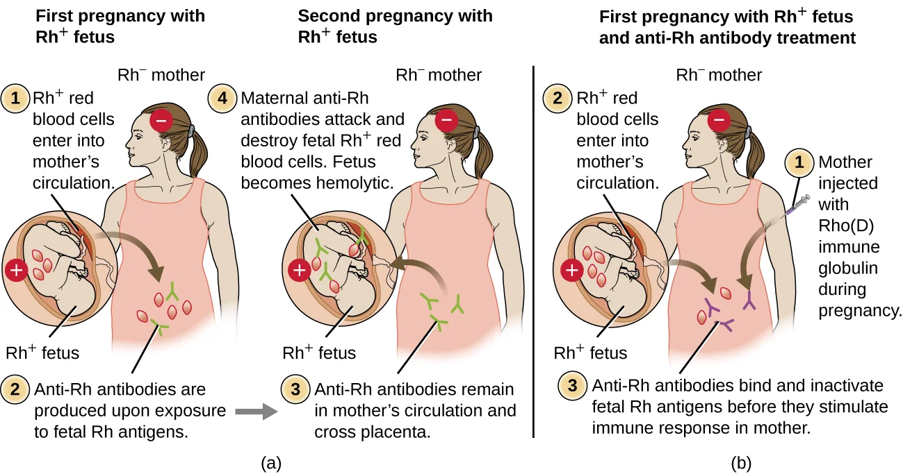
Figure 1OSC Microbio 19 01 HDN
Long description
OpenStax Microbiology figure 19.6 of haemolytic disease of the newborn: panel (a) shows an Rh-negative pregnant person exposed to Rh-positive fetal red cells, with anti-Rh IgG crossing the placenta in a later pregnancy and destroying fetal Rh-positive red cells, and panel (b) shows Rho(D) immune globulin binding fetal Rh-positive cells to prevent the primary immune response.

RhD is highly immunogenic. As little as 0.1 mL of RhD-positive red cells in the maternal circulation can trigger sensitisation. [9]
The sequence is:
- First exposure (usually late in a pregnancy or at delivery) introduces RhD-positive fetal red cells into the mother's circulation.
- The mother mounts a primary response dominated at first by IgM, which is too large to cross the placenta and does not harm the current fetus. B-cell memory is established. This is why the first RhD-incompatible pregnancy is usually unaffected.
- On re-exposure in the next (or later) RhD-positive pregnancy, a rapid secondary response produces IgG anti-D, which does cross the placenta.
- Maternal anti-D coats fetal RhD-positive red cells, which are cleared by macrophages in the fetal liver and spleen and by natural killer cells, producing haemolysis.
- The fetus becomes anaemic; erythropoiesis expands in the bone marrow and liver/spleen (extramedullary), causing hepatosplenomegaly, and the breakdown of haemoglobin raises unconjugated bilirubin. Hydrops fetalis (fetal oedema, ascites, pleural and pericardial effusions) develops in severe cases.
- Because IgG persists for years, the antibody concentration may be boosted by each pregnancy rather than fade.
Mother-neonatal consequences are mirror images: antenatal anti-D destroys fetal red cells, and postnatally the same antibodies persist in the baby's circulation for several months, causing late neonatal anaemia.
The mother is almost always asymptomatic. The condition is found on screening rather than by symptoms.
In an affected pregnancy, the fetus may show reduced movements, growth restriction, polyhydramnios (from hydrops), or frank hydrops on ultrasound. Severity ranges from mild compensated anaemia to stillbirth. [3][9]
In the neonate, look for:
- Jaundice appearing within the first 24 hours (unconjugated hyperbilirubinaemia)
- Pallor and anaemia, a positive direct antiglobulin (Coombs) test on cord blood
- Hepatosplenomegaly, and in severe disease oedema/ascites/hydrops
- Kernicterus (bilirubin encephalopathy) if hyperbilirubinaemia is severe and untreated
No specific maternal physical sign. Routine antenatal examination continues; the important "findings" are laboratory (blood group, antibody screen, antibody concentration) and sonographic (MCA Doppler, signs of hydrops).
A positive antibody screen or a jaundiced, anaemic newborn does not automatically mean RhD disease.
| Condition | Key distinguishing feature |
|---|---|
| ABO incompatibility | Most common cause of HDFN in many countries now; often Group O mother with A/B/AB fetus. Usually milder than RhD disease; may need phototherapy but rarely intrauterine transfusion. [9] |
| Other red cell antibodies (anti-c, anti-K, anti-E etc.) | anti-c and anti-K cause HDFN as serious as anti-D; anti-K causes anaemia with low reticulocytes. Anti-E, anti-e and minor antibodies usually milder. Fetal c/K status is established by genotyping. [4][8] |
| Passive anti-D (from prophylaxis) | Antibody level falls with time, rarely goes above 0.4 IU/mL after a 1500 IU dose, and is detectable for about 8 weeks; no rising trend. [8] |
| Non-immune fetal anaemia (hydrops) | Cardiac defects, chromosomal disorders, parvovirus B19, twin-twin transfusion, alpha-thalassaemia; maternal antibody screen negative. |
| Hereditary haemolytic disease | G6PD deficiency, hereditary spherocytosis and similar disorders; no red cell antibody in the mother. |
| Physiological neonatal jaundice | Appears after 24 hours; infant not anaemic; direct antiglobulin test negative. |
Investigations divide into three groups: maternal status, fetal status, and, at delivery, the volume of feto-maternal haemorrhage.
| Test | When and why | Result that matters |
|---|---|---|
| ABO and RhD typing | Booking and again at 28 weeks, before routine antenatal anti-D is given | Identifies RhD-negative women who need prophylaxis. [4][6] |
| Antibody screen (IAT) | Booking and at 28 weeks, in every pregnant woman regardless of RhD type | Detects clinically significant antibodies early and late in pregnancy. [4][6] |
| Maternal anti-D concentration | Quantified in IU/mL if anti-D is found; monthly to 28 weeks, then every 2 weeks until delivery | Less than 0.2 IU/mL usually needs no action; passive levels rarely exceed 0.4 IU/mL after 1500 IU; immune levels are stable or rise. [8] |
| Immune anti-D thresholds | Concentration interpreted with obstetric history | Less than 4 IU/mL: HDFN unlikely; 4-15 IU/mL: moderate risk, refer to fetal medicine unit; more than 15 IU/mL: high risk, refer. [8] |
| Anti-c thresholds | If anti-c is the antibody | Less than 7.5 IU/mL: unlikely; 7.5-20 IU/mL: moderate risk; more than 20 IU/mL: high risk. [8] |
| Anti-K titre | If anti-K is detected | Refer to fetal medicine unit as soon as antibody is found; titres of 1:32 or higher make significant HDFN more likely. [8] |
| Fetal RHD genotyping (cffDNA from maternal blood) | From 11 weeks in unsensitised RhD-negative women, and from 16 weeks in alloimmunised pregnancies | Avoids unnecessary prophylaxis or fetal sampling; sensitivity for excluding D positivity is about 99.9%. [8] |
| Rosette screen / Kleihauer-Betke test | After a sensitising event at 20 weeks' gestation or more, and at delivery in RhD-negative non-imm sensitised women | Quantifies feto-maternal haemorrhage. The standard 300 microgram (1500 IU) dose of anti-D covers about 15 mL of fetal red cells (30 mL of whole fetal blood). Additional vials are given for larger haemorrhages (more than 30 mL of whole fetal blood), normally guided by Kleihauer-Betke or flow cytometry. [1][9] [9] |
| MCA Doppler (peak systolic velocity) | From about 18-22 weeks in alloimmunised pregnancies, or when a critical antibody titre is reached | A value above 1.5 multiples of the median suggests fetal anaemia; cordocentesis then confirms haemoglobin. [9] |
| Ultrasound signs of hydrops | Throughout the third trimester | Oedema, ascites, pleural/pericardial effusions, placentomegaly; ultrasound alone is a late marker. [9] |
| Cord blood at birth | In babies of mothers with red cell antibodies, and D typing of babies born to RhD-negative mothers | Direct antiglobulin test, haemoglobin and bilirubin; D typing decides whether postpartum anti-D is needed. [8] |
A clinically significant rise is an anti-D concentration increasing by more than 50%. Affected fetuses are usually delivered around 37 weeks to limit further exposure to maternal antibody. [8]
After any potentially sensitising event (PSE): offer anti-D immunoglobulin as soon as possible, and within 72 hours. Each new PSE needs its own dose, independent of routine antenatal prophylaxis. Do not wait for the Kleihauer result before giving the first dose, and, for postpartum prophylaxis, benefit may still be seen up to 28 days later. [9][1] Routine antenatal anti-D prophylaxis (RAADP): check RhD status and an antibody screen at booking; repeat at 28 weeks before the dose is given. [4][6] [9] [10]
Schedules used by major guidelines differ:
| Guideline | Dose and timing | Notes |
|---|---|---|
| ACOG Practice Bulletin No. 181 (2017, reaffirmed 2026) | Single 300 micrograms (1500 IU) at 28 weeks; repeat dose within 72 hours after birth if the baby is RhD positive | Postpartum dose is based on a rosette screen, then Kleihauer/flow cytometry for larger bleeds. [2][9] |
| NICE NG201 (2021, last reviewed 2024) referencing TA156 (2008) | Single 1500 IU at 28-30 weeks, or a two-dose schedule at 28 and 34 weeks | Also advises anti-D for bleeding after 13 weeks if the mother is RhD negative and at risk of isoimmunisation. [4][9] |
| RCOG Green-top Guideline No. 22 (2011, archived) | Single 1500 IU around 28 weeks | Archived: RCOG refers clinicians to the BSH guideline; the BSH 2025 guideline supports targeted prophylaxis using fetal RHD genotyping from 11 weeks. [5][6] [9] |
| BSH red cell antibody guideline (2025) | Testing at booking and 28 weeks; targeted prophylaxis for RhD-negative women carrying an RhD-positive (or unknown) fetus, with titres as in the table above | BSH replaces RCOG Green-top 65 (2014) for the red cell antibody pathway. [6][8] |
| WHO antenatal care guidelines (2016) | RhD typing and antibody screen at booking; anti-D programme at 28 weeks; postnatal anti-D with high-certainty evidence | Routine antenatal anti-D prophylaxis is labelled a context-specific (research) recommendation, and antenatal anti-D is only recommended within rigorous research; postnatal anti-D should still be given where indicated. Programmes in low-resource settings should therefore prioritise delivery-based dosing. [7] |
Weak and partial D: guidelines differ; for example some (such as BSH) treat weak D types 1-3 as RhD positive and avoid prophylaxis, and other variants are managed as RhD negative through the transfusion laboratory. [2] In practice, the local laboratory report should drive the decision, not a bedside label.
Critical early-pregnancy point: most guidelines, including WHO (2016), RCOG, ACOG in its December 2024 clinical practice update, SOGC, SFP and NAF, no longer advise routine RhD testing and anti-D immunoglobulin before 12 weeks' gestation, because evidence of benefit in early pregnancy loss is limited; ACOG still allows case-by-case use after shared decision making. Most experts give the full 300 microgram dose at or beyond 12 weeks. [1][7] [9] Postpartum dose: within 72 hours if the baby is RhD positive, after a D typing of cord blood (or maternal/cord sample if D type unknown). [8] A standard 300 microgram dose covers up to 15 mL of fetal red cells; larger bleeds need extra vials guided by Kleihauer-Betke or flow cytometry. [9] A woman who is confirmed to be alloimmunised (immune, not passive) should not receive anti-D (it no longer helps). [6] [1]
This pathway is for RhD-negative (or any antibody-positive) women in whom immune anti-D has been identified.
Approach to a sensitised pregnancy
- Positive antibody screen or history of alloimmunisationConfirm antibody specificity and maternal RhD status; establish whether anti-D is passive or immune
- Quantify anti-D / antibody and identify any additional antibodiesMonthly to 28 weeks' gestation, then every 2 weeks; use stored aliquots in parallel
- Question: Anti-D level and obstetric history?
Less than 4 IU/mL and no previous HDFN
- Monitor as standardRepeat anti-D every 4 weeks to 28 weeks, then every 2 weeks; start MCA Doppler at the local critical titre
4 IU/mL or more, or any previous HDFN
- Refer to fetal medicine unitWeekly or fortnightly review; MCA Doppler from 18-22 weeks; counsel delivery plan
- Question: MCA peak systolic velocity?
1.5 MoM or lower
- Continue surveillanceRepeat MCA Doppler and ultrasound every 1-2 weeks; no intervention
More than 1.5 MoM, or hydrops on ultrasound
- Cordocentesis to confirm fetal haemoglobinThen intrauterine transfusion of cross-matched, irradiated, leukocyte-depleted red cells between 18 and 35 weeks
- Plan delivery around 37 weeksEarlier if repeat transfusions or severe anaemia; ensure antigen-negative blood and neonatal team available
In general:
- Determine fetal RHD genotype from maternal plasma (from 16 weeks in alloimmunised pregnancies) so an RhD-negative fetus can be spared invasive surveillance. [8]
- Serial antibody monitoring detects a rising trend (more than a 50% increase is significant) and any additional antibodies. [8]
- MCA Doppler surveillance begins at 18-22 weeks, or earlier after a critical titre, and continues every 1-2 weeks; more than 1.5 multiples of the median predicts fetal anaemia with reasonable sensitivity and a false-positive rate of roughly 13-18%. [9] [8]
- Cordocentesis confirms fetal haemoglobin when Doppler is suspicious; a haemoglobin at least 2 standard deviations below the mean (around 0.84 multiples of the median) or a haematocrit of 30% is an indication for intrauterine transfusion (IUT), which is usually performed between 18 and 35 weeks using fresh, cross-matched (to maternal plasma), irradiated, leukocyte-depleted red cells. [9]
- Hydrops on ultrasound is itself an indication for cordocentesis and usually for transfusion. [9]
- Delivery is usually planned at about 37 weeks, or earlier if repeated intrauterine transfusions were needed or severe anaemia develops, with antigen-negative blood available for the neonate. [8][9]
Neonatal follow-up matters as much as the pregnancy: after delivery the baby may need phototherapy, exchange transfusion for severe hyperbilirubinaemia, or later "top-up" transfusion for ongoing haemolysis, and should be monitored for late hyporegenerative anaemia. [1]
- Fetal anaemia, which can progress to hydrops fetalis and stillbirth if untreated.
- Hyperbilirubinaemia and kernicterus in the newborn, with sensorineural deafness, choreoathetosis and cognitive impairment.
- Late neonatal anaemia (hyporegenerative or ongoing haemolysis) weeks after birth.
- Iron overload with multiple intrauterine transfusions.
- Cordocentesis and intrauterine transfusion complications: fetal bradycardia, bleeding, infection, preterm birth.
- Transfusion strategy pitfalls (for example using the wrong Rh phenotype of donor cells in anti-c or anti-e disease) need specialist blood bank input. [9]
- Where a prophylaxis programme exists, RhD alloimmunisation is now rare and perinatal outcomes for affected pregnancies are good, provided surveillance and transfusion are available. [2]
- Alloimmunisation recurrence: a woman who has made anti-D stays sensitised for life; subsequent RhD-positive pregnancies are at risk and usually need surveillance from early in pregnancy. [8]
- After a first intrauterine transfusion, survival and neurodevelopment are good, but pregnancies requiring repeated transfusions have higher complication rates. [9]
- Prognosis in countries without prophylaxis remains poor: stillbirth and neonatal death/brain injury in a large minority. [2]
- Every pregnant woman should have RhD typing and an antibody screen at booking and again at 28 weeks. [4][6]
- RhD-negative, unsensitised women should be offered anti-D after any sensitising event and routine antenatal anti-D prophylaxis under a defined schedule. [4]
- Fetal RHD genotyping from maternal blood allows targeted prophylaxis and reduces the number of women receiving unnecessary anti-D. [6][8]
- Use of Kleihauer/Rosette at delivery to decide whether the postpartum dose given (standard or extra) was enough. [8][9]
- Avoiding RhD-positive transfusion in women under 50 who are RhD negative or of unknown status (relevant for later pregnancies). [8]
- First-degree relatives and familial cases: think of inherited anaemia? Not applicable to RhD.
Pregnancy. This entire topic is pregnancy-focused. Additional points:
- Partner RhD status affects risk; where the father is known to be RhD negative, routine antenatal prophylaxis is unnecessary. Paternity testing should not be used to avoid surveillance in an already sensitised pregnancy. [9]
- Multiple gestation and pregnancy complicated by bleeding need individualised dosing and more frequent Kleihauer testing.
- Breastfeeding does not influence alloimmunisation.
Children. RhD alloimmunisation itself does not occur in children, but neonates with HDFN need bilirubin and haematological monitoring for days to weeks and sometimes "top-up" transfusions; late hyporegenerative anaemia can appear 2-6 weeks after birth. [1]
Older adults. Not directly relevant, although women with a remote history of alloimmunisation may need remembered sensitisation in any future (for example donor) context. RhD incompatibility does not cause disease in non-pregnant adults.
Low-resource settings. ACOG's abstract stresses that haemolytic disease remains a major cause of death and brain injury where prophylaxis programmes are absent. [2] WHO's 2016 guideline describes routine antenatal anti-D as a context-specific (research) recommendation, while affirming postnatal anti-D with high-certainty evidence. [7] Practical priorities where capacity is limited:
- Universal RhD typing and antibody screening at booking, even if nothing else is added. [4]
- Give postpartum anti-D within 72 hours to every RhD-negative woman delivered of an RhD-positive (or untyped) baby, because this single intervention has the strongest evidence and the largest effect. [7][1]
- Prioritise one 1500 IU dose inserted into routine antenatal care (at 28 weeks) where supply and cold chain permit. [9]
- Kleihauer/Rosette can be replaced, where unavailable, by a single standard dose for every delivery, accepting that very large haemorrhages will be under-treated.
- Start a surveillance service (MCA Doppler, IUT) only where fetal medicine expertise exists; otherwise refer complicated pregnancies early to the nearest centre that has it. [9]
See the differential diagnosis table above. In summary: RhD alloimmunisation is antibody-mediated, threatens the current and all future RhD-positive pregnancies, can be predicted by titres and by transplacental Doppler, and is treated antenatally with intrauterine transfusion as well as neonatally. ABO incompatibility is more common but usually milder, while passive anti-D needs to be recognised as not immune.
- RhD-negative women carrying an RhD-positive fetus can become sensitised by delivery, bleeding, miscarriage, abortion, ectopic pregnancy, trauma, ECV or prior transfusion of RhD-positive blood. [1]
- Sensitisation usually spares the first pregnancy; the dangerous illness appears in the next RhD-positive pregnancy, mediated by IgG anti-D crossing the placenta. [9]
- Typing at booking and at 28 weeks (before any anti-D is given), plus anti-D after sensitising events and routine antenatal prophylaxis, reduce alloimmunisation to well under 0.2% of at-risk pregnancies. [2]
- A standard 300 microgram (1500 IU) dose of anti-D protects against up to about 15 mL of fetal red cells (30 mL whole fetal blood); larger bleeds need extra doses guided by Kleihauer-Betke or flow cytometry. [9]
- Unsensitised women need prophylaxis; women with immune (not passive) anti-D must not receive anti-D. [6]
- Passive anti-D rarely exceeds 0.4 IU/mL after a single 1500 IU dose; immune levels are stable or rising and are interpreted against 4 IU/mL and 15 IU/mL thresholds. [8]
- A sensitised pregnancy is managed with monthly-then-fortnightly antibody quantification, fetal MCA Doppler from 18-22 weeks, and intrauterine transfusion when MCA PSV exceeds 1.5 multiples of the median. [8][9]
- Most affected pregnancies are delivered around 37 weeks. [8]
- Hydrops and kernicterus are the feared complications; both are now rare where screening and prophylaxis programmes run. [2]
- WHO's 2016 stance is that routine antenatal anti-D is context-specific (research) while postnatal anti-D rests on high-certainty evidence. [7]
Answer each one in your head first, then open the answer.
A 28-year-old woman whose blood group is A negative books at 10 weeks' gestation. She has had two term pregnancies, both with anti-D given at delivery. What two tests must be sent at booking, and what is given at 28 weeks if she is unsensitised?
Show answerHide answer
Blood group with RhD status and an antibody screen (IAT) at booking and again at 28 weeks, before any anti-D is given. If she remains unsensitised at 28 weeks she is offered routine antenatal anti-D prophylaxis (for example 1500 IU or two 500 IU doses, or a single 300 microgram dose by local protocol). [4] [9]
A woman whose antibody screen shows anti-D at 16 weeks in her second pregnancy. Is this likely to be passive or immune, and what next?
Show answerHide answer
At 16 weeks, before routine antenatal anti-D is given, anti-D is usually immune rather than passive. Quantify it in IU/mL; monitor monthly to 28 weeks then every 2 weeks; refer at 4 IU/mL or above; offer fetal RHD genotyping and MCA Doppler surveillance from 18-22 weeks. [8]
An RhD-negative woman at 30 weeks, known to be unsensitised, has abdominal trauma in a road traffic collision. What do you do about anti-D?
Show answerHide answer
A sensitised mother's anti-D level rose from 2 to 4 IU/mL between 24 and 28 weeks. What does that signify, and how does management change?
Show answerHide answer
A rise of more than 50% counts as a significant increase. She should be referred to (or already be under) fetal medicine, with MCA Doppler surveillance and a planned delivery around 37 weeks (earlier if fetal anaemia develops). [8]
Why is an IgM antibody not able to cause haemolytic disease of the fetus and newborn, while an IgG antibody can?
Show answerHide answer
IgM forms large pentamers that cannot cross the placenta, so it never reaches the fetal circulation; IgG is small enough to cross and, once in the fetus, opsonises RhD-positive red cells for clearance. [9]
A premature 34-week neonate born to a mother with severe anti-D disease has been treated with phototherapy. What complications must you monitor for over the following weeks?
Show answerHide answer
Continue to watch for hyperbilirubinaemia rebound and for late anaemia (both ongoing haemolysis and hyporegenerative anaemia), as well as iron overload if multiple intrauterine transfusions were given. [1]
- Acosta C, Idris I, Romero R, et al. Early Hyporegenerative Anemia Complicating Hemolytic Disease of the Newborn Secondary to Rhesus Alloimmunization. Cureus 2021;13(11):e19603. doi:10.7759/cureus.19603. Accessed .
- ACOG Committee on Practice Bulletins Obstetrics with Silver RM. Practice Bulletin No. 181: Prevention of Rh D Alloimmunization. Obstetrics & Gynecology 2017 (Number 181, August 2017, reaffirmed 2026). Accessed .
- ACOG Committee on Practice Bulletins Obstetrics with Holmgren C and Porter TF. Practice Bulletin No. 192: Management of Alloimmunization During Pregnancy. Obstetrics & Gynecology 2018 (Number 192, March 2018, reaffirmed 2026). Accessed .
- National Institute for Health and Care Excellence. Antenatal care. NICE guideline NG201 (published 19 August 2021; last reviewed 27 December 2024). Accessed .
- Royal College of Obstetricians and Gynaecologists. The Use of Anti-D Immunoglobulin for Rhesus D Prophylaxis (Green-top Guideline No. 22). Originally published 27 April 2011; archived; directs clinicians to BCSH guidance. Accessed .
- British Society for Haematology (BSH). Guideline for the investigation and management of red cell antibodies in pregnancy. Published 6 February 2025. Accessed .
- World Health Organization. WHO recommendations on antenatal care for a positive pregnancy experience. WHO/RHR/16.12, published 28 November 2016. Accessed .
- HaemBase contributors. Red Cell Antibodies in Pregnancy. Educational summary of RCOG Green-top Guideline 65 and the BSH guideline (2025). Accessed .
- Bastian IN, Rose WN. Educational Case: Alloimmunization of pregnancy. Academic Pathology 2022;9:100040. doi:10.1016/j.acpath.2022.100040. Accessed .
- Rodger F and the NHS Borders Hospital Transfusion Team. Anti-D immunoglobulin administration and care of women with anti-red cell antibodies. NHS Borders clinical guideline, maternity, antenatal; last reviewed 29 June 2026. Accessed .
Figure credits
Educational content for medical students and doctors. It is not a substitute for clinical judgement or local guidelines.