Shoulder dystocia
Obstetric emergency in which the fetal head delivers but a shoulder is impacted; recognition, stepwise manoeuvres, complications and prevention.
- Shoulder dystocia is the vaginal birth of a head followed by a shoulder that will not deliver with ordinary traction, most often because the anterior shoulder is stuck against the pubic symphysis.
- It is uncommon but unpredictable: it complicates 0.3% to 3% of vaginal births (about 0.5% to 1% in one midwifery review), and more than half of cases occur with babies weighing less than 4,000 g. [2][3]
- The clock matters: even good management cannot remove the risk of fetal brain injury, maternal bleeding and tearing, so the team must act promptly and systematically. [1][2] [3]
- The first release measures are usually simple and safe: McRoberts manoeuvre plus suprapubic pressure, then internal manoeuvres such as delivery of the posterior arm or internal rotation, then turning the woman to all fours. [2][3]
- Risk factors have poor predictive value; almost no shoulder dystocia can be prevented by prediction. Preparation and training matter more than forecasting. [3][4]
By the end of this page you should be able to:
- Define shoulder dystocia and state the proposed objective definition.
- List the main risk factors and explain why they cannot reliably predict the event.
- Explain the birth mechanics that cause the anterior shoulder to become impacted.
- Recognise the diagnostic features, including the turtle sign.
- Describe the ordered manoeuvres used to deliver a stuck shoulder and the manoeuvres that must be avoided.
- List the major maternal and neonatal complications with approximate rates.
- State what RCOG, ACOG, NICE and WHO-related guidance say about prediction, prevention and management.
- Follow a flowchart of the approach and answer the original Check yourself questions.
Shoulder dystocia is an intrapartum emergency: the head has delivered, but gentle downward traction no longer delivers the shoulders. [2] It cannot reliably be diagnosed before birth. A woman may have several risk factors, none at all, or only an intrapartum course that looked ordinary half an hour earlier. [4] The clinical challenge is therefore mostly one of recognition, calm teamwork and a sequence of release manoeuvres that steadily reduce the distance between the fetal shoulders. [2][4]
Although severe, most cases resolve quickly and the baby is well. Keeping the head-to-body interval short matters, because fetal hypoxia and brachial plexus injury become more likely as time passes; however, even a long interval does not by itself predict asphyxia in an individual baby. [2] Maternal harm is also real: the same mechanical forces can tear the perineum, anal sphincter and rectum, and cause heavy bleeding. [1][2]
A useful clinical picture is that the birth has moved from a normal second stage into a controlled emergency. The team stops the mother from pushing, calls for help clearly, notes the time, and moves through release manoeuvres without lingering on a single ineffective step. [2][3]
A useful teaching definition is that shoulder dystocia is a vaginal cephalic birth in which additional obstetric manoeuvres are required to deliver the fetal shoulders after the head has been born, because routine traction has not been enough. [1][2] It usually arises when the anterior shoulder is impacted behind the maternal symphysis pubis; less commonly the posterior shoulder is caught against the sacral promontory. [1][2]
Classification is clinical rather than staged:
- Anterior shoulder impaction makes up the great majority of cases: the anterior shoulder lodges behind the pubic symphysis after restitution has not carried it past the pubis.
- Posterior shoulder impaction occurs when the posterior shoulder is arrested against the sacral promontory; this is much less common but is managed with the same ordered approach.
- Severity is graded by the difficulty of release, not by a separate disease stage. The more manoeuvres used, and the longer the head-to-body interval, the higher the complication risk and the stronger the need for clear documentation and neonatal assessment.
An objective definition sometimes used is a head-to-body delivery interval of more than 60 seconds, because longer intervals are associated with more birth injuries and lower Apgar scores. The data are not routinely collected in many systems, so this threshold is used for teaching and audit more than for moment-to-moment diagnosis. [1][2] In practice, the diagnosis is made when gentle downward traction fails to deliver the shoulders, or when the head retracts against the perineum after delivery. [2]
Shoulder dystocia is one of the least predictable intrapartum emergencies. It complicates 0.3% to 3% of all vaginal deliveries, while one narrative midwifery review uses 0.5 to 1% of vaginal births. [2][3] The true incidence is hard to establish, because the diagnosis is subjective, settings define it differently, and there are no universally agreed diagnostic criteria. [2] A sensible way to remember the variation is that incidence rises steeply with fetal size, but large babies do not account for the whole problem.
Fetal size matters, but not in a simple way:
- Shoulder dystocia occurs in about 1% of fetuses weighing less than 4 kg, about 5% of those weighing 4 to 4.5 kg, and about 10% of those weighing more than 4.5 kg. [3]
- One review cited in AAFP found that half of babies delivered after shoulder dystocia weighed less than 4,000 g and only 20% of mothers had diabetes. [2]
- More than half of all cases occur in pregnancies with a normal birth weight fetus. [3]
Maternal complications are well described regardless of how many manoeuvres are needed. RCOG quotes an 11% incidence of postpartum haemorrhage and a 3.8% incidence of obstetric anal sphincter injury in shoulder dystocia births. [1] The AAFP summary gives the same maternal figures. [2]
Neonatal injury is the feared outcome. Brachial plexus injury complicates 2.3% to 16% of shoulder dystocia births in the RCOG material. [1] The AAFP source states that transient brachial plexus injury may occur in up to 20% of deliveries complicated by shoulder dystocia, most resolving without permanent disability, although approximately 10% may result in permanent neurologic injury. [2] These figures come from different series and are not meant to be merged into a single number. [1][2]
No prenatal or intrapartum risk factor, alone or in combination, predicts shoulder dystocia reliably. ACOG describes prepregnancy, antepartum and intrapartum risk factors as having extremely poor predictive value. [4] One midwifery review estimated that only 50% to 70% of risk factors predict the event, with gestational diabetes the notable exception, and notes that macrosomia is the most significant risk factor. [3] This poor predictability has a strong practical implication: every vaginal delivery should be conducted with the possibility in mind, and the team should be able to respond at once. [2][3]
The commonly quoted risk factors are:
- Fetal macrosomia: macrosomia is the commonest and strongest single predictor, with an odds ratio of 16.1 in the analysis cited by AAFP. [2]
- Previous shoulder dystocia: odds ratio 8.25 in that analysis. [2]
- Diabetes (pregestational or gestational): odds ratio 1.8; a large fetus in a diabetic woman has different body proportions, with relatively larger shoulders. [2][3]
- Maternal obesity and large maternal weight gain: associations repeated across studies, with meta-analytic evidence for prepregnancy obesity. [2]
- Intrapartum factors: induction of labour, oxytocin augmentation, prolonged first or second stage, assisted vaginal delivery with forceps or vacuum, male fetal sex, multiparity and pregnancy beyond 42 weeks. [2][3]
- Pelvic factors, such as a small pelvic outlet, are mentioned in one review, although pelvic shape alone is rarely predictive. [3]
Macrosomia is difficult to identify before labour. Fetal sonography at term has at least a 10% margin of error. [2] WHO and many national protocols therefore do not recommend inducing labour simply because macrosomia is suspected. [6] As summarised in AAFP, ACOG instead recommends considering caesarean delivery for a non-diabetic woman carrying a fetus estimated at 5,000 g or more, and for a diabetic woman carrying a fetus estimated at 4,500 g or more. [2] NICE lists "shoulder dystocia" as a previous complication that suggests birth in an obstetric unit, and a history of a baby more than 4.5 kg among factors to consider when planning place of birth. [5] Management and prevention are discussed later. [3]
Understanding the mechanism makes the manoeuvres logical. After the fetal head is delivered, the shoulders should enter the pelvis in the oblique diameter, the largest available pelvic diameter, and rotate as they descend. In shoulder dystocia the shoulders fail to complete this movement. The anterior shoulder lodges behind the pubic symphysis, or the posterior shoulder is caught at the sacral promontory, and the shoulders cannot rotate into the oblique diameter. [1][2]
Several mechanical features matter:
- The bony obstruction is the pubic symphysis for the anterior shoulder, and the sacral promontory for the posterior shoulder. The anterior shoulder usually impacts because it is larger, or because restitution has not occurred. [1]
- The bisacromial diameter is the distance between the outer ends of the fetal clavicles. The manoeuvres aim to reduce this diameter, turn the shoulders into a better diameter, or bring the posterior shoulder out so that the anterior shoulder can follow.
- Position changes pelvic geometry: flexing the hips and knees toward the chest (McRoberts manoeuvre) straightens the sacrum and rotates the symphysis, increasing the available outlet; turning the woman on to all fours can increase the pelvic outlet in another axis. [3]
- Traction must be axial: traction in line with the fetal spine is appropriate. Lateral deviation or downward traction stretches the brachial plexus and does not relieve the obstruction. [1][2]
- Fundal pressure makes matters worse: it drives the shoulder harder into the pubis and increases the risk of uterine rupture. It is specifically identified as a manoeuvre to avoid. [2][3]
Persistent obstruction means the baby's chest cannot be born. The umbilical cord can be compressed between the fetal head and shoulder, oxygenation falls, and the baby may become acidotic if birth is delayed. The mother may bleed because the lower segment and perineum are stressed, and the fetal brachial plexus is put under traction as the head is dragged away from the impacted shoulder. [2]
Shoulder dystocia is recognised during labour, but the background is important. A woman may have had a previous shoulder dystocia, gestational diabetes or a suspected large baby. In practice, many cases occur in women with no risk factors at all, including babies weighing less than 4,000 g, so no one should be reassured solely because the risk list is empty. [2][3] Antenatal clues are best treated as prompts to prepare for an emergency, not as a diagnosis. Women with a suspected large-for-gestational-age baby should be given balanced information about risks and options, and those with a previous shoulder dystocia should share the decision about mode of birth. [1][5]
The diagnosis is made at birth, when the sequence of normal delivery does not occur. Key signs are:
- The turtle sign: the fetal head delivers, then retracts against the perineum, as though trying to withdraw back inside. [2]
- Failure of restitution and shoulder descent: the usual turning of the head and descent of the shoulders do not occur, or are delayed, after the head is out. [2]
- Difficulty delivering the chin and shoulders after the head.
- Diagnostic traction: gentle traction does not deliver the baby and the usual descent does not occur. This traction is a diagnostic test, not a treatment. [3]
- The more objective cut-off, head-to-body delivery longer than 60 seconds, is not routinely measured on every delivery but is a useful teaching and audit threshold. [1][2]
A helpful bedside habit is for the accoucheur to announce delivery of the head and start a timer, or ask an assistant to do so. This keeps the team aware that the interval is becoming prolonged. [2]
| Finding | What it means | Caveat |
|---|---|---|
| Turtle sign | Head delivers then retracts; the chin cannot rotate out | Most reliable practical clue. |
| Delayed restitution | Head does not turn back to the transverse position as usual | May be subtle in fast deliveries. |
| No shoulder descent | Gentle traction does not bring down either shoulder | Diagnostic traction must be gentle. |
| Head-to-body over 60 seconds | Proposed objective definition of shoulder dystocia | Used for audit; not routinely recorded. [1][2] |
The differential is usually practical: why has the baby not been born after the head? The table helps exclude alternative delivery emergencies.
| Condition | Typical situation | How shoulder dystocia differs |
|---|---|---|
| Breech delivery | Usually known before the head is born; delivered first by assisting breech extraction. | Shoulder dystocia is a vertex birth with normal head delivery followed by an impacted shoulder. [2] |
| Compound presentation | A hand may present alongside the head; the cord can be wrapped. | Identified before the head is out. [2] |
| Cephalopelvic disproportion before the head | Failure to progress while the head is still in the pelvis. | In shoulder dystocia the head is already born, so a different immediate cause should be sought. [2] |
| Cord prolapse/compression | Sudden fetal bradycardia, palpable cord. | Can occur together with shoulder dystocia; always relieve compression while preparing to deliver. [2][6] |
| Large baby without shoulder dystocia | Birth weight high, but head and shoulders deliver normally. | Macrosomia is a risk factor, not a diagnosis; most large babies are born without shoulder dystocia. [2][3] |
Shoulder dystocia itself is not a laboratory diagnosis. In practice the investigations are those used for risk assessment, preparation and aftercare. Investigations should help the woman, the baby and future pregnancies rather than simply label the event.
| Investigation or cut-off | What it measures and the threshold | Interpretation |
|---|---|---|
| Head-to-body interval | Objective criterion more than 60 seconds proposed as an objective definition of shoulder dystocia. [1][2] Earlier than this it is diagnosed by failed gentle traction or the turtle sign. | Used for audit and learning; usually timed by an assistant. |
| Fetal weight estimation late in pregnancy | Ultrasound has at least a 10% margin of error for macrosomia. [2] | Too blunt to predict individual shoulder dystocia. [2][3] |
| Diabetes screening and control | Confirm and treat diabetes, because fetal shoulder and macrosomia risk differ from non-diabetic pregnancy. [2][3] | Obstetric review and birth planning in a unit that can deal with the complication. [5] |
| Birth plan after previous shoulder dystocia | NICE: previous shoulder dystocia suggests an obstetric unit birth; history of a baby more than 4.5 kg is a "consider" factor. [5] | RCOG: shared decision-making about mode of birth, not automatic caesarean section. [1] |
| Estimated fetal weight caesarean thresholds | ACOG, summarised in AAFP: consider caesarean birth for non-diabetic women with estimated fetal weight 5,000 g or more, and diabetic women with estimated fetal weight 4,500 g or more. [2] | Macrosomia alone is a poor predictor of shoulder dystocia. [2] [3] |
| Sonographic or pelvic anatomy measures | No reliable single measure predicts shoulder dystocia. [2][3] | Routine antenatal screening is not recommended as a stand-alone test. [3] |
The first aim is to prevent further descent and impaction. The accoucheur should clearly announce that there is a shoulder dystocia, stop the mother from pushing, call for additional qualified help (obstetric, midwifery, paediatric and anaesthetic), and note the time. [2] One assistant should act as timekeeper; another can record manoeuvres, names and timing. [2][3] Good documentation matters later for the baby's paediatric assessment and for the next pregnancy. [2][3]
An AAFP-cited shoulder dystocia programme achieved a reduction in neonatal brachial plexus injury at discharge from 7.6% to 1.3% when the team routinely made an unequivocal announcement, called for additional qualified help, and had an assistant announce time from delivery of the fetal head every 30 seconds. [2] Training and simulation therefore have the best evidence base in this field. [2][4]
The WHO operational protocol emphasises that shoulder dystocia cannot be predicted and that the answer is to shout for help, mobilise staff, encourage the woman, and avoid forcing or pulling the head; this is the basis of low-resource training curricula. [6]
Figure 1McRoberts maneuver
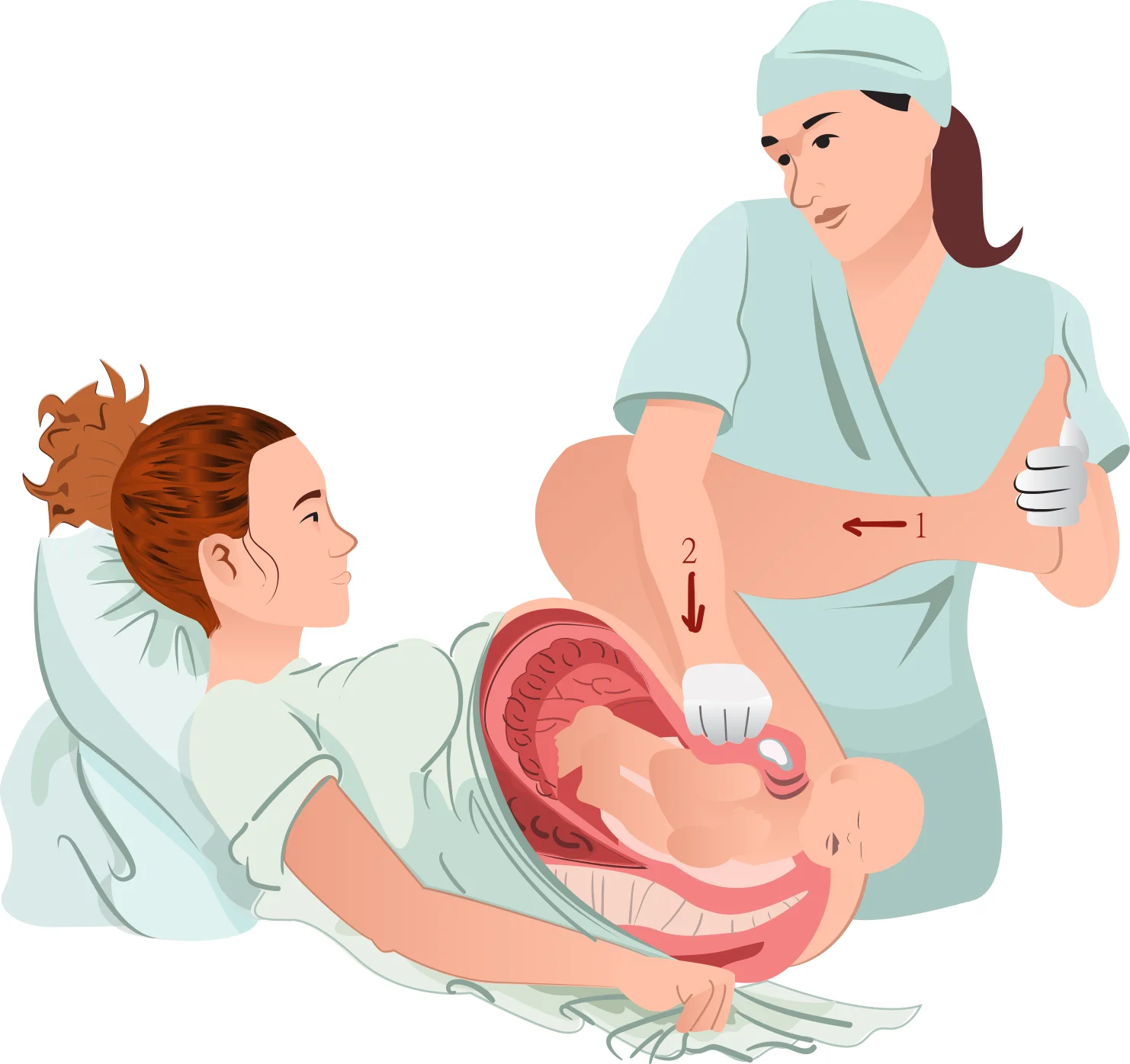
Long description
An illustration titled "McRoberts maneuver", described on Wikimedia Commons as an obstetrical maneuver used to assist in childbirth.

No randomised trial compares the individual manoeuvres, and most evidence is retrospective, so the order represents clinical consensus designed to use the simplest and safest move first. [2][3] AAFP, RCOG and the Advanced Life Support in Obstetrics programme recommend McRoberts first, then suprapubic pressure if necessary. [2] The AAFP list separates initial, secondary and catastrophic manoeuvres. [2]
Between manoeuvres, the mother is asked not to push, because maternal pushing without release of the shoulder adds traction and risk. [2][3]
- McRoberts manoeuvre: flex both hips and bring both knees toward the chest. This rotates the symphysis cephalad, straightens the lumbosacral angle and opens the pelvic outlet. [2] As the sole manoeuvre it succeeds in up to 42% of cases. [2]
- Suprapubic pressure: performed concurrently with McRoberts, an assistant applies firm downward or oblique pressure just above the pubic symphysis, toward the side the fetus is facing. This narrows the bisacromial diameter and may move the anterior shoulder past the symphysis. [2] A midwifery review notes that the combination is recommended and that McRoberts plus suprapubic pressure succeeds in 24% to 62% of cases. [3]
- Delivery of the posterior arm: if the first two fail, try to bring out the posterior arm. The operator's hand travels along the fetal chest (not the back) to grasp the wrist or elbow, flex it across the fetal chest and deliver it; this reduces the bisacromial diameter and is associated with less brachial plexus stretch in modelling studies. [2] A retrospective review found that McRoberts, suprapubic pressure and posterior arm delivery succeeded within four minutes in 95% of cases. [2]
- Menticoglou (posterior shoulder) manoeuvre: if the posterior arm is tight against the vaginal wall and cannot be reached, one finger from each hand is placed under the posterior axilla to apply traction gently along the pelvic curve. [2]
- Gaskin all-fours manoeuvre: turn the woman, if she can move and consent, to hands and knees and repeat traction and manoeuvres. This had an 83% success rate as the sole manoeuvre in one series, but is difficult with exhaustion or dense neuraxial anaesthesia. [2]
An episiotomy may be necessary for access, but is not mandatory for every manoeuvre. It should be performed when the operator needs room for posterior arm or rotational release, not as a routine first step. [2][3]
- Rubin II manoeuvre: two fingers in the vagina push the posterior aspect of the anterior shoulder toward the fetal face, aiming to rotate the shoulders about 30 degrees and reduce the bisacromial diameter. [2][3]
- Woods (corkscrew) manoeuvre: pressure on the front of the posterior shoulder to rotate the fetus 180 degrees, often combined with Rubin II. [2][3]
- Reverse Woods manoeuvre: rotate in the opposite direction, using the front of the anterior and the back of the posterior shoulder.
- Posterior axillary sling traction: a suction catheter or urinary catheter placed under the posterior axilla can apply downward traction, or be used to rotate 180 degrees like a Woods manoeuvre. [2]
These are uncommon but important, because a team that knows the entire ladder does not waste time hesitating:
- Intentional clavicular fracture: fracture one or both clavicles to collapse the shoulders inward. Performed bluntly, never with sharp instruments, because of the risk to vessels and lung. [2]
- Zavanelli (cephalic replacement): replace the delivered head into the vagina, using uterine relaxation, and deliver by emergency caesarean section. [2][3]
- Abdominal rescue (hysterotomy under general anaesthesia): a hysterotomy to disimpact the shoulders in an institution that can prepare for general anaesthesia quickly.
- Symphysiotomy: surgical division of the pubic symphysis. This is a last resort and can leave permanent pelvic instability and incontinence. [3]
One source notes that after all first- and second-line manoeuvres the team may need to start again: take the woman out of McRoberts and re-enter the manoeuvre, or roll her. This is preferable to repeating a failing manoeuvre idly. [3]
The four named guidance systems agree on the emergency nature of the condition, avoidance of fundal pressure, the first-line role of McRoberts (often combined with suprapubic pressure) and the value of training. They differ mainly in depth and in prevention recommendations.
| Source and edition | Main points from the opened page |
|---|---|
| RCOG Green-top Guideline No. 42, 3rd ed. 2026 | Defines shoulder dystocia, reports brachial plexus injury in 2.3% to 16%, postpartum haemorrhage 11% and sphincter injury 3.8%, states that the condition is largely unpredictable, notes that practical manoeuvres require practical training and are not described in full here, supports at least annual shoulder-dystocia training for all maternity staff, and supports shared decision-making about mode of birth after previous shoulder dystocia. [1][2] |
| ACOG Practice Bulletin No. 178, 2017 (reaffirmed 2024) | States shoulder dystocia is unpredictable and unpreventable, that prepregnancy, antepartum and intrapartum risk factors have extremely poor predictive value, and supports a systematic approach and simulation training to improve outcomes and documentation. [4] |
| NICE guideline NG235, intrapartum care (2023, updated 2026) | Uses previous shoulder dystocia to recommend birth in an obstetric unit and lists a history of a baby more than 4.5 kg as a factor to consider when planning place of birth. [5] |
| WHO labor and delivery protocol | Defines shoulder dystocia as the situation after the head delivers if restitution does not happen naturally and the anterior shoulder becomes stuck; states it cannot be predicted; advises shouting for help, mobilising staff, encouraging the woman, and not forcing or pulling the head. [6] |
This is a summary of the opened pages; full guideline recommendations require access to the complete texts.
- Fundal pressure, because it worsens impaction and increases the risk of uterine rupture. [2]
- Excessive or downward traction on the head and neck, because it causes brachial plexus stretch without relieving obstruction. [2]
- Twisting or bending of the neck. [3]
- Manual manipulations in a birth pool or with the woman standing: move her out of the pool if shoulder dystocia is suspected. [3]
- Waiting or repeating a failing manoeuvre without progressing.
Maternal complications are mostly mechanical:
- Postpartum haemorrhage (about 11%), uterine atony and, rarely, uterine rupture. [1][2]
- Genital tract lacerations, including third- and fourth-degree tears; soft-tissue injuries are common. [1][3]
- Bladder or rectal injury, symphyseal separation and lateral femoral cutaneous neuropathy. [2]
- Psychological distress and later anxiety in subsequent pregnancies. [1][3]
Neonatal complications:
- Brachial plexus injury in 2.3% to 16% of cases, transient in many (up to 20% overall), permanent in roughly one in ten of those injured. [1][2]
- Clavicular or humeral fracture and, rarely, diaphragmatic or facial nerve paralysis. [2][3]
- Hypoxic-ischaemic encephalopathy, neonatal asphyxia and, rarely, hypovolaemia from cord compression and blood sequestration in the placental circulation. [2][3]
- Risk of neonatal death, although rare when the team is trained. [3]
The interval from head delivery to body delivery is not a reliable predictor of fetal asphyxia or death in an individual case, but delay still increases risk; a systematic approach therefore remains essential. [2]
Most babies are born without injury when manoeuvres are performed promptly. Many brachial plexus injuries recover fully in the first few months, but roughly 10% may leave a permanent disability. [2] The future of both mother and child is best secured by documentation, rehabilitation and follow-up: examine the baby carefully for nerve and bone injury, review shoulder and arm function, and arrange physiotherapy or neurology referral if recovery is not rapid. [2][3]
For the woman, the next pregnancy becomes the focus of concern. Women with a previous shoulder dystocia should be counselled about recurrence, about planned options, and have their future births planned in an obstetric unit with a documented plan. [1][5] Correctable factors such as diabetes control should be identified, and women should be reassured honestly: most subsequent deliveries are uncomplicated, but certainty is not possible. [1]
Shoulder dystocia is a poor candidate for screening in the usual sense: no test predicts it, ultrasound weight estimates are inaccurate, and induction for suspected macrosomia is not recommended. [2][3][6] Birth planning changes mainly when the estimated weight is very high. RCOG states that the guideline does not include primary prevention of large-for-gestational-age fetuses, because that is covered by NICE guideline NG3 on diabetes in pregnancy. [1]
Evidence-based preventive points from the sources:
- Diabetes care: treat maternal hyperglycaemia to reduce macrosomia and shoulder dystocia risk. [3][5]
- Careful birth planning: if a baby may be over 4,500 g in a diabetic woman or over 5,000 g in a non-diabetic woman, consider caesarean birth. [2]
- Operative birth discipline: shoulder dystocia cannot be prevented by avoiding instrumental birth alone; instrumental delivery is one of several intrapartum risk factors. [2][3]
- Drills prevent harm rather than the event itself: annual simulation training improves team performance and documentation, and one programme reduced neonatal brachial plexus injury at discharge from 7.6% to 1.3%. [2][4]
- Team training: RCOG requires all maternity staff to participate in shoulder dystocia training at least annually; ACOG and the Advanced Life Support in Obstetrics programme recommend regular multidisciplinary drills. [1][2]
- Shared decision-making: women with a previous shoulder dystocia should be offered options, and women with a suspected large baby should receive balanced information. [1][5]
Preparing for shoulder dystocia resembles preparing for other obstetric emergencies: value is found in the drill, the team and the recorded plan, not in an unreliable forecast. [2][4]
Women with diabetes, obesity or a previous large baby are over-represented, but as ACOG states, these risk factors are poorly predictive. [3][4] Management should include antenatal counselling and a written birth plan in an obstetric unit when previous shoulder dystocia or strong suspicion exists. [1][5] Water birth remains possible, but if shoulder dystocia is suspected in water the woman should be advised to exit the pool so manoeuvres can be performed safely and efficiently. [3]
Here the "children" are the babies. Neonatal assessment is mandatory after any shoulder dystocia: brachial plexus function, clavicles, humeri, facial nerve, tone and respiration. [2][3] Many brachial plexus injuries recover; arrange physiotherapy and paediatric follow-up, and refer early if recovery is not evident. [2]
Most mothers are of reproductive age, but later in life or with comorbidities, factors such as diabetes and obesity become more common and the threshold for obstetric unit birth and written planning falls. [5] There are no age-specific manoeuvres; the same stepwise approach applies. [2]
The WHO protocol and international teaching manuals place special emphasis on recognition and avoidance of harmful actions when advanced help is limited. [6] Practical adaptations include:
- Arrange for skilled help before delivery if macrosomia or diabetes is suspected.
- Announce the emergency loudly and stop pushing. [6]
- Use the simplest effective manoeuvres first: McRoberts, then suprapubic pressure, then posterior arm or internal rotation, then all-fours. [2][3]
- Avoid fundal pressure and downward traction. [2][3][6]
- Prepare for neonatal resuscitation and hypovolaemia after the birth. [3]
- If a baby with suspected macrosomia is delivered at home, follow local protocols for urgent transport.
The key distinguishing feature is that the head has been delivered, but the shoulders have not. Conditions confused with it usually involve problems before delivery of the head, or a completely different mechanism.
| Condition | Typical situation | How shoulder dystocia differs |
|---|---|---|
| Breech delivery | Usually known before the head is born; delivered first by assisting breech extraction. | Shoulder dystocia is a vertex birth with normal head delivery followed by an impacted shoulder. [2] |
| Compound presentation | A hand may present alongside the head; the cord can be wrapped. | Identified before the head is out. [2] |
| Cephalopelvic disproportion before the head | Failure to progress while the head is still in the pelvis. | In shoulder dystocia the head is already born, so another immediate cause should be sought. [2] |
| Cord prolapse/compression | Sudden fetal bradycardia, palpable cord. | Can occur together with shoulder dystocia; always relieve compression while preparing to deliver. [2][6] |
| Large baby without shoulder dystocia | Birth weight is high, but head and shoulders deliver normally. | Macrosomia is a risk factor, not a diagnosis; most large babies are born without shoulder dystocia. [2][3] |
Recognition and management of shoulder dystocia
- Head delivers and stallsGentle traction fails or the turtle sign appears
- Announce shoulder dystocia and stop pushingCall qualified help, record the time
- McRoberts manoeuvre plus suprapubic pressureFlex hips and knees toward chest; firm downward pressure above the pubis
- Question: Shoulder released?
Yes
- Complete delivery carefullyAvoid further traction; be ready for PPH and neonatal resuscitation
No
- Internal manoeuvresPosterior arm, Rubin II, Woods, reverse Woods or sling; consider all-fours
- Question: Shoulder released?
Yes
- Complete delivery and document every manoeuvre
No
- Catastrophic manoeuvresConsider clavicular fracture, Zavanelli, abdominal rescue or symphysiotomy
- Restart once if neededMove out of McRoberts and re-enter, or roll the woman
- Shoulder dystocia is unpredictable and occurs in 0.3% to 3% of vaginal births; even babies weighing less than 4,000 g are often affected. [2][3]
- Diagnosis rests on the turtle sign, poor restitution, failed gentle traction and, on audit, a head-to-body interval over 60 seconds. [1][2]
- Fundal pressure and downward traction must be avoided; only axial traction is appropriate. [2]
- First-line manoeuvres are McRoberts and suprapubic pressure; delivery of the posterior arm and Gaskin all-fours are the next strong options. [2]
- Catastrophic options include intentional clavicular fracture, Zavanelli, abdominal rescue and symphysiotomy. [2][3]
- Maternal complications include postpartum haemorrhage (about 11%) and obstetric anal sphincter injury (about 3.8%). [1][2]
- Neonatal risk includes brachial plexus injury in 2.3% to 16% of cases, usually with good recovery but permanent disability in roughly one in ten of those. [1][2]
- Training and simulation have the best evidence base; birth plans should be written after previous shoulder dystocia or suspected macrosomia. [1][2][4]
- RCOG No. 42 (3rd ed., 2026), ACOG No. 178 (2017, reaffirmed 2024), NICE NG235 and the WHO labor protocol all stress unpredictability, immediate help and a systematic stepwise approach. [1][4][5][6]
Answer each one in your head first, then open the answer.
A woman is in the second stage and a vertex baby is born up to the chin, after which the head retracts against the perineum. Gentle traction does not bring the shoulders. What is the first manoeuvre to perform?
How does the McRoberts manoeuvre alter pelvic geometry?
Which two actions must be avoided, and why?
A retrospective review found that three combined manoeuvres resolved 95% of cases within four minutes. Which three?
Show answerHide answer
McRoberts manoeuvre, suprapubic pressure and delivery of the posterior arm. [2]
Which two women described below both need preparation for shoulder dystocia?
How does a neonatal brachial plexus injury after shoulder dystocia differ from a clavicular fracture?
Show answerHide answer
The brachial plexus injury is a traction neuropathy of the arm nerves, often transient; the clavicular fracture is a mechanical fracture, usually healing without treatment. Transient brachial plexus injuries may occur in up to 20% of shoulder dystocia deliveries, and permanent injury in roughly 10% of those. [1][2]
Plain-text pointers: diabetes in pregnancy; antepartum fetal surveillance; obstetric exam and fetal heart rate monitoring; obstetric analgesia and anaesthesia; neonatal resuscitation; fetal growth restriction and macrosomia; planning place of birth; neonatal seizures and hypoxic-ischaemic encephalopathy; paediatric orthopaedic injuries; obstetric anal sphincter injury repair; postpartum haemorrhage.
- Lattey K, Winter C, Ellis V, Crofts JF, et al. Shoulder Dystocia. Royal College of Obstetricians and Gynaecologists Green-top Guideline No. 42, 3rd ed. (2026). BJOG 2026;133:e165-e186.. Accessed .
- Hill DA, Lense J, Roepcke F. Shoulder Dystocia: Managing an Obstetric Emergency. American Family Physician 2020;102(2):84-90.. Accessed .
- Bothou A, Apostolidi DM, Tsikouras P, et al. Overview of techniques to manage shoulder dystocia during vaginal birth. European Journal of Midwifery 2021;5:48.. Accessed .
- Committee on Practice Bulletins--Obstetrics, American College of Obstetricians and Gynecologists. Practice Bulletin No. 178: Shoulder Dystocia. Obstetrics & Gynecology 2017;129(5):e123-e133, reaffirmed 2024.. Accessed .
- National Institute for Health and Care Excellence. Intrapartum care (NICE guideline NG235). NICE guideline, published 2023, last updated 9 June 2026.. Accessed .
- World Health Organization. Labor and Delivery Protocol. WHO operational guidance, shoulder dystocia section.. Accessed .
Figure credits
Educational content for medical students and doctors. It is not a substitute for clinical judgement or local guidelines.