Nephritic syndrome and glomerulonephritis
Blood and protein in the urine with pressure and swelling: what nephritic syndrome is, its glomerular causes, and how it is investigated and treated.
- Nephritic syndrome is kidney inflammation (glomerulonephritis) that leaks blood into the urine and injures filtration enough to raise blood pressure, cause swelling and raise the creatinine slightly. The signature finding is red cells in the urine, often as red cell casts, together with hypertension, oedema, mild proteinuria (usually under 3 g per day) and a raised creatinine.
- It is the mirror image of nephrotic syndrome (heavy proteinuria, low albumin, high lipids): nephritic syndrome is dominated by inflammation and bleeding, nephrotic syndrome by leaky membranes and protein loss.
- Causes run from mild and self-limiting post-infectious disease in children to rapidly progressive glomerulonephritis that can destroy kidneys within weeks. The urgent question is whether there are crescents on biopsy, because that changes treatment from supportive care to immunosuppression and sometimes plasma exchange.
- Complement levels give a quick steering clue: C3 is low in post-infectious, immune-complex and complement-mediated disease, while C3 and C4 are both usually normal in IgA nephropathy, ANCA disease and anti-GBM disease.
- Treatment is therefore two-layered: control blood pressure, volume and any dangerous complications first, then match immunosuppression to the specific cause found on biopsy and serology.
By the end of this page you should be able to:
- Define nephritic syndrome in plain words and distinguish it from nephrotic syndrome and from acute kidney injury without glomerular disease.
- Explain how post-infectious, IgA, ANCA, anti-GBM and lupus injury produce the same clinical picture through different immunological mechanisms.
- Read the urinalysis, complement pattern and biopsy and use them to separate the causes.
- State the principles of blood-pressure and RAAS-based support, the indications for immunosuppression, plasma exchange and dialysis, and how these differ by cause.
- Outline the KDIGO glomerular disease approach: biopsy, timed urine collection, supportive care and risk-stratified immunosuppression.
- Describe management and precautions in pregnancy, children and low-resource settings.
DefinitionNephritic syndrome
Nephritic syndrome is a pattern of glomerular injury in which inflammation of the glomerular capillaries (glomerulonephritis) makes them leak red blood cells and a modest amount of protein, and reduces filtration enough to raise blood pressure, cause salt and water retention, and raise serum creatinine. It is a clinical syndrome, not a single disease, and is confirmed or classified by urinalysis, serology and usually kidney biopsy.[2][3]
Kidney biopsy remains central to the diagnosis whenever the cause is not otherwise clear, and an internationally validated scoring system should be used when reporting it.[2]
Five features, remembered in many textbooks, make up the syndrome:
- Haematuria is the defining feature. The dipstick is positive for blood and microscopy shows dysmorphic red cells and red cell casts. Red cell casts are the clue that bleeding is glomerular rather than from the lower urinary tract.
- Hypertension, often mild to moderate and sometimes severe.
- Oedema, usually mild periorbital or ankle swelling rather than the massive oedema of nephrotic syndrome.
- Proteinuria, but of modest degree. In practice this is usually under 3 g per day; heavy proteinuria suggests either nephrotic syndrome or a mixed (nephritic-nephrotic) picture.
- Renal impairment, a rise in creatinine from a fall in glomerular filtration rate.
These five features share one mechanism. Inflammation swells and damages the glomerular capillary wall. Red cells and some protein leak through it; filtration falls because the capillary loops are partly occluded by cells and immune deposits, and because reduced filtration raises pressure inside the glomerulus. The kidney senses reduced effective perfusion and retains salt and water through the renin-angiotensin-aldosterone system, which drives both the hypertension and the oedema.
A useful distinction is between nephritic and nephrotic syndrome:
| Feature | Nephritic syndrome | Nephrotic syndrome |
|---|---|---|
| Blood in the urine | Present, often with red cell casts | Absent or only microscopic |
| Proteinuria | Mild to moderate, usually under 3 g per day | Heavy, 3.5 g per day or more |
| Serum albumin | Usually normal or near normal | Low |
| Oedema | Mild | Marked, often anasarca |
| Lipids | Usually normal | Raised |
| Blood pressure | Often raised | Often normal unless fluid overloaded |
| Underlying problem | Glomerular inflammation | Glomerular permeability without prominent inflammation |
Nephritic syndrome should also be separated from plain acute kidney injury. Infection, hypoperfusion, obstruction and nephrotoxic drugs can all raise creatinine without haematuria or red cell casts. The combination of glomerular blood plus a falling filtration rate points instead to a glomerulonephritis and usually warrants serology and biopsy.
In the KDIGO approach, nephritic syndrome is one of the clinical syndromes of glomerular disease, alongside nephrotic syndrome, asymptomatic haematuria or proteinuria, and rapidly progressive renal failure. The syndrome labels the presentation; the final diagnosis is a specific glomerular disease, and the proportions differ from other presentations: when biopsy is performed because of nephritic syndrome, IgA nephropathy is by far the commonest finding, followed by lupus nephritis and Henoch-Schonlein purpura (IgA vasculitis).[3]
A second distinction separates acute from rapidly progressive disease:
- Acute glomerulonephritis runs over days to a few weeks. The filtration fall is modest, urine output may be reduced, and it is usually reversible with treatment. Post-infectious disease is the classic example.
- Rapidly progressive glomerulonephritis (RPGN) is defined by a rapid loss of kidney function over days to weeks, with extensive crescents on biopsy. Crescentic glomerulonephritis needs urgent immunosuppression. Anti-GBM disease, ANCA-associated vasculitis and some severe immune-complex diseases present this way.
Nephritic syndrome is a common indication for kidney biopsy. In a pooled analysis of 26,414 nephritic patients from 47 studies in 23 countries, nephritic syndrome accounted for about 21% of all biopsies, ranging from about 8% in South Asia to 36% in East Asia. In that same analysis, IgA nephropathy was the single most common diagnosis (about 38%), followed by lupus nephritis (about 8%) and Henoch-Schonlein purpura (about 7%), with marked regional and age differences.[3] IgA nephropathy is the most frequent finding in East Asia and in adults; lupus is proportionally more common in children and young women.
The relative frequency also varies with age. Children with a recent infection and nephritic syndrome most often have post-infectious disease, which is often not biopsied because it resolves. Young women are more likely to have lupus nephritis. Older adults more often have pauci-immune ANCA disease, membranoproliferative patterns or paraprotein-related disease. [2]
The glomerular inflammation can be produced by four broad mechanisms. Each mechanism predicts the presentation, the complement pattern and the biopsy.
Post-infection glomerulonephritis classically follows a streptococcal throat or skin infection, but many organisms can trigger it (viruses such as hepatitis B and C, bacteria, and malaria and other parasites). It is immune-complex disease: antibody-antigen complexes deposit in glomeruli and activate complement. CARI's summary of KDIGO-related guidance notes that post-streptococcal disease is more common than the global average among Aboriginal, Torres Strait Islander, Pasifika and Maori peoples, particularly in children under 14, and often follows skin infection (about 65%) or throat infection (about 27%).[2]
- IgA nephropathy (Berger disease) and IgA vasculitis (Henoch-Schonlein purpura). Abnormally glycosylated IgA1 forms immune complexes that deposit in mesangial areas.
- Lupus nephritis. Autoantibodies (anti-dsDNA and others) form complexes against nuclear antigens and deposit in multiple glomerular sites.
- Membranoproliferative patterns can be immune-complex mediated, for example after hepatitis C infection or cryoglobulinaemia.
Anti-GBM disease is an autoimmune attack on type IV collagen in the glomerular basement membrane. When it also involves pulmonary capillaries it is called Goodpasture syndrome, with haemoptysis and alveolar haemorrhage. Renal involvement is typically rapidly progressive.
Antineutrophil cytoplasmic antibodies (ANCA) activate neutrophils that damage small vessels, producing a pauci-immune (few immune deposits) necrotising crescentic glomerulonephritis. The three main clinical forms are:
- Granulomatosis with polyangiitis (GPA), often with respiratory tract disease.
- Microscopic polyangiitis (MPA).
- Eosinophilic granulomatosis with polyangiitis (EGPA), with asthma and eosinophilia.
- Alport syndrome, an inherited defect in type IV collagen, produces persistent haematuria, sensorineural deafness and, in males, lenticonus of the eye. Renal function declines over years to decades.
- Thin basement membrane disease (benign familial haematuria) is a milder inherited collagen defect that causes lifelong microscopic haematuria and rarely progresses.
- C3 glomerulopathy / dense deposit disease, a complement-mediated pattern that can masquerade as membranoproliferative disease.
Risk factors worth remembering include a recent infection, systemic lupus erythematosus and other autoimmune disease, hepatitis B or C, HIV, certain drugs (for example, hydralazine, propylthiouracil and allopurinol can produce ANCA), and a family history of Alport syndrome or thin basement membrane disease.
The common pathway is glomerular capillary inflammation. Immune complexes, autoantibodies or complement activation deposit in or bind to the glomerulus. Neutrophils and monocytes are recruited, capillary loops swell and may rupture, and crescents (proliferating parietal epithelial cells and macrophages) form in Bowman's space in severe disease. A crescent physically compresses the tuft and is the histological mark of rapidly progressive disease.
Each cause has a signature lesion:
- Post-infectious gives an endocapillary (inside the capillary) proliferative picture, with neutrophils and large subepithelial immune deposits on electron microscopy (the "humps").
- IgA nephropathy gives mesangial IgA deposits and mesangial proliferation; crescents occur in aggressive disease.
- Anti-GBM gives a linear ribbon of IgG along the basement membrane on immunofluorescence, with necrotising crescents.
- ANCA disease gives necrotising crescents with very little immune staining ("pauci-immune").
- Lupus gives deposits in mesangial, subendothelial and subepithelial sites, producing "full house" immunofluorescence (IgG, IgA, IgM, C3 and C1q together) and the "wire loop" lesions of membranous lupus.
- Alport and thin basement membrane disease show structural basement-membrane defects rather than immune deposits.
- Membranoproliferative / C3 disease shows mesangial and endocapillary proliferation with split basement membranes ("tram track" or double contours) and thickened loops.
Patients or carers describe:
- urine that is cola- or tea-coloured, or visibly bloody (often the first symptom);
- foamier urine or swelling around the eyes on waking;
- headache, blurred vision or nosebleeds from hypertension;
- fatigue, nausea or breathlessness if renal impairment is significant;
- joint pains, rash, mouth ulcers or hair loss (lupus);
- haemoptysis, sinus disease, deafness or asthma (ANCA, anti-GBM or vasculitis clues);
- a recent sore throat or skin infection weeks earlier (post-infectious);
- hearing loss or a family history of kidney disease (Alport).
A 24-hour urine collection is preferred over a spot test when protein is heavy, because timed collection is more accurate and less variable; routine assessment of haematuria matters especially in IgA nephropathy and IgA vasculitis.[2]
- Blood pressure, including lying and standing, and fundoscopy for hypertensive retinopathy.
- Volume status: periorbital puffiness, sacral or ankle oedema, raised jugular venous pressure, pulmonary crackles.
- Rash (purpura on the legs and buttocks suggests IgA vasculitis), arthritis, oral ulcers.
- Signs of chronic disease: deafness, eye changes (Alport lenticonus), livedo, neuropathy.
- Abdominal tenderness, especially in IgA vasculitis with gut involvement, which can occur after the rash.
Red flags that demand same-day assessment include anuria or rapidly falling urine output, severe hypertension (for example above 180 systolic or symptoms of hypertensive emergency), gross haematuria with clots, and haemoptysis.
The differential is usually one of mechanism rather than a long list of diseases. Table 1 summarises the causes, their clues, the complement pattern, the characteristic biopsy appearances and the usual treatment approach. All units below are SI.
| Cause | Typical clues | Complement (C3/C4) | Light / IF / EM | Treatment and outlook |
|---|---|---|---|---|
| Post-infectious GN | Child or young adult; 1-3 weeks after throat or skin infection; self-resolving | Low C3 (often C4 normal) | Endocapillary proliferative / granular C3, IgG / subepithelial humps | Supportive: salt and fluid control, BP control, treat infection; dialysis if needed. Usually good recovery. |
| IgA nephropathy (Berger) | Young adult with synpharyngitic haematuria; episodic macroscopic haematuria with infection | Normal C3/C4 | Mesangial proliferation / mesangial IgA / mesangial deposits | BP and RAAS blockade; risk-stratified glucocorticoids; excellent in mild disease, guarded with crescents. |
| IgA vasculitis (Henoch-Schonlein) | Palpable purpura, arthritis, abdominal pain; child or young adult | Normal C3/C4 | Mesangial similar to IgA / IgA-dominant / mesangial deposits | Supportive; immunosuppression when RPGN or significant renal involvement. |
| Anti-GBM (Goodpasture) | Rapid renal failure; pulmonary haemorrhage or haemoptysis; smokers | Normal C3/C4 | Necrotising crescents / linear IgG on GBM / no deposits | Plasma exchange plus cyclophosphamide and glucocorticoids; better when started early. |
| Pauci-immune ANCA (GPA, MPA, EGPA) | RPGN; sinus, lung, skin, nerve or ear disease; older adults | Normal C3/C4 | Necrotising crescents / pauci-immune / no significant deposits | Induction with glucocorticoids plus cyclophosphamide or rituximab; maintenance after remission. |
| Lupus nephritis | Young woman; rash, arthritis, serositis; any ISN/RPS class | Low C3 (and often C4) | Class varies; full-house IF / subendothelial-subepithelial deposits | Induction with glucocorticoids plus mycophenolate or cyclophosphamide; maintenance; hydroxychloroquine. |
| Alport syndrome | Lifelong haematuria, deafness, eye changes; male predominant, family history | Normal | No immune deposits; basket-weave basement membrane on EM (split lamina densa) | No immunosuppression; ACEI/ARB, audiology and eye follow-up. |
| Thin basement membrane disease | Lifelong mild haematuria; family history; normal renal function | Normal | Thin basement membrane on EM; no deposits | Reassurance; no specific treatment. |
| MPGN / C3 glomerulopathy (incl. dense deposit disease) | Nephritic or mixed picture; may follow infection or monoclonal gammopathy | Low C3; C4 variable | Proliferation with double contours / immune complex or C3-only IF / subendothelial-subepithelial or dense deposits | Treat underlying cause; complement blockade under study; supportive care; prognosis variable. |
Other masqueraders include IgA-dominant post-infectious glomerulonephritis (particularly in older adults with diabetes and staphylococcal infection), C1q nephropathy, cryoglobulinaemic disease and drug-induced interstitial nephritis. These are separated by infection history, monoclonal studies and the biopsy pattern.
Figure 1Post-infectious glomerulonephritis - high mag
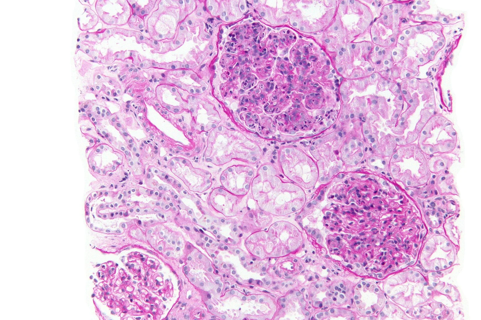
Long description
Kidney biopsy stained with PAS at high magnification: crowded, hypercellular glomeruli with tubules around them, as in post-infectious glomerulonephritis. Two large glomeruli and part of a third fill the field; each tuft is packed with nuclei and fills the capillary spaces.

Figure 2Immunofluorescence staining for IgG along glomerular capillary loops
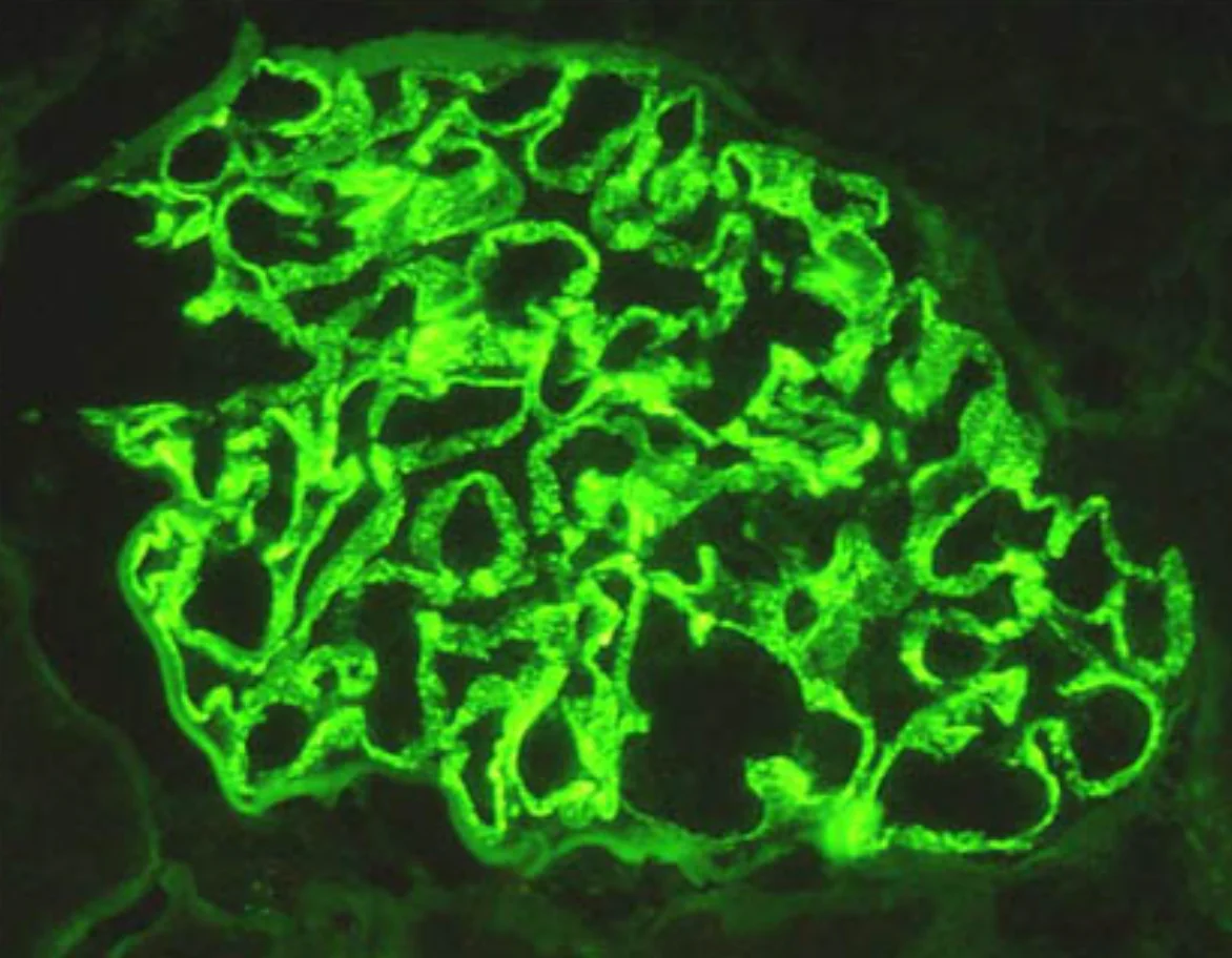
Long description
Immunofluorescence image of a renal glomerulus showing bright green linear-to-granular staining along the glomerular basement membranes, described as consistent with immune complex or antibody deposition.

Figure 3Red blood cell cast in urine sediment
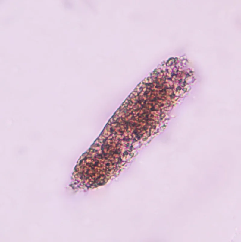
Long description
A urine sediment microscopy image captioned "Red blood cell cast in urine sediment" (800 x 801 px) from a StatPearls urinalysis article.

- Urinalysis: microscopy for red cells, casts, white cells and bacteria; dipstick protein. Dysmorphic red cells and red cell casts point to glomerular bleeding.
- Renal function and electrolytes: urea or BUN, creatinine, eGFR, sodium, potassium, bicarbonate.
- Protein estimation: protein-to-creatinine ratio on a spot sample, or a 24-hour collection when heavy or when KDIGO-style accuracy matters; timed collection is more accurate in marked proteinuria.[2]
- Full blood count and coagulation before biopsy.
- Blood pressure measured properly, including both arms if indicated.
- Complement C3 and C4 (see Table 1).
- ANCA (indirect immunofluorescence plus anti-PR3/anti-MPO) for pauci-immune disease.
- Anti-GBM antibodies when renal failure is rapid or there is haemoptysis.
- ANA and anti-dsDNA, C3 and C4 for lupus.
- Antistreptolysin O and anti-DNase B for recent streptococcal infection.
- Hepatitis B and C serology, and HIV testing when risk factors exist, because they change both the diagnosis and the treatment.
- Cryoglobulins and rheumatoid factor where indicated.
CARI's KDIGO commentary recommends that all glomerular disease patients be screened for tuberculosis, syphilis, hepatitis and HIV, and that vaccination status be checked before immunosuppression.[2]
- Renal tract imaging: ultrasound assesses kidney size, echogenicity and obstruction. Small kidneys suggest chronic disease; normal or large kidneys favour an active glomerulonephritis.
- Chest imaging when haemoptysis, GPA, MPA or lupus is suspected.
- Renal biopsy to establish the diagnosis, to distinguish inflammation from scarring, and to direct treatment. Biopsy is usually performed unless it would be unsafe or unless a clear monogenic diagnosis (such as Alport) is already proved by genetic testing.[2] For typical active disease, validate the result with an internationally recognised scoring system (ISN/RPS for lupus nephritis, Oxford MEST-C for IgA nephropathy).[2]
There are no numerical diagnostic criteria for nephritic syndrome in the same way there are for chronic kidney disease staging. The diagnosis is clinical (haematuria with red cell casts, hypertension, oedema, mild proteinuria and raised creatinine) and is confirmed by biopsy. Severity is described by:
- estimated filtration rate and its change over days;
- degree of proteinuria;
- extent of crescents on biopsy (focal versus diffuse);
- serum creatinine at presentation, which is the strongest predictor of renal survival in anti-GBM and ANCA disease;
- for IgA nephropathy at high risk of progression, proteinuria above 0.75 to 1 g per day after at least 90 days of supportive care.[2]
The creatinine rises should be staged sensibly: nephritic syndrome can coexist with acute kidney injury, and many of the AKI principles (avoid nephrotoxins, volume control) still apply.
Approach to suspected nephritic syndrome
- Confirm glomerular bleedingDipstick blood with dysmorphic red cells or red cell casts on microscopy; check blood pressure, oedema and creatinine
- Stabilise immediate threatsSevere hypertension, pulmonary oedema or hyperkalaemia; reduce salt, and use diuretics or dialysis as needed
- Baseline bloodsCreatinine and eGFR, electrolytes, full blood count, complement C3/C4, ANA, ANCA, anti-GBM, ASO, hepatitis B and C, HIV
- Assess severity and speedCreatinine trend, urine output, red flags; consider acute versus rapidly progressive disease
- Question: Is renal function falling rapidly or are there crescents expected?
Yes
- Urgent nephrology and biopsyAnti-GBM and ANCA serology; plasma exchange and immunosuppression as indicated
No
- Supportive careRAAS blockade, blood pressure and volume control; treat infection or trigger
- Biopsy if cause unclear or persistentDirect long-term treatment and prognosis
- Start cause-specific treatmentPost-infectious: supportive; IgA/IgAV: supportive with risk-stratified glucocorticoids; lupus: induction and maintenance; Alport/TBM: supportive
- Follow-upBlood pressure, proteinuria, eGFR, drug toxicity and long-term renal and extrarenal outcomes
The general rule is to treat the reversible complications first and the glomerular inflammation second. Two cautions follow from KDIGO's glomerular disease guidance: do not rush immunosuppression before infection is excluded, and tailor supportive therapy to the histology (for example, RAAS blockade is inappropriate in steroid-responsive minimal change disease, although that belongs to nephrotic rather than nephritic syndrome).[2]
- Blood pressure and RAAS blockade: target a systolic blood pressure below 120 mmHg where tolerated; use renin-angiotensin-aldosterone system inhibitors (ACE inhibitor or ARB) plus other antihypertensives, and titrate with regard to creatinine and potassium.[2]
- Volume and oedema: salt restriction, loop diuretics when overloaded; thiazides and combinations where resistant.
- Diet: sodium under 2 g (90 mmol) per day, moderate protein intake (0.8 to 1.9 g per kg per day), heart-healthy fat intake and weight control; avoid very high protein intake.[2]
- Fluid intake: encourage steady hydration, and avoid dehydration, NSAIDs and unnecessary nephrotoxins.
- Treatment of precipitants: eradicate streptococci, treat hepatitis B or C where present, and review all drugs.
- Thrombosis prophylaxis when nephrotic features coexist (heavy proteinuria, low albumin), using heparin or a heparin derivative, or warfarin; online risk tools can guide the decision.
- Infection prevention: all patients with glomerular disease should receive pneumococcal and influenza vaccine, should be screened for tuberculosis, syphilis, hepatitis and HIV, and should receive Pneumocystis prophylaxis when given high-dose steroids or other immunosuppressants.[2]
CARI's commentary notes that, where immunosuppression is planned in a woman of childbearing age, cumulative cyclophosphamide dose matters because it can cause infertility, and that RAS inhibitors are teratogenic.[2]
Treatment is supportive. Penicillin (or an appropriate macrolide where penicillin allergy) eradicates residual streptococci, salt and fluid are restricted, hypertension controlled, and diuretics given for overload. Most children recover fully within weeks. Dialysis is reserved for the unusual patient with dangerous hyperkalaemia, severe acidosis or pulmonary oedema. If adult disease is slow to resolve, repeat the biopsy or reassess for a second diagnosis such as IgA nephropathy, because late or persistent disease in adults is less benign.
Supportive care (RAAS blockade, blood pressure, cardiovascular risk management, smoking cessation) is first-line for everyone. For patients at high risk of progression, defined as proteinuria above 0.75 to 1 g per day after at least 90 days of supportive care, a 6-month course of glucocorticoids is recommended, with caution when eGFR is below 50 mL per min per 1.73 m2 because high-dose methylprednisolone carries a higher infection risk than placebo.[2] SGLT2 inhibitors are increasingly standard add-on therapy for chronic kidney disease and are supported for IgA nephropathy where available. Targeted-release budesonide, sparsentan and other endothelin antagonists and complement inhibitors are emerging options still awaiting approval in many regions.[2]
IgA vasculitis with rapidly progressive glomerulonephritis is treated similarly to ANCA-associated vasculitis (see below). Patients with crescentic IgA nephropathy and acute kidney injury are treated with cyclophosphamide and glucocorticoids, as for ANCA disease.[2] Children with IgA nephropathy or vasculitis should be co-managed with a paediatric nephrologist.[2]
This is a nephrology emergency. Treatment combines:
- plasma exchange to remove circulating antibody;
- cyclophosphamide and glucocorticoids to stop new antibody production. KDIGO has dedicated anti-GBM disease guidance and CARI's commentary endorses that guidance.[1][2] Renal recovery is most likely when treatment starts before creatinine is very high; once oliguric renal failure is established with a high proportion of crescents, recovery is unlikely and treatment may be limited to controlling pulmonary haemorrhage. Avoiding smoking and managing iron are relevant because pulmonary haemorrhage is the immediate threat. Plasma exchange details, antibody titres and cyclophosphamide dosing are managed by the nephrology team and local protocols.
Induction of remission is achieved with glucocorticoids plus cyclophosphamide or rituximab, followed by maintenance therapy to prevent relapse (typically rituximab or azathioprine). KDIGO has separate guidance for ANCA-associated vasculitis (2024) and CARI endorses it.[1][2] Avacopan, a complement C5a receptor antagonist, is an emerging steroid-sparing option in some regions. Dialysis is given when needed, and plasma exchange is used in selected severe presentations such as severe kidney failure or pulmonary haemorrhage according to local protocol.
Induction therapy usually combines glucocorticoids with mycophenolate mofetil or cyclophosphamide, followed by maintenance with mycophenolate (or azathioprine), plus hydroxychloroquine for systemic lupus and renal protection. Belimumab and voclosporin are newer additions in some regions.[1][2] The biopsy class (ISN/RPS classes I to VI) determines intensity: mesangial (I-II) needs little or no immunosuppression, focal or diffuse proliferative disease (III-IV) needs induction, membranous (V) is treated according to proteinuria, and sclerotic (VI) is usually beyond reversal. KDIGO's separate lupus guidance and the ISN/RPS biopsy classification provide the standard framework.[2]
There is no immunosuppression for these structural diseases. ACE inhibitors or ARBs slow progression in Alport, blood pressure is controlled, and patients need audiology and eye follow-up. Genetic counselling is offered where Alport is confirmed. Thin basement membrane disease needs reassurance only.
Management targets the underlying trigger (infection such as hepatitis C, monoclonal gammopathy, complement dysregulation). Supportive care is standard; complement-directed agents (for example, iptacopan, eculizumab) are emerging and were not standard at the time of the KDIGO 2021 guidance.[2]
Dialysis is not started from a single creatinine. It is started when a complication (dangerous hyperkalaemia, fluid overload or pulmonary oedema, severe acidosis or uraemic symptoms) does not respond to medical treatment, or when there is severe, progressive oliguric failure despite cause-directed therapy.
- Hypertensive emergency and hypertensive encephalopathy from severe nephritic hypertension.
- Acute kidney injury and fluid overload, sometimes needing dialysis.
- Electrolyte problems: hyperkalaemia and metabolic acidosis in severe reduction of filtration.
- Pulmonary haemorrhage in anti-GBM disease and, less commonly, in ANCA disease.
- Nephrotic overlap: proteinuria can rise and albumin fall, adding thrombosis and infection risk.
- Progression to chronic kidney disease if inflammation is prolonged or incompletely treated.
- Drug toxicity from immunosuppressants: infection, cyclophosphamide-related bladder toxicity and infertility, steroid metabolic effects, rituximab-related infection.
Prognosis varies more by cause than by syndrome:
- Post-infectious disease in children usually resolves fully.
- IgA nephropathy is generally slow, but high-risk disease progresses; crescents and a rising creatinine at biopsy worsen outlook.
- ANCA and anti-GBM diseases are life-threatening without treatment but outcomes have improved with modern induction; early treatment of anti-GBM disease protects kidneys, especially if dialysis is not yet established.
- Lupus nephritis remits and relapses; long-term renal survival depends on achieving and maintaining remission.
- Alport syndrome progresses to end-stage renal disease in males in middle age, later or not at all in many female carriers.
- Thin basement membrane disease is essentially benign.
Most of the prognostic gains in modern practice come from measuring proteinuria and creatinine serially and stepping up treatment when targets (for example, blood pressure under 120 mmHg systolic and proteinuria reduction) are not met.
There is no vaccine or specific prevention for most glomerulonephritis. Sensible measures are:
- prompt treatment and skin care for streptococcal throat and skin infections, especially in high-risk communities;
- vaccination (pneumococcal, influenza, hepatitis B) where appropriate, particularly before immunosuppression;
- early recognition of lupus and vascular disease;
- family screening (urinalysis, and genetic testing with counselling) when Alport or thin basement membrane disease is found.
Pregnancy. Glomerular disease in pregnancy is high-risk and should be managed by a multidisciplinary team (nephrologist, obstetrician and maternal-fetal medicine). RAS inhibitors and mycophenolate are teratogenic; blood pressure should be aimed below 140/90 mmHg; low-dose aspirin, calcium and vitamin D are often used; and prophylactic anticoagulation considered when nephrotic or with prior clots. Cyclophosphamide must be weighed against irreversible infertility.[2]
Children. Post-infectious disease is common and usually self-limiting. IgA nephropathy, vasculitis, Alport and thin basement membrane disease present in childhood. Children with suspected monogenic disease (Alport, familial cases) may be tested genetically, sometimes before biopsy.[2]
Older adults. Pauci-immune ANCA disease and paraprotein-related disease become more common; treatment intensity must balance benefit with infection and bone marrow suppression.
Where laboratory, serology or biopsy are limited:
- Make the diagnosis clinically from haematuria with red cell casts, hypertension, oedema and rising creatinine, then stratify by speed of decline and red flags.
- Check urine dipstick and do microscopy where possible; a basic urine sediment distinguishes glomerular red cells from lower-tract bleeding.
- Use ultrasound to exclude obstruction and to judge whether kidneys are small and chronic.
- Control blood pressure with the available agents (avoid NSAIDs); diuretics and salt restriction manage overload.
- Treat infection when suspected, and send or store serum for later ANCA, anti-GBM, complement, ANA and hepatitis serology if samples can be transported.
- Start immunosuppression for rapidly progressive or pulmonary-haemorrhage presentations only where it can be monitored; refer early.
- Dialysis may not be available; focus on treating dangerous hyperkalaemia and acidosis, restricting potassium and, if absolutely necessary, using whatever renal replacement (including peritoneal dialysis) the setting allows.
- Plan follow-up for blood pressure and proteinuria with the resources that exist.
- Nephritic syndrome is glomerular inflammation presenting with haematuria (often red cell casts), hypertension, oedema, mild to moderate proteinuria and raised creatinine.
- It is a syndrome, not a disease; the biopsy and serology determine the cause.
- The urgent division is acute, treatable inflammation versus rapidly progressive crescentic disease, because the latter needs immunosuppression and sometimes plasma exchange.
- Complement patterns steer the diagnosis: C3 low in post-infectious, complement-mediated and lupus disease; C3 and C4 usually normal in IgA, ANCA and anti-GBM disease; C4 also low in lupus and cryoglobulinaemia.
- On biopsy, post-infectious disease shows endocapillary proliferation with humps; IgA shows mesangial deposits; anti-GBM shows linear IgG; ANCA shows pauci-immune necrotising crescents; lupus shows full-house deposits.
- Supportive care (salt and volume control, RAAS blockade, blood-pressure target below 120 mmHg systolic, infection prevention) underlies every cause.
- Immunosuppression is cause-specific: cyclophosphamide with glucocorticoids for anti-GBM disease plus plasma exchange; glucocorticoids with cyclophosphamide or rituximab for ANCA disease; risk-stratified glucocorticoids and RAAS blockade for IgA nephropathy; glucocorticoids with mycophenolate or cyclophosphamide for lupus nephritis.
- Alport and thin basement membrane disease have no immunosuppression; Alport needs ACE inhibitor or ARB and follow-up for hearing and eyes.
- KDIGO glomerular disease guidance underpins modern care: biopsy with validated scoring, timed urine collection, cardiovascular and renal protection, infection screening and vaccination, and risk-stratified immunosuppression.
- Pregnancy and childhood need specialist, multidisciplinary management.
Answer each one in your head first, then open the answer.
A 9-year-old boy has dark urine two weeks after impetigo. Blood pressure is 130/85 mmHg, there is ankle swelling, creatinine is 88 micromol/L and C3 is low. What is the most likely diagnosis, and what would you expect on biopsy?
Show answerHide answer
Post-infectious (streptococcal) glomerulonephritis. Biopsy, if performed, shows endocapillary proliferation with neutrophils and large subepithelial electron-dense humps; immunofluorescence is granular for C3 and IgG. Management is supportive; most children recover within weeks.
A 24-year-old woman has haematuria with red cell casts, proteinuria 0.6 g per day and a creatinine of 96 micromol/L. ANA is positive, anti-dsDNA is high, and both C3 and C4 are low. What diagnosis is most likely, and which biopsy class drives treatment intensity?
Show answerHide answer
Lupus nephritis. The ISN/RPS class determines intensity: classes III and IV (focal or diffuse proliferative disease) need induction with glucocorticoids plus immunosuppression, while classes I and II need little or none.
A 60-year-old man presents with haemoptysis, creatinine rising over days and red cell casts. Anti-GBM antibody is positive and C3 and C4 are normal. What is the emergency treatment plan?
Show answerHide answer
Plasma exchange to remove antibody, combined with cyclophosphamide and glucocorticoids, plus nephrology support. Renal recovery is best when treatment starts before severe oliguric renal failure.
A patient has heavy macroscopic haematuria with each upper respiratory infection, persistent microscopic haematuria between episodes and a normal creatinine. What is the likely diagnosis, and do C3 and C4 help?
Show answerHide answer
IgA nephropathy (synpharyngitic haematuria). C3 and C4 are normally normal, so they do not separate it from normal; the persistence of haematuria between infections and the biopsy do.
Which complement pattern would you expect in ANCA-associated glomerulonephritis, and why does a pauci-immune biopsy matter?
Show answerHide answer
C3 and C4 are usually normal. The pauci-immune biopsy (few immune deposits despite necrotising crescents) points to ANCA-driven neutrophil injury rather than immune-complex deposition, which changes the immunosuppressive induction to glucocorticoids with cyclophosphamide or rituximab.
In pregnancy with glomerular disease, what is the blood pressure target and which two drug classes must be avoided?
Show answerHide answer
[2]Aim for blood pressure below 140/90 mmHg. Renin-angiotensin system inhibitors and mycophenolate are teratogenic and must be stopped or avoided; multidisciplinary care is essential.
KDIGO recommends a glucocorticoid course of what duration for IgA nephropathy at high risk of progression, after how long of supportive care, and at what proteinuria threshold?
Show answerHide answer
[2]A 6-month glucocorticoid course for proteinuria above 0.75 to 1 g per day that persists after at least 90 days of supportive care, with caution when eGFR is below 50 mL per min per 1.73 m2.
Nephrotic syndrome; Acute kidney injury; Chronic kidney disease; Hypertension; Systemic lupus erythematosus; IgA (Berger) nephropathy; ANCA-associated vasculitis; Alport syndrome; Complement; Glomerular diseases in pregnancy. (Plain-text names only; not all yet have MedGrasp topics.)
- Kidney Disease: Improving Global Outcomes (KDIGO) Glomerular Diseases Work Group. KDIGO 2021 Clinical Practice Guideline for the Management of Glomerular Diseases. Kidney International. 2021;100(4S):S1-S276. doi:10.1016/j.kint.2021.05.021. Accessed .
- Bose B, Carter SA, Abdy ME, et al. CARI Guidelines Commentary on the KDIGO Clinical Practice Guideline for the Management of Glomerular Diseases. Nephrology (Carlton). 2025;30(9):e70119. doi:10.1111/nep.70119. Accessed .
- Taheri S. Renal biopsy reports in nephritic syndrome: Update. World Journal of Nephrology. 2022;11(2):73-85. doi:10.5527/wjn.v11.i2.73. Accessed .
Figure credits
Educational content for medical students and doctors. It is not a substitute for clinical judgement or local guidelines.