Scrotal swelling, testicular torsion and epididymitis
Approach to scrotal swelling, testicular torsion and epididymitis: causes, red flags, examination, tests, treatment and low-resource care.
- The acute scrotum is a medical emergency until proved otherwise: a man with new scrotal pain, swelling or tenderness needs rapid evaluation.
- Testicular torsion is the diagnosis that must not be missed: in adolescents it presents with sudden severe pain, a high-riding transversely lying testis, loss of the cremasteric reflex, and the testis is at risk after about six hours.
- Torsion needs emergency scrotal exploration without waiting for, or being delayed by, imaging.
- Epididymitis is the common mimic; it is usually accompanied by urethritis or a urinary infection, and treatment depends on whether the cause is sexually transmitted or enteric.
- Chronic scrotal swelling usually has a non-emergency cause (hydrocele, varicocele, hernia, tumour), but a new or hard mass, or a right-sided or sudden varicocele, must be treated as malignant or retroperitoneal until ruled out.
By the end of this page you should be able to:
- Work through acute and chronic scrotal swelling by urgency, not by memorising lists.
- Recognise testicular torsion from its history and signs, and say why imaging must not delay surgery.
- Describe the bell-clapper deformity and the management of torsion, including detorsion and bilateral orchidopexy.
- Differentiate epididymitis from torsion, know the causes by age, and prescribe first-line regimens.
- Describe torsion of the appendix testis, hydrocele, varicocele, inguinal hernia and testicular tumour, including red flags.
- Choose and interpret Doppler ultrasound and explain its pitfalls.
- Outline management that is realistic in a low-resource setting.
A scrotal swelling is any visible or palpable enlargement of the scrotal contents. It may be acute or chronic, painful or painless, and it may lie inside the testis, around it, or in the cord or scrotal skin. The first task is to decide how urgent it is, because two of the causes, testicular torsion and an incarcerated hernia, are surgical emergencies that threaten the testis or the bowel.
This page is a presentation topic: it covers the whole front-door problem of scrotal swelling and then takes each important cause in turn. Anatomy is brief but needed. Each testis is a roughly 3 to 5 cm ovoid organ enclosed in the tunica albuginea, and the tunica vaginalis is the potential space around it where fluid can collect. The epididymis sits posterolaterally. The cremasteric reflex is the contraction of the cremaster muscle that lifts the testis when the inner thigh is stroked. These structures explain most of the conditions below.[1]
DefinitionAcute scrotum
An acute scrotum is new-onset pain, swelling or tenderness of the intrascrotal contents that develops over minutes to a couple of days. It is a working diagnosis, not a final one, and it obliges you to exclude testicular torsion.[1]
Think of the acute scrotum in three groups: surgical emergencies, urological infection, and the benign or self-limiting conditions that still cause great anxiety.
| Cause | Typical patient | Onset and pain | Examination clues | Immediate action |
|---|---|---|---|---|
| Testicular torsion | Adolescents and young men; neonates | Sudden, severe, often waking from sleep | High-riding, transverse lie; absent cremasteric reflex; tender, firm testis | Emergency scrotal exploration; do not delay for imaging |
| Torsion of the appendix testis | Prepubertal boys (most common cause of acute scrotal pain in this age) | Sudden focal pain, usually milder than torsion | Tender nodule at superior pole; "blue dot sign"; normal testicular lie | Reassure if torsion excluded; analgesia; resolve over days to weeks [6] [4] |
| Epididymitis / epididymo-orchitis | Young men after sexual debut; older men with urinary tract disease | Gradual over hours to days; pain less dramatic | Swollen, tender epididymis; hydrocele; urethritis or cystitis; cremasteric reflex usually present | Antibiotics guided by age and risk plus analgesia; exclude torsion first |
| Incarcerated inguinal hernia | Any age; more often infants and older men | Sudden pain with a groin or scrotal mass | Mass extends from groin; may not transilluminate; may be irreducible, tender | Urgent surgical review |
| Trauma, including rupture | Any age | Follows injury | Bruising, haematocoele, abnormal contour | Urgent ultrasound and urology review |
| Systemic oedema, filariasis | Endemic areas; nephrotic, heart failure | Bilateral, painless, often chronic | Pitting oedema; normal testis; overnight improvement possible | Treat underlying condition |
Red flagRed flags in scrotal swelling
Sudden severe pain in an adolescent, a high-riding or transversely lying testis, loss of the cremasteric reflex, a hard intratesticular mass, a varicocele that is right-sided or appears suddenly, a non-transilluminating or irreducible scrotal mass, fever with severe pain, and an infant with an acute scrotum all justify same-day specialist review.[9]
Chronic swelling is much more common than acute torsion and is usually not dangerous, but the underlying cause still matters.
- Hydrocele: a fluid collection between the layers of the tunica vaginalis. It is the commonest cause of painless scrotal swelling and usually transilluminates.[7]
- Varicocele: dilated pampiniform veins, feeling like a "bag of worms", more often on the left, with sudden or right-sided occurrence raising concern for a retroperitoneal or renal mass.[8][9]
- Spermatocele or epididymal cyst: a cystic swelling arising from the epididymis, usually above and behind the testis, and often transilluminating.
- Inguinal hernia: bowel or omentum entering the scrotum via a patent processus vaginalis.
- Testicular tumour: a firm, non-tender intratesticular mass that does not transilluminate. Most common in young men.
- Chronic epididymitis / orchialgia: discomfort lasting six weeks or more, infectious (for example tuberculosis) or non-infectious; specialist territory.[4]
- Systemic causes: cardiac, renal or hepatic oedema; filarial (lymphatic) hydrocele in endemic areas.
The discussion of each of these follows below.
A good history already separates most causes.
- Onset and tempo. Torsion is sudden, often during sleep or exercise, and reaches maximum intensity within minutes. Epididymitis builds over hours to days. A hydrocele or varicocele may be noticed incidentally.
- Pain. Torsion and appendix torsion are painful; hydrocele and varicocele are usually painless; a painful varicocele is uncommon.
- Age. Torsion of the testis peaks in adolescents and young adult men and also occurs in neonates; appendix torsion is most common in prepubertal boys; tumour is most common in young men; enteric epididymitis is commoner in older men.
- Fever, urinary symptoms, discharge. Dysuria, urethral discharge or fever point toward epididymitis or a urinary infection.
- Sexual history. New or multiple partners, and unprotected sex, increase the likelihood of a sexually transmitted cause of epididymitis.
- Trauma. A direct blow can cause haematoma or testicular rupture and can also precipitate torsion.
- Bowel symptoms or groin lump. Suggest an inguinal hernia.
- Fertility concerns. A varicocele may be relevant in a man trying to conceive.
- Travel or residence in an endemic area. Filariasis can cause massive hydrocele.
- Previous surgery or instrumentation. Prostate biopsy, catheterisation or vasectomy predispose to enteric epididymitis.
Examine the patient standing and lying, in a warm room, with good lighting.
- Inspection. Look at the lie of the testes. A torsed testis often rides higher and lies horizontally. The scrotal skin may be red or oedematous in epididymitis or late torsion.
- Palpation of the testis and epididymis. In torsion the whole testis is tender and firm. In epididymitis tenderness starts at the tail of the epididymis and spreads; the epididymis is swollen and the testis may be secondarily involved.
- Cremasteric reflex. Stroke the inner thigh and watch for testicular elevation. It is usually present in epididymitis and absent in most cases of testicular torsion in children. It is a useful clue, not an absolute rule.[9]
- High-riding, transverse lie. A shortened spermatic cord makes the testis sit higher and rotate horizontally; this is a classic but not invariable sign of torsion.[9]
- Prehn sign. Easing of pain when the scrotum is lifted is classically described in epididymitis, but it is unreliable and must never be used to exclude torsion.
- Transillumination. A hydrocele transilluminates with a bright glow; a solid mass or hernia does not. Always darken the room and use a light from behind.
- Valsalva and standing. A varicocele becomes more obvious on standing or straining, then decompresses when the patient lies down. Cremasteric relaxation is helped by a warm room.
- Inguinal canal. Palpate for a hernia or cord thickening.
- Systemic signs. Fever, lymph nodes, oedema of the legs.
Clinical pearlWhy torsion hurts the way it does
The spermatic cord twists, first obstructing venous drainage so the testis becomes swollen and congested, then arterial inflow, so the testis becomes ischaemic. Venous obstruction explains the sudden pain and nausea; arterial ischaemia is what makes the six-hour window matter.[2]
- Urinalysis and urine culture. Pyuria or bacteriuria supports a urinary cause of epididymitis, especially in older men, but is often absent in sexually transmitted cases.
- Nucleic acid amplification tests (NAAT) for Chlamydia trachomatis and Neisseria gonorrhoeae on first-void urine for any sexually active man with acute epididymitis.
- HIV and syphilis serology in all men with sexually transmitted epididymitis.[4]
- Full blood count and inflammatory markers if the patient is systemically unwell or the diagnosis is unclear.
Doppler (colour) ultrasonography is the most useful test in the acute scrotum, but its role is to answer specific questions, not to replace clinical judgement.
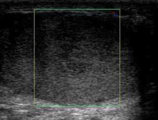
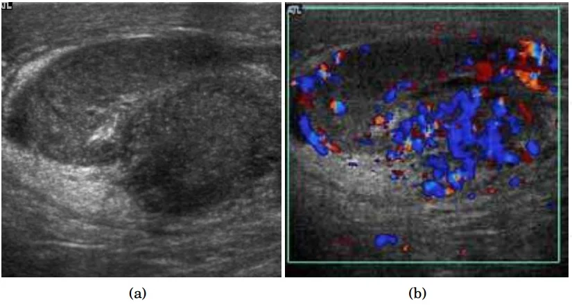
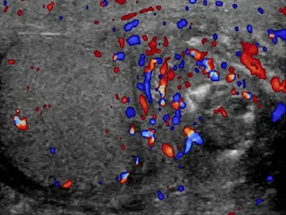
- In testicular torsion, colour Doppler usually shows absent or reduced flow in the testis compared with the other side. In epididymitis it shows hyperaemia of the epididymis and often a reactive hydrocele.
- It can reliably exclude torsion in many cases, but in early or partial torsion some perfusion may be preserved, giving a false-negative result. If you still suspect torsion, explore.[10]
- Ultrasound should be used primarily to rule out torsion when the history and examination are equivocal; it provides little added value in a clinically straightforward epididymitis and a negative study does not change management.[4]
- In partial or intermittent torsion, imaging can mislead; clinical suspicion wins.
- Scrotal ultrasound for a chronic or complex mass determines whether it is cystic or solid and where it sits; a solid intratesticular mass is treated as a tumour until proved otherwise.
- Tumour markers (alpha-fetoprotein, beta-human chorionic gonadotrophin, lactate dehydrogenase) are part of staging once a testicular cancer is suspected, but they do not make the diagnosis.
- Do not biopsy a testicular mass through the scrotum. Transcrotal biopsy can spread tumour in the lymphatics of the scrotum. The diagnosis is made by radical inguinal orchidectomy.[11]
- Do not give antibiotics "just in case" when torsion is possible: they do not treat torsion and may delay surgery.
Approach to acute scrotal pain or swelling
- Acute onset of scrotal pain, swelling or tendernessTreat as emergency until torsion excluded
- Question: Is testicular torsion suspected on history and examination?
Yes (sudden pain, high-riding / transverse testis, absent cremasteric reflex, adolescent)
- Emergency scrotal exploration with detorsion and orchidopexyDo not delay for ultrasound or other tests; manual detorsion if surgery not immediately possible
- Exploration findingsIf the testis is viable, detorse and fix both testes; if not, orchidectomy plus fixation of the contralateral testis
No, but torsion not confidently excluded
- Colour Doppler ultrasound nowAbsent flow or equivocal result with ongoing suspicion -> explore
- If torsion excluded, treat the cause found
Features point to epididymitis
- Exclude torsion firstHigh-riding or transverse testis, absent cremasteric reflex or sudden onset override the diagnosis
- Antibiotics by age and sexual riskSTI regimen or enteric regimen; analgesia, rest, elevation
- Reassess within 72 hoursPersistent swelling suggests tumour, abscess, infarction or tuberculosis
Appendix torsion likely
- Confirm normal testicular flowDoppler if any doubt about torsion
- Conservative managementAnalgesia and rest; resolution usually over days to weeks
Torsion is a time-critical emergency. Testicular viability falls significantly after about six hours from the onset of symptoms, which is why the operation should not wait for imaging and should not be delayed while analgesia is given.[2][9]
- Emergency surgical exploration is the definitive treatment, with detorsion and orchidopexy of both sides. A bell-clapper deformity in which the tunica vaginalis surrounds the testis and part of the cord (incidence up to 12%) allows the testis to twist freely; once one side has torsed, the other is at risk, so both are fixed.[1][2]
- If surgery cannot be performed immediately, manual detorsion may be tried as a temporising measure, turning clockwise on the left and anticlockwise on the right, then explore anyway.[9]
- If the testis is not viable at exploration, orchidectomy is performed and the contralateral testis fixed.
- Analgesia and anti-emetics help, but must not push the patient down a queue: treat as a priority case in triage.
- There is no role for antibiotics, anticoagulation or thrombolysis in acute torsion.
Epididymitis and epididymo-orchitis (infection or inflammation of the epididymis, which can spread to the testis)[3]
- Presumptive antibiotics are started in sexually active men at the first visit, before laboratory results return.[4]
- Cause by age and risk:
- Young, sexually active men: usually Chlamydia trachomatis or Neisseria gonorrhoeae. Regimen: ceftriaxone 500 mg intramuscularly as a single dose plus doxycycline 100 mg by mouth twice daily for 10 days.[4]
- Men who practise insertive anal sex: ceftriaxone 500 mg intramuscularly as a single dose plus levofloxacin 500 mg by mouth once daily for 10 days.[4]
- Older men, or men after prostate biopsy, vasectomy or urinary instrumentation: enteric organisms (for example Escherichia coli). Regimen: levofloxacin 500 mg by mouth once daily for 10 days (monotherapy if gonorrhoea has been excluded).[4]
- For persons weighing 150 kg or more, ceftriaxone 1 g is used.[4]
- Adjuncts: bed rest, scrotal elevation and non-steroidal anti-inflammatory drugs until fever and inflammation subside; complete resolution may take weeks.[4]
- Repeated assessment: if there is no improvement within 3 days (72 hours), rethink the diagnosis and therapy; persistent swelling needs evaluation for abscess, infarction, tumour, tuberculosis or fungal infection.[4]
- Aftercare: abstain from sex until treated, test partners within the previous 60 days, test for HIV and syphilis, and trace contacts.[4]
- Usually in boys aged 7 to 14 years, the commonest cause of acute scrotal pain in prepubertal boys. The appendix (hydatid of Morgagni) is a Müllerian remnant present in about 76 to 83 percent of testes.[5][6]
- Examination shows a tender nodule at the upper pole of the testis and the blue dot sign, a cyanotic spot visible through the scrotal skin over the non-viable appendage.[6]
- Management is conservative (analgesia, rest), because the torsed appendage infarcts and atrophies; surgery is reserved for persistent pain. Ultrasound must first exclude testicular torsion if there is any doubt.[5][6]
- Congenital hydroceles in children usually resolve by one to two years and are repaired if persistent; communicating hydroceles continue to fill.[7]
- In adults, primary hydroceles arise from an imbalance of fluid production and reabsorption; secondary hydroceles follow infection, trauma, surgery or a tumour.
- Management is expectant for a small painless collection; surgery (hydrocelectomy or sclerotherapy) for discomfort, cosmetic concern or a secondary cause. Always look for a testicular tumour in an adult with a new hydrocele.
- Varicoceles occur in about 15 to 20 percent of all men and in about 40 percent of infertile men.[8]
- Most are on the left because the left testicular vein drains into the high-pressure left renal vein.[8]
- A right-sided, sudden-onset or non-decompressing varicocele is a red flag for a retroperitoneal or renal mass.
- Surgical repair in infertile men with abnormal semen improves counts, motility, morphology and pregnancy rates, and repair in adolescents may normalise testis size and sperm parameters.[8][9]
A scrotal swelling that reduces with gentle pressure, gurgles, or changes with coughing is likely bowel in the scrotum. An incarcerated hernia causes sudden pain and a tense, non-transilluminating mass and needs urgent surgery. In general, asymptomatic hernias are repaired electively and incarcerated ones urgently.
- Testicular cancer is the commonest cancer in young men and typically presents as a painless, firm intratesticular lump that does not transilluminate. Back pain, gynaecomastia or metastatic disease can dominate.
- Germ cell tumours make up over 95 percent of cases and over half are seminomas; beta-human chorionic gonadotrophin or lactate dehydrogenase rise in only 10 to 25 percent of seminomas, and alpha-fetoprotein does not rise in pure seminoma.[11]
- Do not delay orchidectomy to stage; radical inguinal orchidectomy is both the diagnosis and the primary treatment. Testis-sparing surgery with frozen section is for small or indeterminate masses in selected men.[11]
Figure 1Scrotal ultrasonography of testicular torsion
Long description
Colour Doppler ultrasound of a testis with no detectable blood flow inside the colour box, the finding in testicular torsion.

Figure 2Scrotal ultrasonography of epididymo-orchitis
Long description
Scrotal ultrasound from an open-access scrotal ultrasonography chapter showing an enlarged heterogeneous epididymal head and adjacent testis in epididymo-orchitis, with arrows marking the structures.

Figure 3Doppler ultrasound of epididymitis
Long description
Doppler ultrasonography of a testicle (left in image) and an epididymis (right in image), the latter swollen with increased blood flow, which are typical findings in epididymitis.

Colour Doppler is the imaging backbone of the acute scrotum, but it is an adjunct.
- What it does well: confirms absent testicular perfusion in torsion, and hyperaemia of the epididymis in epididymitis; separates cystic from solid masses; stages tumours; maps a varicocele.
- Its pitfalls: early or partial torsion can retain some flow, so a normal study does not rule out torsion in the first hours. If the story and signs point to torsion, operate.[10]
- Do not let imaging delay surgery. The indication for exploration is clinical suspicion; ultrasound is done on the way if, and only if, time allows.
Many of the steps above assume a laboratory and an ultrasound. Where these are not available:
- Suspect torsion clinically. A young man with sudden severe pain, a high testis with horizontal lie and loss of the cremasteric reflex needs urgent surgery whether or not ultrasound is available. Imaging is helpful but never a precondition for exploration.[2][9]
- Experienced clinicians can perform colour Doppler if it exists; a single absent or reduced-flow study in a strongly suspicious case should lead to exploration.
- Presumptive antibiotics for epididymitis can be chosen by age and risk without NAAT where tests are unavailable, and azithromycin or doxycycline with a single ceftriaxone dose is widely available. Urine microscopy and culture are still worth obtaining.
- Testicular salvage is poor once presentation is delayed. Series from low-resource settings report that many torsed testes needed orchidectomy, and presentation after 48 hours was associated with much higher odds of orchidectomy than presentation within 12 hours (exact figures vary by series).[12]
- Sensitise young men and families to the symptoms of torsion and to seek care immediately.
- Hydrocele and varicocele surgery is often available at district level; do not let a hydrocoele mask a tumour, palpate carefully.
- Refer early to a centre with surgical capability, because testicular fixation or orchidectomy needs an operating theatre.
- Send every excised testis or mass for histology if pathology is available.
- Neonates. Both extravaginal and (rarely) intravaginal torsion occur, as does torsion of the appendix testis. An acute neonatal scrotum still warrants urgent surgical assessment.
- Children. Appendix torsion is the commonest cause of acute scrotal pain; still exclude testicular torsion.
- Older men. Think of tumour and of enteric epididymitis from bladder outlet obstruction; a new hydrocele in an adult deserves careful testicular examination.
- Pregnancy partners. Epididymitis is not dangerous to a pregnancy in itself, but treat the sexually transmitted infection and trace partners.
- Scrotal swelling is common; testicular torsion is uncommon but the one diagnosis that must not be missed.
- Torsion presents with sudden severe pain, a high-riding, transversely lying testis, an absent cremasteric reflex and nausea; it peaks in adolescents but occurs at any age.
- Testicular viability falls markedly after about six hours, so exploration should not wait for imaging.[2]
- A bell-clapper deformity is found in up to 12 percent of testes and fixes both sides at risk.[1]
- Detorsion, then bilateral orchidopexy; orchidectomy plus contralateral fixation if the testis is dead.
- Epididymitis is the usual mimic. Young men get chlamydia and gonorrhoea; older men get enteric organisms.[4]
- First-line regimens are ceftriaxone plus doxycycline for sexually transmitted infection and levofloxacin for enteric infection, given for ten days.[4]
- Prehn sign is unreliable and must not be used to exclude torsion.
- Torsion of the appendix testis is the commonest acute scrotal pain in prepubertal boys, shows a blue dot, and is managed conservatively after real torsion is excluded.[5][6]
- Hydrocele transilluminates and is usually painless; it is secondary until proved otherwise in an adult.
- Varicocele is usually left-sided and can impair fertility; a right-sided or sudden varicocele needs investigation for a retroperitoneal mass.
- A firm intratesticular mass is cancer until proved otherwise; radical inguinal orchidectomy, never scrotal biopsy.[11]
- Colour Doppler is essential but can be falsely reassuring in early or partial torsion.
- In low-resource settings, clinical diagnosis, early exploration and sensitisation save testes.
Answer each one in your head first, then open the answer.
A 16-year-old wakes at 03:00 with sudden severe pain in the left testis. The testis is high and lies horizontally, and the cremasteric reflex is absent. The emergency department can arrange an ultrasound in 45 minutes. What should happen next, and why?
Show answerHide answer
Emergency scrotal exploration now. The story and signs are classic for testicular torsion; testicular viability falls markedly after about six hours, and ultrasound should not delay surgery. Manual detorsion may be attempted if the theatre is not immediately available.[2]
How does the bell-clapper deformity cause torsion, and why are both testes fixed at operation?
Show answerHide answer
The tunica vaginalis surrounds the testis and part of the spermatic cord, so the testis is not fixed to the scrotal wall and can twist freely (incidence up to 12 percent). Both sides share the same anatomy, so the contralateral testis is at risk and is fixed at the same operation.[1]
A 24-year-old man has had three days of gradually worsening unilateral scrotal pain. He has urethral discharge. What is the diagnosis and most appropriate first-line treatment?
Show answerHide answer
Acute epididymitis, probably sexually transmitted. Empiric ceftriaxone 500 mg intramuscularly once plus doxycycline 100 mg by mouth twice daily for ten days, with testing of the man and his partners for HIV and syphilis and abstinence until cleared.[4]
A 9-year-old boy develops sudden mild pain at the top of the testis. Examination shows a blue spot on the scrotal skin and a normal testicular position; colour Doppler shows normal testicular flow. What is likely and how is it managed?
A 40-year-old man has a large painless swelling that brightly transilluminates. What are the likely cause and the important caveat?
Show answerHide answer
A hydrocele. It often transilluminates because it is fluid. In an adult a new hydrocele must be examined carefully, because it can be secondary to a testicular tumour, infection or trauma.
A man has a left-sided bag of worms that decompresses on lying down and reduces after a warm shower. He and his partner are trying to conceive. What is the condition and when might treatment help?
Show answerHide answer
A varicocele. It is more common on the left because the left testicular vein drains into the left renal vein. Surgical repair may help semen parameters and fertility when the couple is infertile with abnormal semen.[8]
A 28-year-old notices a firm, non-tender lump inside the testis that does not transilluminate. What is the correct first step and what must not be done?
Show answerHide answer
Urgent urology referral for radical inguinal orchidectomy, which is diagnostic and therapeutic. Do not biopsy the mass through the scrotum.[11]
Why can colour Doppler ultrasound miss testicular torsion in the first few hours?
Show answerHide answer
In early or partial (incomplete) torsion some arterial perfusion may still be present, so the scan can look normal. The decision to explore rests on clinical suspicion, not on the scan alone.[10]
- Acute scrotum and urological infection topics elsewhere in this library, for example male infertility, testicular cancer, inguinal hernia repair, chronic pelvic pain and tropical infections.
- Surgical topics such as scrotal exploration and hydrocelectomy as they become available.
End of topic.
- Velasquez J, Boniface MP, Mohseni M. Acute Scrotum Pain. StatPearls [Internet]. Treasure Island (FL): StatPearls Publishing; 2023.. Accessed .
- Schick MA, Sternard BT. Testicular Torsion. StatPearls [Internet]. Treasure Island (FL): StatPearls Publishing; 2023.. Accessed .
- Rupp TJ, Leslie SW. Epididymitis. StatPearls [Internet]. Treasure Island (FL): StatPearls Publishing; 2023.. Accessed .
- Centers for Disease Control and Prevention. Sexually Transmitted Infections Treatment Guidelines, 2021. Epididymitis. CDC. Atlanta (GA); last reviewed 2021-07-22.. Accessed .
- Mo MB, Leslie SW. Appendix Testis Torsion. StatPearls [Internet]. Treasure Island (FL): StatPearls Publishing; 2026.. Accessed .
- Krishnan A, Rich MA, Swana HS. Torsion of the Appendix Testis in a Neonate. Case Reports in Urology 2016;2016:9183196. doi:10.1155/2016/9183196. Accessed .
- Conjeevaram R, Huzaifa M, Leslie SW. Hydrocele. StatPearls [Internet]. Treasure Island (FL): StatPearls Publishing; 2026.. Accessed .
- Leslie SW, Sajjad H, Siref LE. Varicocele. StatPearls [Internet]. Treasure Island (FL): StatPearls Publishing; 2023.. Accessed .
- Gordhan CG, Sadeghi-Nejad H. Scrotal pain: Evaluation and management. Korean Journal of Urology 2015;56(1):3-11. doi:10.4111/kju.2015.56.1.3. Accessed .
- Agrawal AM, Tripathi PS, Shankhwar A, Naveen C. Role of ultrasound with color Doppler in acute scrotum management. Journal of Family Medicine and Primary Care 2014;3(4):409-412. doi:10.4103/2249-4863.148130. Accessed .
- Okoye E, Hammoud A, Leslie SW, Saikali SW. Radical Orchiectomy and Testis-Sparing Surgery for Testicular Neoplasms. StatPearls [Internet]. Treasure Island (FL): StatPearls Publishing; 2025.. Accessed .
- Mohamed MA, Abraha D, Olasinde AA, Kiswezi A, Molen SF, Muhumuza J, Lule H. Testicular salvageability and its predictors among patients with testicular torsion in a resource limited setting: a multicentre longitudinal study. BMC Surgery 2023;23:248. doi:10.1186/s12893-023-02118-z. Accessed .
Figure credits
- Figure 1: Chee-Wai Mak and Wen-Sheng Tzeng, via Wikimedia Commons, CC BY 3.0, CC BY 3.0.
- Figure 2: Chee-Wai Mak and Wen-Sheng Tzeng, via Wikimedia Commons, CC BY 3.0, CC BY 3.0.
- Figure 3: Alfredo D’Andrea, Francesco Coppolino, Elviro Cesarano, Anna Russo, Salvatore Cappabianca, Eugenio Annibale Genovese, Paolo Fonio and Luca Macarini, via Wikimedia Commons, CC BY 2.5, CC BY 2.5.
Educational content for medical students and doctors. It is not a substitute for clinical judgement or local guidelines.