Acute abdomen and intestinal obstruction
Approach to the acute abdomen: peritonitis, obstruction, perforation, mesenteric ischaemia, work-up, imaging, resuscitation and when to operate.
- An acute abdomen is a clinical syndrome, not a diagnosis: sudden, severe abdominal pain that often brings a patient to the emergency department, sometimes with peritonitis, vomiting, fever or shock.
- Between 5% and 12% of patients presenting to an emergency department have acute abdominal pain, and 15% to 20% of them need an intervention or operation; emergency abdominal surgery still carries a mortality of 2% to 12%.[1]
- The first job is to decide whether the patient is unstable, has peritonitis or a suspected catastrophe (obstruction, perforation, mesenteric ischaemia, ruptured aneurysm or ectopic pregnancy) and to act at the same time as investigating.
- Pain character, exact site and its change over time, together with peritoneal signs, place the diagnosis before any scan is done.
- Contrast-enhanced computed tomography (CT) is the emergency imaging study of highest sensitivity and specificity; plain films and ultrasound each have a narrower, well-defined role.
By the end of this page you should be able to:
- Define the acute abdomen and list its common surgical and non-surgical causes.
- Interpret abdominal pain by its character, site, onset and progression, and recognise peritonitis.
- Take a history and perform an examination that reliably detects a surgical abdomen.
- Choose and interpret the appropriate first-line and confirmatory investigations, including plain film, ultrasound and CT.
- Resuscitate an unstable patient and list the indications for emergency laparotomy.
- Recognise small- and large-bowel obstruction, perforation with free air, and mesenteric ischaemia, and outline their initial management.
DefinitionAcute abdomen
A clinical syndrome of severe abdominal pain of sudden onset, possibly with guarding (involuntary contraction of the abdominal wall to limit pain), reduced general well-being and, at times, circulatory collapse or shock. It is not a single diagnosis. The spectrum runs from a subacute abdomen to a fulminant acute abdomen.[1]
An acute abdomen carries a mortality of 2% to 12%, which rises with every hour of delay until effective treatment is given.[1] Five to twelve percent of emergency department attendees have acute abdominal pain, up to one-third of whom need emergency treatment.[1] Patients should be triaged as high priority (Emergency Severity Index 1 or 2) and seen by a physician within 10 minutes, with surgical consultation sought within 60 minutes when a surgical cause is possible.[1]
This page covers the adult non-trauma presentation. Traumatic and obstetric emergencies are handled elsewhere; the acute abdomen can, however, be mimicked by purely medical conditions such as diabetic ketoacidosis, hypercalcaemia, Addison disease and sickle cell crisis.[2]
The commonest categories among patients presenting with acute abdominal pain, with the proportions reported in one large review, are: nonspecific abdominal pain (30% to 41%), acute appendicitis (8% to 30%), acute cholecystitis (9% to 11%), ileus (4% to 5%), gynaecological causes (1% to 4%), acute pancreatitis (2% to 4%), urological causes (2% to 4%), peptic ulcer including perforation (1% to 3%), oncological causes (1% to 2%) and diverticulitis with or without perforation (1% to 2%). Abdominal ischaemia accounts for 0% to 1%.[1]
| Group | Typical conditions |
|---|---|
| Gastrointestinal (surgical) | Appendicitis, cholecystitis, cholangitis, pancreatitis, perforated peptic ulcer, diverticulitis, bowel obstruction, volvulus, intussusception, incarcerated hernia, mesenteric ischaemia, splenic or hepatic injury, abscess |
| Gynaecological | Ectopic pregnancy, ovarian torsion, pelvic inflammatory disease, ruptured cyst or abscess, degenerating fibroid |
| Urological | Ureteric colic, pyelonephritis, urinary retention, papillary necrosis |
| Vascular | Ruptured abdominal aortic aneurysm or dissection, mesenteric ischaemia, splenic infarction |
| Medical and metabolic mimics | Diabetic ketoacidosis, hypercalcaemia, Addison disease, sickle cell crisis, uremia, lead poisoning, acute intermittent porphyria, hereditary angioedema, methanol toxicity |
| Thoracic (referred) | Myocardial infarction, pulmonary embolism, aortic dissection, basal pneumonia or pleurisy, oesophageal rupture |
Cancers of the colon, stomach, pancreas and ovary present more often subtly in older adults, and a condition that appears "medical" at first can still be a surgical abdomen.
| Region | Common causes |
|---|---|
| Right upper quadrant | Acute cholecystitis, cholangitis, hepatitis, liver abscess, right lower lobe pneumonia or pleurisy, pyelonephritis, herpes zoster, peptic ulcer |
| Left upper quadrant | Acute pancreatitis, splenic infarction or rupture, gastric or duodenal ulcer, colonic (splenic flexure) diverticulitis, left lower lobe pneumonia, renal colic |
| Right lower quadrant | Appendicitis, terminal ileitis or Crohn disease, caecal diverticulitis, right-sided colonic cancer, ectopic pregnancy, ovarian torsion, renal or ureteric colic, incarcerated hernia, psoas abscess |
| Left lower quadrant | Sigmoid diverticulitis, colitis, left-sided colonic cancer or obstruction, ectopic pregnancy, ovarian torsion or cyst, renal/ureteric colic, incarcerated hernia |
| Central or periumbilical | Early appendicitis, small-bowel obstruction or ischaemia, mesenteric adenitis, gynaecological causes, aortic aneurysm |
| Epigastrium | Peptic ulcer (including perforation), pancreatitis, biliary colic, myocardial ischaemia, oesophageal or aortic disease |
| Back or flank | Renal or ureteric colic, pyelonephritis, abdominal aortic aneurysm, pancreatitis (boring through to the back), musculoskeletal pain |
Pain should be understood from what is happening to the organ. Visceral pain comes from hollow organs and capsules: it is dull, deep, diffuse and hard to point to, because the viscera are poorly localised. It is triggered by distension, spasm or sudden tension on a capsule (biliary colic, ureteric colic, liver capsule stretch). Somatic pain, from the parietal peritoneum, is sharp, burning and well localised; it follows inflammation or injury (appendicitis spreading to the peritoneum, perforation, peritonitis).[2]
Site often mirrors embryological origin, at least early in the illness:[2]
- foregut (stomach, pancreas, liver, biliary system, proximal duodenum) - epigastrium;
- midgut (small bowel, appendix, proximal colon) - periumbilical region;
- hindgut and pelvis (distal colon, bladder, pelvic organs) - suprapubic region;
- retroperitoneal structures (aorta, kidneys) - back pain.
Character that usually matters:
- Colicky, intermittent pain suggests a hollow tube trying to push contents past a blockage: small- or large-bowel obstruction, ureteric or biliary colic.
- Steady, constant pain suggests inflammation or ischaemia of a solid organ or wall (appendicitis, cholecystitis, pancreatitis, mesenteric ischaemia).
- Pain that reaches maximal intensity within seconds to a minute raises the possibility of a catastrophe: perforated viscus, ruptured aneurysm or dissection, torsion, volvulus or mesenteric ischaemia. In older adults a perforated ulcer is reported to begin abruptly in only about 47% of cases, so onset alone cannot rule one out.[2]
- Pain that wakes the patient from sleep should be treated as serious until proven otherwise.[2]
- Pain that worsens with movement, coughing or bumps in the road implies peritoneal irritation.[2]
- Pain relieved by eating points to duodenal ulcer; pain exacerbated by eating to gastric ulcer; pain precipitated by fatty food to biliary disease; post-prandial pain to intestinal angina from mesenteric ischaemia.[2]
Severity alone is unreliable, especially in older patients who may under-report pain. A useful rule of thumb is that serious causes present early; benign causes declare themselves and then improve. Gallbladder pain averages 5 to 16 hours and almost never lasts less than 1 hour, while small-bowel obstruction classically changes from intermittent colic to constant pain as distension develops.[2]
Referred pain is common and predictable: diaphragmatic irritation (free blood, pleurisy, subphrenic abscess) can produce shoulder pain; biliary disease can refer to the right scapula. Always look for a chest or retroperitoneal cause when the abdomen "should" hurt but the examination is unrevealing.[2]
Peritonitis is inflammation of the peritoneum. It may be localised, around one organ, or generalised when pus, blood, intestinal contents or urine have spread throughout the cavity. Peritoneal irritation is the central physical sign of a surgical abdomen.
Signs to elicit: guarding (increased muscular tone that does not relax on inspiration, unlike the "voluntary guarding" of an anxious patient), rebound tenderness (pain worse on sudden release), rigidity (board-like), pain on coughing or heel-drop, percussion tenderness ("pain on vibration") and percussion rebound. Indirect tests such as the cough test have a sensitivity of about 78% and specificity about 79% for peritonitis in one prospective study, while traditional rebound testing has sensitivity near 80% but specificity of only 40% to 50%, so negative peritoneal signs never exclude disease, particularly in older adults.[2]
Other supporting clues: absent bowel sounds later in the illness (high-pitched tinkling early in obstruction), fever, tachypnoea, tachycardia, hypotension, altered mental state in the elderly, and free air on imaging. Guarding may be absent in older adults because of laxity of the abdominal wall; only about 21% of patients over 70 with a perforated ulcer have epigastric rigidity.[2]
Red flagPeritonitis is an indication to operate
Generalised peritonitis from perforation, strangulated obstruction or mesenteric infarction does not improve with antibiotics alone. The patient needs resuscitation and surgical source control. Delay in abdominal sepsis raises mortality from up to 26% to as high as 43%.[1]
- An "ill-looking" patient with abdominal pain, especially an older adult, whose mild complaints hide serious disease.
- Abdominal pain with unexplained hypotension or shock (bleeding, third spacing, sepsis, ruptured aneurysm).
- Pain out of proportion to the physical findings (classic, though now less common, for early mesenteric ischaemia).
- An acute onset reaching peak intensity within seconds.
- Guarding, rigidity or rebound tenderness.
- Free air on an upright chest film or CT.
- Abdominal pain in a pregnant, immunocompromised or neutropenic patient, in whom signs may be muted.
The history is the strongest diagnostic tool: a careful history and physical examination reach a correct diagnosis with about 88% sensitivity and 41% specificity, often reducing unnecessary testing.[1] Ask in a way that lets the story unfold, then fill gaps with direct questions.
- Where exactly is the pain, and has it moved? Migration (for example from periumbilical to right-lower-quadrant pain) is characteristic of appendicitis.
- What does it feel like (colicky, burning, gnawing, sharp) and is it constant or comes in waves?[2]
- When did it start and how fast did it peak? Sudden versus gradual, and the time of onset, separates a catastrophe from an inflammatory process.[2]
- How bad is it now and has it changed? Persistent worsening pain is worrisome; improving pain is favourable.[2]
- Does it go anywhere else (shoulder, back, scrotum, groin)?
- What makes it worse or better (movement, cough, food, position, antacids, defecation)?
- Associated symptoms: fever, anorexia, nausea, vomiting (pain usually precedes vomiting in surgical conditions), diarrhoea, constipation, inability to pass flatus, jaundice, dysuria, haematuria, vaginal bleeding, syncope, chest pain or breathlessness.[2]
- Past history: prior abdominal or gynaecological surgery (adhesions), peptic ulcer, inflammatory bowel disease, pancreatitis, alcohol, smoking, vascular disease, diabetes, medications (non-steroidal anti-inflammatory drugs, steroids, anticoagulants, narcotics), allergies and last menstrual period.
- Social history: alcohol, cocaine (intestinal as well as cardiac ischaemia), domestic violence in trauma.
- In women of childbearing age, a pregnancy test is mandatory even with a normal menstrual history, because pregnancy can be present and complicates diagnosis.[2]
An important trap: diarrhoea does not exclude a surgical diagnosis. It occurs in about one-fifth of colonic obstruction, in mesenteric ischaemia, in appendicitis and in early small-bowel obstruction as the distal bowel empties. Absence of flatus is a more reliable sign of obstruction than constipation, because gas is cleared faster than fluid.[2]
Begin with the patient's general appearance, position, breathing pattern and speech. An "ill-looking" adult with abdominal pain is a serious patient; conversely, a calm elderly person may still have a perforation, because ageing blunts signs.
Vital signs: fever (absent in more than 30% of appendicitis and most cholecystitis), tachypnoea (chest disease or metabolic acidosis from ischaemic bowel or diabetic ketoacidosis), tachycardia and hypotension.[2] With hypotension and no obvious blood loss, think third spacing, which can shift enormous volumes into the bowel lumen or peritoneum in obstruction or other catastrophes.[2]
- Inspect for scars, distension, skin changes (herpes zoster, caput medusae), Cullen (periumbilical) and Grey Turner (flank) bruising, and hernias; inspect the patient standing if no hernia is found supine.[2]
- Auscultate last and only briefly: prolonged listening is a poor use of time. High-pitched tinkling suggests early small-bowel obstruction; silence suggests ileus or late catastrophe.[2]
- Percuss to distinguish gaseous distension (drum-like tympany, as in large-bowel obstruction) from ascites, and to elicit pain on vibration.[2]
- Palpate gently from non-tender areas towards the painful area with the knees and hips flexed. Localised tenderness is a reliable pointer to the organ; generalised tenderness is harder to interpret. Always reconsider appendicitis in any patient with right-lower-quadrant tenderness who still has an appendix.[2]
- Assess for an expansile, tender pulsatile mass (abdominal aortic aneurysm); unequal femoral pulses suggest dissection.[2]
- Pelvic and rectal examination as indicated: a tender pelvic mass, cervical excitation or blood; rectal examination is of limited value but may detect ischaemia, an obstructing rectal mass, high prostate disease, or blood. It adds little in children.[2]
- Genital and hernial orifices: in men, the testes (torsion can present as abdominal pain); in women with lower abdominal pain, a pelvic examination is usually necessary.[2]
- Adjacent areas: back for costovertebral-angle and spinal tenderness, chest and heart (virtually any chest disease can present with abdominal pain), groins for hernias, and a full neurological assessment when spinal disease is suspected.[2]
- Rebound and cough tests as above; gentle percussion is kinder and at least as informative.
- Guarding and rigidity as above.
- Carnett sign (tenderness worse with the abdominal wall tensed) suggests an abdominal-wall source such as haematoma.
- Murphy sign (inspiratory arrest on right-upper-quadrant palpation) points to cholecystitis, with sensitivity about 65%.[2]
- Psoas and obturator signs indicate an inflammatory process adjacent to those muscles (appendicitis, pelvic abscess, psoas abscess, pancreatitis). They are sensitive only 15% to 35% but quite specific (85% to 95%) for appendicitis.[2]
- Rovsing sign (right-lower-quadrant pain on left-sided pressure and release) is another indirect marker of appendicitis.
Investigations run in parallel with resuscitation. All tests have false-negative rates; a negative result never excludes a high-probability diagnosis.[2]
- Arterial or venous blood gas: acid-base status, anion gap, lactate, glucose and electrolytes. Lactate is raised in bleeding or ischaemia.[1]
- Full blood count: anaemia, leukocytosis (but the count may be normal in serious infection).[2]
- C-reactive protein and procalcitonin: inflammatory markers. Procalcitonin has been reported as 75% sensitive and 100% specific for the need for surgical treatment. C-reactive protein above 75 mg/L and white cell counts above 10,000 per mm3 can suggest peritoneal inflammation, though neither is sensitive nor specific on its own.[1][3]
- Amylase and lipase: lipase is preferred for pancreatitis.[1]
- Urea and electrolytes, creatinine: dehydration, renal impairment, vomiting alkalosis, hypokalaemia.
- Liver function tests and bilirubin, alkaline phosphatase and gamma-glutamyltransferase: biliary and hepatic disease.
- Coagulation profile and group and save: potential transfusion.
- Urine dipstick and culture and serum or urine pregnancy test in women who could be pregnant.
- Troponin if cardiac ischaemia is suspected.
Figure 1Free air2010
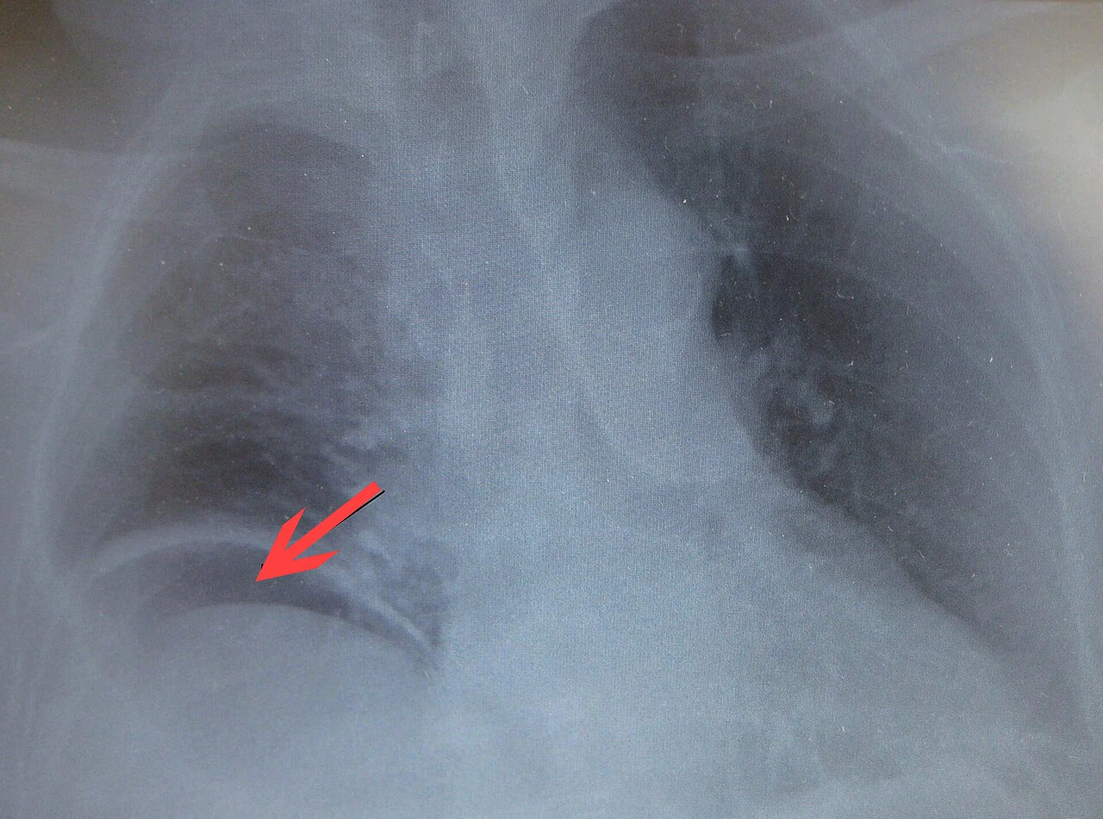
Long description
Free air under the right hemidiaphragm from a bowel perforated.

Plain abdominal films are of limited but real value.[2] An erect chest (or left lateral decubitus abdominal) film is the classic test for free air under the diaphragm, and may show air-fluid levels in obstruction; calcified aortic aneurysm or radio-opaque stones. A standard upright film will not show free air in up to 40% of perforated ulcers, and plain films in general have low sensitivity (47% to 56%) for tiny amounts of free air.[1][2] Plain films are also insensitive for the early signs of strangulation and do not distinguish the causes of obstruction. Many authors would abandon them in the work-up of adult acute abdominal pain, but they remain a pragmatic test for perforation and high-grade obstruction when CT is unavailable or delayed.[1][2]
Ultrasound is the imaging study of first choice for the acute abdomen: widely available, rapid, bedside, radiation-free and especially useful in pregnancy.[1] Combined with history and examination it gives the correct diagnosis in 53% to 83% of cases, and classic causes such as cholecystitis, appendicitis and sigmoid diverticulitis are diagnosed reliably in up to 70% of cases.[1] Focused assessment with sonography for trauma (FAST) is a rapid, point-of-care screen for free fluid, haemoperitoneum and aortic aneurysm, and is of greatest value in the unstable patient.[1]
Limitations: obesity, gaseous distension, free intraperitoneal air, an uncooperative or very tender patient, and operator dependence.
Contrast-enhanced abdominal CT has the highest sensitivity and specificity of all imaging for the acute abdomen, and intravenous contrast should generally be given; native (non-contrast) CT is roughly 30% less accurate.[1] In acute appendicitis, CT sensitivity and specificity are about 91% and 90%, versus 78% and 83% for ultrasound.[1] For mesenteric ischaemia, CT angiography is the study of choice (see below). CT also guides drainage of abscesses and can reveal the site and cause of a perforation. CT is not needed in every patient, but it is indispensable when the diagnosis is uncertain, the patient is unstable, or an intra-abdominal catastrophe cannot be excluded clinically. Oral contrast rarely helps in acute disease and delays imaging; it is reserved for situations such as assessing intestinal peristalsis or a rectovesical fistula.[1]
MRI has a smaller role because of availability and scan time. It is useful in a stable pregnant patient to limit fetal radiation exposure, usually without contrast, and nursing mothers may safely undergo iodinated or macrocyclic gadolinium imaging with breastfeeding continued.[1]
When history, examination, laboratory and imaging leave an acute abdominal problem undiagnosed (about a quarter of cases), diagnostic laparoscopy has 75% to 100% sensitivity, specificity and accuracy, permits treatment in more than 75% of cases and reduces unnecessary laparotomy.[1] It is contraindicated in diaphragmatic rupture and in the haemodynamically unstable patient.
Resuscitation runs in parallel with diagnosis, and should never be deferred to await imaging.[2]
- Place the patient on a cardiac monitor, obtain large-bore intravenous access and resuscitate with crystalloid (with blood products if there is haemorrhage), correcting electrolyte abnormalities.
- Give adequate analgesia early, including opioids; analgesia does not obscure diagnosis or examination.[2]
- Prescribe antiemetics, keep the patient nil by mouth, and insert a nasogastric tube for vomiting or obstruction.
- Give broad-spectrum antibiotics covering Gram-negative enteric organisms when infection, peritoneal soiling or sepsis is suspected; cover anaerobes for colonic or perforated sources.[2][4]
- Consider venous thromboembolism prophylaxis once bleeding is controlled.
- Take blood for typing and cross-match in anyone who might need transfusion.[1]
- Monitor urine output, lactate and acid-base status; repeat examination is invaluable because the abdomen changes over time.[2]
Analgesia is part of good care, not an enemy of diagnosis.
There is no universal list, but the following situations demand operative source control without waiting for complete certainty:
- generalised peritonitis;
- strangulated or ischaemic bowel, including closed-loop obstruction;
- proven or strongly suspected perforation of a viscus (free air with peritonitis);
- obstruction that fails to resolve, or any obstruction in which strangulation cannot be excluded;
- intra-abdominal haemorrhage that is not controlled (ruptured aneurysm, splenic or hepatic injury, ruptured ectopic pregnancy);
- mesenteric ischaemia with peritonitis or non-viable bowel;
- acute abdomen in a patient who is deteriorating despite resuscitation and treatment;
- diagnostic uncertainty after appropriate imaging, where laparoscopy or laparotomy is both diagnostic and therapeutic.[1][4]
In mesenteric arterial occlusion, exploratory laparotomy may be needed both to revascularise and to resect non-viable bowel.[4]
Approach to a patient with an acute abdomen
- Arrival and triageAssess airway, breathing, circulation; assign high priority (ESI 1 or 2); monitor; large-bore intravenous access; resuscitate; control pain
- Brief history and examinationPain: site, character, onset, radiation, progression; prior surgery; drugs; pregnancy; vital signs; peritoneal signs; hernias; FAST or bedside ultrasound
- Question: Is the patient unstable, shocked, or peritonitic?
Yes
- Resuscitate and investigate in parallelElectrocardiogram, blood tests, bedside ultrasound; CT only if it will not delay life-saving care
- Call the surgeon earlyConsider direct transfer to theatre for ruptured aneurysm, uncontrolled bleeding, perforation with peritonitis, strangulation or AMI
No
- Question: Is the diagnosis clear clinically?
Yes
- Targeted treatmentAntibiotics, fluids, drainage, endoscopy or surgery as indicated; repeated reassessment
No or uncertain
- First imagingBedside ultrasound first; then contrast-enhanced CT (or CT angiography if AMI suspected). Plain film for free air or high-grade obstruction
- Reassess after resultsIf still uncertain, observe with serial examination, repeat imaging, or diagnostic laparoscopy
- Definitive treatment and reassessmentSource control (operation, drainage or antibiotics), organ support, senior review, and re-examination; document bowel viability and length if operated
Obstruction is a mechanical or functional block to the passage of contents. Ileus (mechanical or functional obstruction) accounts for 4% to 5% of acute abdominal pain presentations.[1] Adhesions from previous surgery are the leading cause of small-bowel obstruction, accounting for about 60% of cases; the rest are mostly incarcerated hernias, malignant or benign tumours, volvulus and, less often, Crohn strictures, bezoars, gallstone ileus and intussusception.[3] Large-bowel obstruction is more often malignant and, in the caecum, may be caused by a closed-loop volvulus.
Ileus (paralytic ileus) is a functional failure of propulsion without a mechanical obstruction; it is a differential, not a cause, of obstruction, and sounds are diminished or absent rather than high-pitched.
Classic features: colicky pain, vomiting, distension and constipation or inability to pass flatus. Vomiting appears early in proximal obstruction and may be absent in large-bowel obstruction or partial small-bowel obstruction. Watery diarrhoea can occur in incomplete obstruction. Pain becomes constant when strangulation or distension supervenes. Sensitivity of physical examination for strangulation is only about 48%, even in experienced hands, so vigilance is required.[3]
Investigation: plain film may show air-fluid levels, dilated small-bowel loops and absent colonic gas, but sensitivity is only about 70% and signs of strangulation are missed early. CT (often with water-soluble contrast) is the preferred imaging, with roughly 90% accuracy in predicting strangulation and the need for urgent surgery, and helps distinguish causes.[3] Useful pointers to peritoneal inflammation are C-reactive protein above 75 mg/L and a white cell count above 10,000 per mm3, although both are insensitive and nonspecific.[3]
Management principles (adhesive small-bowel obstruction, which is the commonest variety):[3]
- if there are signs of peritonitis, strangulation or ischaemia, operate;
- otherwise start a trial of non-operative management (nil by mouth, nasogastric or long-tube decompression, intravenous fluids and electrolyte correction), which succeeds in about 70% to 90% of patients; [3]
- most authors consider 72 hours a safe and appropriate limit for a non-operative trial, with surgery sooner for deterioration; [3]
- surgery is required in an estimated 20% to 30% of cases;
- a water-soluble contrast study (with a film at 24 hours) predicts the need for surgery and may shorten stay; [3]
- laparotomy or, in selected simple adhesions, laparoscopy, with careful assessment of bowel viability; temporary closure and planned re-look in doubtful cases.
In children the causes are different (atresia, intussusception, malrotation with midgut volvulus, incarcerated hernia, Meckel diverticulum). In pregnancy, use ultrasound and MRI first to avoid radiation, and manage as a team.
Free intraperitoneal air usually means a perforated hollow viscus (peptic ulcer, diverticulitis, appendicitis, obstruction with ischaemia or trauma). It may be detected on an erect chest radiograph, left lateral decubitus film, or CT. CT is far more sensitive and can locate the perforation and its cause; a plain film misses many small collections (see above).[1][2]
Perforation in the presence of peritonitis mandates operation. Even without peritonitis, free air with a surgical cause (diverticular perforation, perforated ulcer with an ongoing leak, or perforation complicating obstruction or cancer) usually needs source control, although a few sealed perforations can be observed with antibiotics. The decision is clinical and repeated, not a single scan.
A chest radiograph is also worth taking in the febrile, unwell patient with abdominal pain, because basal pneumonia can masquerade as an acute abdomen.[2]
Mesenteric ischaemia means inadequate blood supply to the bowel, which progresses from mucosal injury (3 to 6 hours) to full-thickness infarction. It is uncommon, estimated at 0.09% to 0.2% of all acute surgical admissions, and the incidence rises steeply with age; in patients 75 years or older it is a more common cause of acute abdomen than appendicitis, and the incidence in an 80-year-old is roughly ten times that in a 60-year-old.[4] Mortality exceeds 50% overall, and can be higher for arterial occlusion and for non-occlusive disease.[4] Significant delay is lethal: mortality can rise to over 90% within 12 hours, and every 6 hours of delay roughly doubles mortality.[1][4]
Aetiology (acute mesenteric ischaemia): mesenteric arterial embolism about 50% (often from atrial fibrillation, left ventricular thrombus, endocarditis), mesenteric arterial thrombosis 15% to 25% (atherosclerosis, often with a history of post-prandial pain and weight loss), non-occlusive ischaemia about 20% to 25% (low flow, vasopressors, cardiac failure), and mesenteric venous thrombosis less than 10%.[4]
Presentation and bedside clues:
- sudden, severe, poorly localised pain that is "out of proportion" to a relatively quiet abdomen: the classic, though now less common, presentation;
- risk factors of atrial fibrillation, recent myocardial infarction, valvular disease, diffuse atherosclerosis, heart failure and shock (especially when vasopressors are used);
- gastrointestinal symptoms: nausea (about 44%), vomiting (about 35%), diarrhoea (about 35%), blood per rectum (about 16%); about one-third of patients have the triad of abdominal pain, fever and occult blood; [4]
- signs of peritonitis once infarction has occurred.[4]
Diagnosis: suspicion, not a single test. Plain radiograph is not recommended and a negative film does not exclude the diagnosis. Laboratory tests are nonspecific but supportive: leukocytosis in more than 90% of cases, metabolic acidosis with elevated lactate in about 88%; a lactate level above 2 mmol/L is associated with irreversible ischaemia, and raised D-dimer (above 0.9 mg/L) has a specificity of about 82%, sensitivity of about 60% and accuracy of about 79%. Elevated amylase occurs in roughly half of patients and can mislead toward pancreatitis. CT angiography is the study of choice, with sensitivity about 93%, specificity 100%, positive predictive value 100% and negative predictive value 94%; it should be performed without delay, even in the presence of renal impairment, because a missed diagnosis is worse.[4]
Initial measures (revascularise early):[4]
- aggressive fluid resuscitation with crystalloid and blood products, correcting acidosis and electrolytes (hyperkalaemia may be severe), with haemodynamic monitoring;
- nasogastric decompression;
- broad-spectrum antibiotics immediately, and for at least 4 days in immunocompetent stable patients; [4]
- anticoagulation with intravenous unfractionated heparin, unless there is a clear contraindication;
- use vasopressors cautiously (they can worsen splanchnic perfusion);
- if there is peritonitis or non-viable bowel, operate without delay to re-establish perfusion and resect necrotic bowel, using damage-control principles, and reassess viability at a planned second look;
- endovascular revascularisation (aspiration embolectomy, stenting) is increasingly first-line where expertise and anatomy permit, especially in the absence of peritonitis.
The threshold for imaging should be low: in an elderly patient with vascular risk factors and severe pain, a pain-free interval followed 3 to 6 hours later by deterioration is itself a warning.[1]
- Children: causes are different (atresia, malrotation with volvulus, intussusception, incarcerated hernia, Meckel diverticulum); midgut volvulus is a surgical emergency. Signs can be subtle and a high index of suspicion is needed.
- Pregnancy: ultrasound first, then MRI; operate when indicated on obstetric and surgical criteria together.
- Immunocompromised and neutropenic patients: signs may be muted; CT is usually appropriate; involve the multidisciplinary team early.
- Elderly patients: pain may be mild despite serious disease; mortality is high (11% to 14% for acute abdomen at all ages in one series, higher when unstable). Under-reporting is common.[2]
- Low-resource settings: bedside ultrasound is the single most useful test, with careful clinical assessment; where CT is unavailable, plain film plus targeted ultrasound, and water-soluble contrast studies for obstruction, carry much of the diagnosis. Surgical source control and referral should not wait for technology.[3][1]
East Africa
In settings where CT is rarely available, plain abdominal radiography plus clinical assessment performs a large share of the diagnosis; an Ethiopian small-bowel obstruction study reported that CT was almost never performed. The principles of resuscitation, antibiotics and source control are unchanged, and early transfer or teleconsultation for mesenteric ischaemia is valuable.
- An acute abdomen is a syndrome, not a diagnosis, and it carries a 2% to 12% mortality that rises with delay.[1]
- 5% to 12% of emergency department patients have acute abdominal pain; 15% to 20% need an intervention or operation.[1]
- Triage as high priority (ESI 1 or 2) with a physician assessment within 10 minutes, and involve surgery early.[1]
- Visceral pain is dull and diffuse; somatic (peritoneal) pain is sharp and localised; colicky pain suggests obstruction.
- Guarding that does not relax on inspiration, rebound, rigidity and pain on coughing indicate peritonitis; these signs may be absent in older adults.
- Serum studies should include blood gas, full blood count, urea and electrolytes, liver function, lipase, C-reactive protein, procalcitonin, lactate, coagulation and a pregnancy test; leukocyte and C-reactive protein thresholds are suggestive but not diagnostic.[3]
- Plain radiograph is of limited utility but is used for free air and high-grade obstruction; CT is the imaging study of highest sensitivity and specificity; ultrasound is first-line and safest in pregnancy.[1]
- Resuscitate, give analgesia and antibiotics early and in parallel with diagnosis; analgesia does not obscure the examination.[2]
- Operate for peritonitis, strangulation, uncontrolled haemorrhage, perforation, failure to resolve, deterioration or diagnostic uncertainty.
- Adhesions cause about 60% of small-bowel obstructions; 70% to 90% resolve with a controlled non-operative trial, usually limited to about 72 hours.[3]
- Mesenteric ischaemia is uncommon but lethal: look for pain out of proportion in vascular-risk patients, diagnose with CT angiography without delay, anticoagulate and revascularise early.[4]
- Analgesia is standard care; treat the patient and not the test, and reassess repeatedly.
Answer each one in your head first, then open the answer.
A 74-year-old man attends with sudden central abdominal pain that began 2 hours ago and is now maximal. He looks grey, is tachycardic and hypotensive. The abdomen is distended and tympanitic with absent bowel sounds. What are the three most likely catastrophic diagnoses and your immediate actions?
Show answerHide answer
Mesenteric ischaemia (arterial embolus or thrombosis), perforated viscus with peritonitis, and strangulated bowel obstruction. Immediate actions: airway, oxygen, large-bore intravenous access and crystalloid resuscitation, bloods including lactate, cross-match and upright chest film, broad-spectrum antibiotics, analgesia, early surgical and anaesthetic involvement, and CT or CT angiography only if it will not delay life-saving care. If peritonitis, shock or generalised rigidity is present, proceed to laparotomy or laparoscopy without waiting for perfect imaging.
Why can peritoneal signs be absent in older adults despite a perforated ulcer, and what is the sensitivity of rebound testing for peritonitis?
Show answerHide answer
Ageing reduces abdominal-wall tone and the inflammatory response can be blunted by comorbidity or drugs, so only about 21% of patients over 70 with a perforated ulcer show epigastric rigidity. Rebound testing has a sensitivity of about 80% but a specificity of only 40% to 50%, so a negative result never excludes peritonitis; use indirect tests such as the cough test and repeated examination.
Which imaging study has the highest accuracy for the acute abdomen, which is safest in pregnancy, and what is the role of the plain radiograph?
Show answerHide answer
Contrast-enhanced CT has the highest sensitivity and specificity (native CT is roughly 30% less accurate). Ultrasound is safest and is the study of first choice, especially in pregnancy. Plain abdominal and erect-chest films have a limited, specific role: detecting free air, air-fluid levels in high-grade obstruction and calcified aneurysm; an upright film misses free air in up to 40% of perforated ulcers, and CT is more sensitive. [2]
A stable patient with a suspected adhesive small-bowel obstruction has no peritoneal signs and a normal lactate. How long can non-operative management be continued, and what are the two findings that push you to operate sooner?
Show answerHide answer
Most authors consider 72 hours a safe and appropriate limit for a non-operative trial. Operate sooner for signs of peritonitis, strangulation or ischaemia, deteriorating clinical status, or failure of a water-soluble contrast study to reach the colon by about 24 hours.
What is the classic clinical clue to early mesenteric ischaemia, and what investigation should be ordered without delay?
Show answerHide answer
Severe abdominal pain that is out of proportion to the physical examination, often in a patient with atrial fibrillation, atherosclerosis or heart failure, with relatively few signs at first. CT angiography should be performed without delay; plain radiography is not recommended and a negative film does not exclude the diagnosis.
A patient with free air on an erect chest film also has generalised peritonitis. Is a CT scan required before going to theatre?
Show answerHide answer
No. Free air with generalised peritonitis is an indication for resuscitation and source control without waiting for CT. CT can be very useful when the diagnosis is uncertain or there are no peritoneal signs, but it should not delay operation in an acute abdomen.
Small-bowel obstruction; large-bowel obstruction; acute appendicitis; acute cholecystitis; peptic ulcer disease; acute diverticulitis; acute pancreatitis; acute mesenteric ischaemia; abdominal aortic aneurysm; ectopic pregnancy; shock; peritonitis; abdominal examination.
- Börner N, Kappenberger AS, Weber S, Scholz F, Kazmierczak P, Werner J. The Acute Abdomen: Structured Diagnosis and Treatment. Dtsch Arztebl Int 2025;122(5):137-143. Accessed .
- Macaluso CR, McNamara RM. Evaluation and management of acute abdominal pain in the emergency department. Int J Gen Med 2012;5:789-797. Accessed .
- ten Broek RPG, et al. Bologna guidelines for diagnosis and management of adhesive small bowel obstruction (ASBO): 2017 update. World J Emerg Surg 2018;13:24. Accessed .
- Bala M, et al. Acute mesenteric ischemia: updated guidelines of the World Society of Emergency Surgery. World J Emerg Surg 2022;17:54. Accessed .
Figure credits
Educational content for medical students and doctors. It is not a substitute for clinical judgement or local guidelines.