Burns
Burns: depth, surface area, resuscitation, inhalation, chemical and electrical injuries, wound care, referral and rehabilitation.
- A burn is an injury caused by heat, chemicals, electricity or radiation; heat (thermal) burns and scalds are by far the most common, and the first job is always to stop the burning process and then cool the skin safely. [2] [3]
- What matters clinically is depth (how far through the skin) and how much of the body surface is involved, expressed as percentage total body surface area (TBSA). [1]
- Small, cool, superficial burns can be managed at home; burns involving the face, hands, feet, genitalia, a major joint, airway or inhalation, chemicals, electricity, or a large TBSA need specialist (burn centre) care. [1]
- The first 24 hours are dominated by fluid loss through the injured skin: large burns need planned intravenous fluid resuscitation, usually guided by the Parkland approach and adjusted to urine output. [5]
- Scarring, stiffness and psychological recovery are the long game: rehabilitation begins on day one and continues for months. [8]
By the end of this page you should be able to:
- Classify a burn by depth (superficial to fourth degree) and say what each looks and feels like, how painful it is, and how it heals. [1]
- Estimate TBSA in an adult with the rule of nines and the palmar method, and say when to use a Lund-Browder chart. [1]
- Give safe immediate emergency care for thermal, chemical and electrical burns. [2] [3]
- Recognise inhalation injury and decide when airway protection is needed. [5] [6]
- Plan fluid resuscitation for a large burn, including the formula, the crystalloid, the endpoints and the groups in which guidelines disagree. [5]
- State the indications for referral to a burn centre. [1] [8]
- Describe wound care, tetanus prophylaxis, nutrition, scar prevention and rehabilitation. [1]
Figure 11st, 2nd, and 3rd degree burns
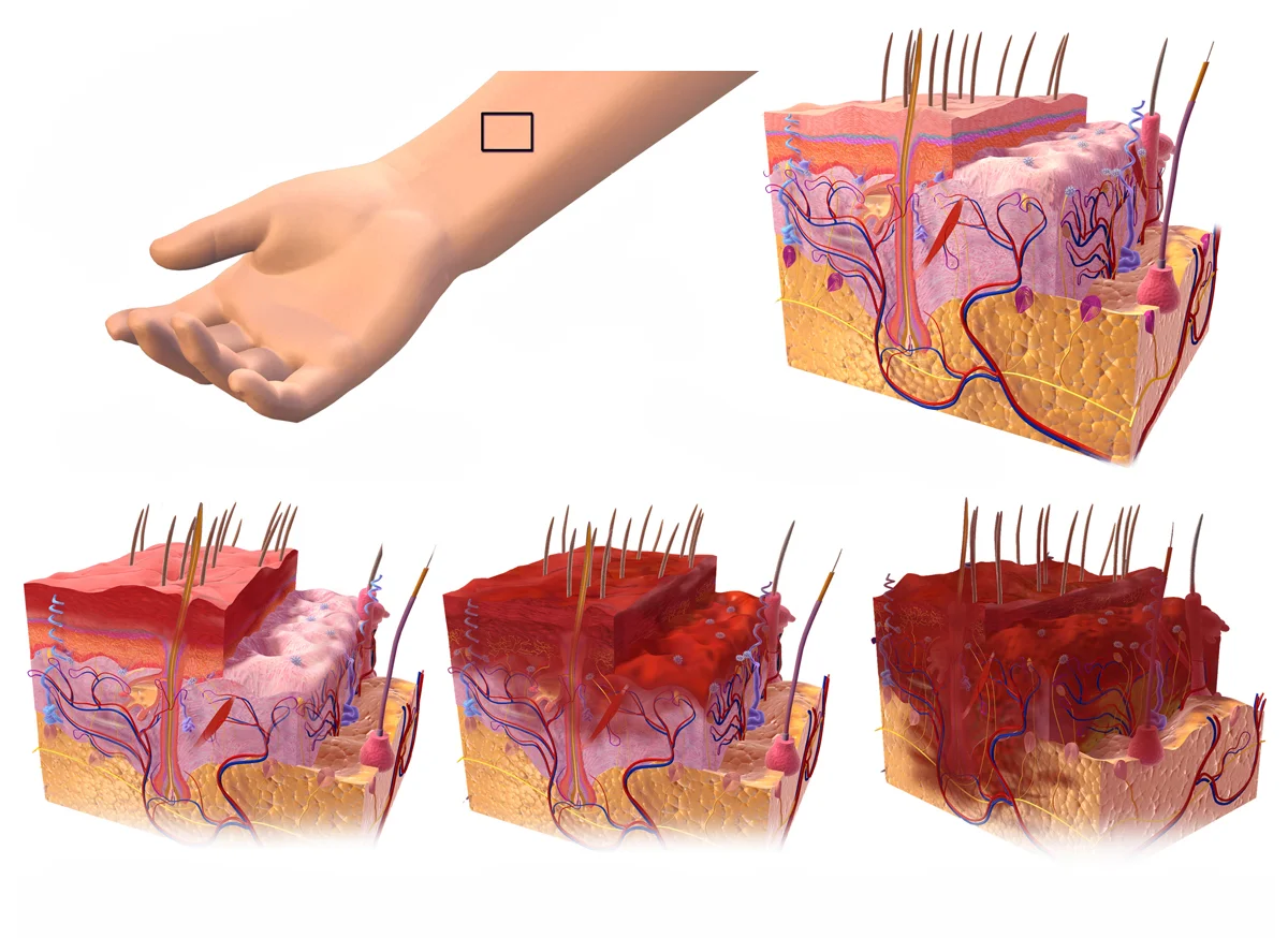
Long description
1st, 2nd, and 3rd degree burns affecting the epidermis, dermis, and hypodermis levels of the skin.

DefinitionBurn
A burn is tissue injury caused by thermal (heat), chemical, electrical or radiation energy. Scalding is a thermal burn caused by hot liquid or steam. [3]
Depth is described in layers. The skin has an outer epithelial layer (the epidermis) over a deeper layer (the dermis) containing blood vessels, nerves, hair follicles, sweat glands and collagen. The deeper the injury, the fewer skin structures survive to regenerate the surface.
The American Burn Association groups burns into four depths. Superficial burns are dry, red, blanch with pressure and are sometimes painful; they do not count towards TBSA. The other three depths are counted. [1]
| Depth | Layers injured | Appearance | Pain | Healing |
|---|---|---|---|---|
| Superficial (1st degree) | Epidermis only | Dry, red, blanches easily | Mild, like sunburn | Short period, no scar |
| Superficial partial thickness (2nd) | Epidermis and upper dermis | Moist, red, blisters, blanches | Very painful, sensitive to air | Usually heals without scarring |
| Deep partial thickness (2nd) | Epidermis and deeper dermis | Drier, paler, less blanching, sluggish capillary return | Less pain (nerve endings damaged) | May need grafting; scar likely |
| Full thickness (3rd) | Epidermis and entire dermis | Dry, leathery, white, brown or black; no blanching | Painless (nerves destroyed) | Will not heal without grafting |
| Fourth degree (4th) | Skin plus underlying fat, muscle or bone | Charred or black, hard | Painless | Surgical debridement and reconstruction |
Fourth-degree burns extend into subcutaneous fat, muscle or bone and are classed with full-thickness injury in most referral schemes, but they are mentioned separately because they change the operation needed (deeper debridement, possible amputation). [1]
A useful bedside rule: pain that disappears as a burn deepens is a warning sign, not reassurance; it means the nerves have been destroyed.
Burns are a global problem. In 2023, about 163,000 people died worldwide from exposure to fire, heat and hot substances, and children under 15 accounted for an estimated 35,600 of these deaths. [2] Females are affected more than males in most low- and middle-income regions (84,600 versus 78,800 deaths in 2023). [2] Two-thirds of deaths occur in the African (about 52,100) and South-East Asian (about 56,300) regions. [2] Non-fatal burns cause pain, prolonged treatment, scarring, disability and psychological distress. [2]
In the United States, about 435 children aged 0 to 19 are treated in emergency departments each day for burn-related injuries, and 2 die. [4] Children under 5 are more likely to be scalded; older children more often have flame burns. [4]
Most burns are accidental and occur at home or work. Risk is higher with poverty, crowded housing, open fires and unsafe cooking (paraffin/kerosene stoves, open flames), faulty electrical wiring, workplace chemicals, smoking and alcohol, and lack of working smoke alarms. [2] [4] Children and the elderly are most at risk because their skin is thinner, so a scald is deeper, and because they cannot remove themselves from the heat quickly. [3]
A burn does three things. First, direct heat denatures proteins and kills cells in the zone of greatest injury. Second, heat causes blood vessels to leak, so plasma pours out into the tissues, producing oedema and blistering and, in a large burn, depleting the circulation. Third, dead tissue (eschar) becomes a haven for bacteria while the inflammatory response makes the patient hypermetabolic for weeks.
The centre of a burn is coagulated; around it is a zone of stasis where cells may recover if perfusion and resuscitation are adequate; and around that is a zone of hyperaemia that heals spontaneously. This is the rationale for "cool, don't freeze": cooling limits the zone of stasis, while ice damages it further. [2] [3]
Symptoms depend on depth. Superficial burns sting; superficial partial-thickness burns are among the most painful injuries seen in emergency care; deep partial-thickness burns hurt less; full-thickness burns are painless in the centre but the edges (which are partial thickness) hurt. Patients with large burns also show signs of shock (tachycardia, low urine output, thirst) and, with inhalation, respiratory distress.
General examination should record: time and mechanism of injury, agent and (for chemicals) concentration and duration of contact, site and size of burn, circumferential burns and distal circulation, associated trauma (falls, explosion), and signs of inhalation (hoarseness, stridor, soot, carbonaceous sputum). [1] [5]
Treat a major burn like any major trauma: airway, breathing, circulation, disability, exposure. Remove the patient from the burning environment, stop the burning process (stop, drop and roll; douse flames), remove clothing and jewellery that is not stuck, and cover the wound loosely with clean cling film (after cooling) to reduce pain and contamination. [3]
Initial management of a major burn
- Stop burning and ensure safetyMove away from heat source; stop, drop, roll; remove non-stuck clothing and jewellery
- AirwayAssess patency; look for inhalation, facial burn, soot, hoarseness, stridor; intubate early if at risk
- BreathingOxygen, auscultation, carboxyhaemoglobin if fire in an enclosed space
- CirculationLarge-bore IV access; calculate % TBSA; start resuscitation if over about 15 to 20% TBSA
- Exposure and temperatureCool smaller burns up to about 20 minutes; warm the patient to prevent hypothermia
- Assess depth and TBSA; photographRule of nines, palmar method or Lund-Browder chart
- Tetanus, analgesia, wound coverUpdate tetanus; give multimodal analgesia; cover with sterile non-adherent dressings
- Decide referral to burn centreApply ABA referral criteria; arrange transfer early if met
Cool thermal burns with cool running water for about 20 minutes; this reduces pain, depth of injury and need for surgery. Run the water over the burn, not ice. Do not apply ice, butter, oils, creams, sprays, pastes or traditional remedies. Do not pop blisters or pull off clothing stuck to the wound. After cooling, cover loosely with cling film or a clean cloth and keep the patient warm overall to prevent hypothermia (core temperature below 35 degrees Celsius). [2] [3]
Burns above the neck can affect breathing and vision and need urgent assessment. Call for emergency help if a child is burnt, or if there is a large burn area, airway injury, white or charred skin, shock, a chemical or electrical burn, or a burn on the hands, face, feet or groin. [3] [4]
Figure 2Wallace Rule of Nines
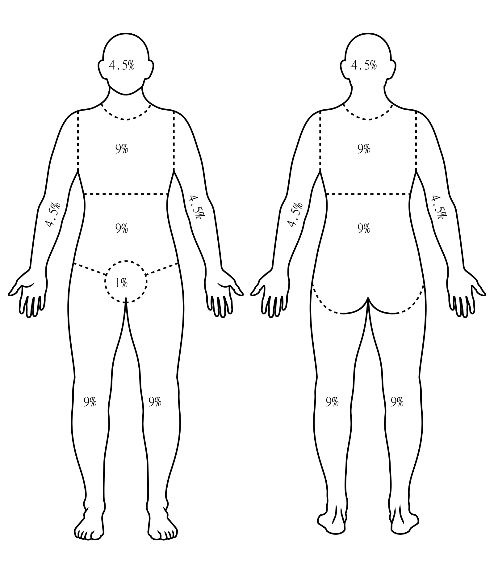
Long description
A total burns surface area estimation model approximating the percentage of body surface area burned using multiples of 9, presented as two annotated human figures (anterior on the left, posterior on the right).

Figure 3Wallace rule of nines-en
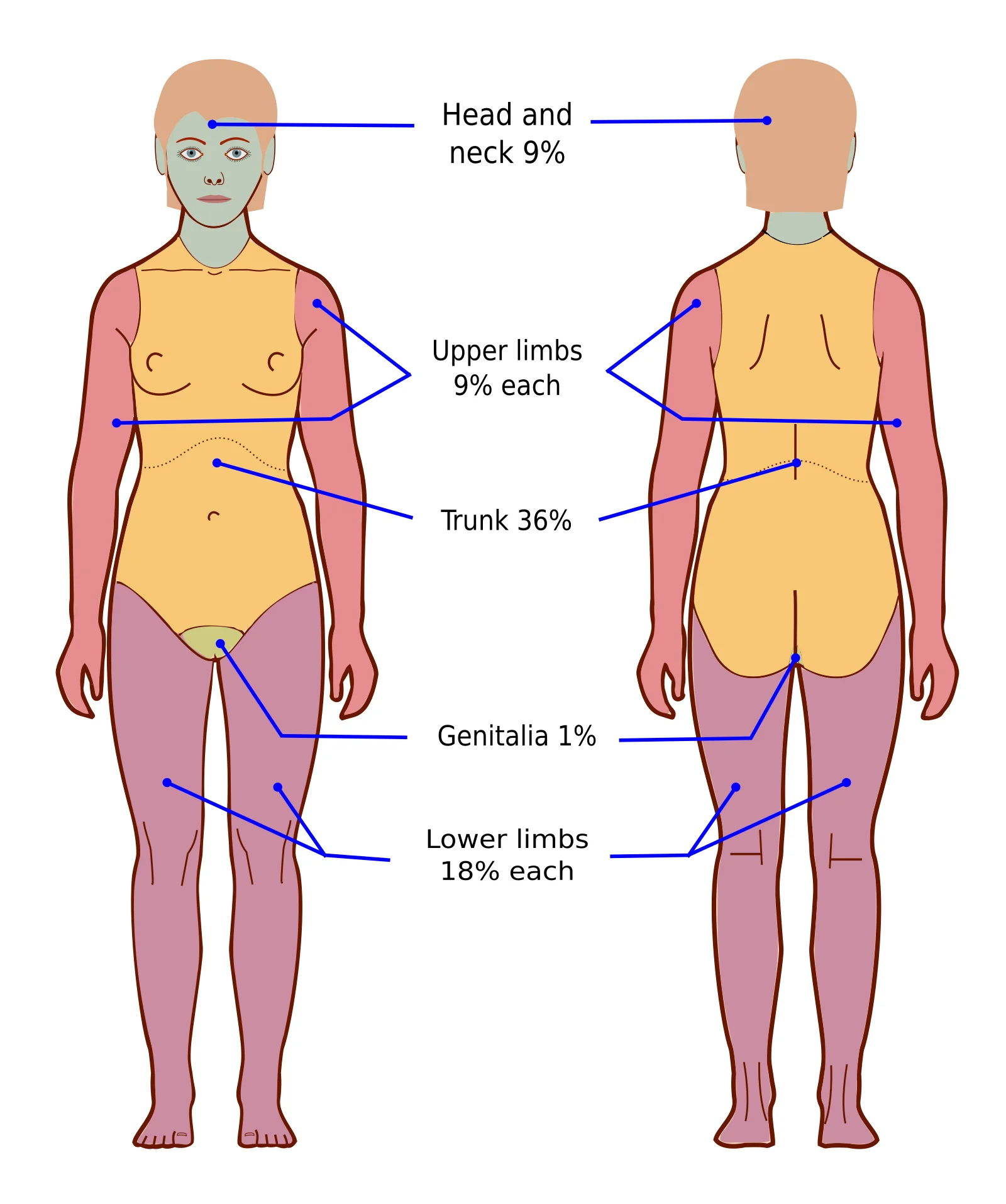
Long description
A front-facing human body diagram of the Wallace rule of nines, with percentage regions used to estimate burn surface area.

TBSA is the proportion of body surface that is partial or full thickness. Superficial burns are excluded. [1]
The rule of nines is the rapid adult estimate: head and neck 9%; each arm 9%; each leg 18%; front of torso 18%; back of torso 18%; perineum 1%. [1]
The palmar method uses the patient's own palm (including fingers) as about 1% of TBSA and is used for small, scattered or irregular burns, and when total involvement is under about 15 to 20%. [1]
The Lund and Browder chart divides the body into smaller regions whose proportions change with age (the infant's head is a larger fraction of surface area than the adult's) and is more accurate, especially in children. Use it whenever a precise figure is needed for resuscitation planning or referral. [1]
| Method | Best for | Notes |
|---|---|---|
| Rule of nines | Rapid adult estimate | Quick, approximate; overestimates in children |
| Palmar method | Small or scattered burns under about 15 to 20% TBSA | Patient's own palm as about 1% |
| Lund and Browder chart | Children and precise planning | Accounts for age-related body proportions |
Children's skin is thinner, so children cool faster and burns are deeper for the same exposure: plan early transfer for any significant paediatric burn. [3]
Inhalation injury is damage to the upper and/or lower airway and lungs from breathing hot gases, smoke or chemical irritants, and is a major cause of death in burns. Suspect it with any burn in an enclosed space, or if there is facial or neck burn, singed nasal hairs, soot on the face, hoarseness, cough, carbonaceous sputum, stridor, wheeze or dyspnoea. [5] [6]
Smoke carries carbon monoxide (and sometimes hydrogen cyanide), which can poison the patient before any burn is visible. Measure carboxyhaemoglobin in any patient burnt in an enclosed space; high levels or confusion warrant 100% oxygen and hyperbaric consideration per local protocol.
Decide to intubate when there is impending airway obstruction, reduced consciousness, haemodynamic instability, significant facial or oral burns, trouble clearing secretions, or for safe transport. Early intubation is usually wise because airway oedema peaks over the first day and an intubation that is easy now can become very difficult later. [5] [6]
The hardest decision is the patient with suspected but not proven inhalation injury. Physical signs are highly specific but not sensitive: the presence of dyspnoea, stridor or hoarseness strongly suggests airway injury, while their absence does not exclude it. Superficial markers such as singed nasal hairs and facial burns are unreliable alone. Combine several findings, examine serially, and use bronchoscopy where available as the reference standard. [6]
Resuscitation is needed when partial- or full-thickness burns exceed about 15 to 20% TBSA in adults. Guidelines differ on the threshold: 20% TBSA for EBA/ABA/DGV/ISBI, 15% for BBA/JSBI/IAEM/MSF. [5]
The aim is to replace the plasma leaking from the burn wound and keep organs perfused, judged primarily by urine output. The classic Parkland approach is 4 mL of crystalloid per kilogram body weight per percent TBSA burned, half given in the first 8 hours from the time of injury and the rest over the next 16 hours. Guidelines now quote a range: ABA recommends 2 mL/kg/%TBSA, EBA and others 2 to 4, IAEM 4, MSF 2 mL/kg/%TBSA after an initial 20 mL/kg bolus. [5] Use a balanced crystalloid (most use Ringer's lactate or Hartmann's; DGV advises against Ringer's lactate because of concerns about lactate metabolism, and JSBI still recommends Ringer's lactate). [5]
- Endpoints: urine output 0.5 to 1.0 mL/kg/h in most guidelines (ABA/IAEM), 0.3 to 0.5 mL/kg/h (DGV/ISBI), 1 to 2 mL/kg/h (MSF), combined with heart rate under 110 per minute, mean arterial pressure above 65 mmHg, lactate below 2 mmol/L and, where available, invasive haemodynamic measures. [5]
- Avoid over-resuscitation: "fluid creep" worsens oedema and complications (abdominal/chest compartment, airway swelling). Titrate the rate down every 2 to 3 hours rather than running it blind. [5]
- Adjuncts: albumin may be considered after 12 to 24 hours as a rescue in patients failing resuscitation; fresh frozen plasma shows promise in animal work but lacks clinical evidence. Vasopressors are not routine; reserve for life-threatening hypotension despite adequate volume. [5]
Pre-existing fluid (beverages, water) is not part of the formula. Reassess volumes in children (different formulas and endpoints) and in the elderly, who are prone to fluid overload.
Chemical burns behave differently from thermal burns: the injury continues until the agent is removed or inactivated. Delayed ophthalmic and skin injury can occur hours after exposure (for example, hydrofluoric acid).
Immediate care:
- Remove and discard contaminated clothing and, for dry powders, brush the powder off the skin before any water touches it.
- Irrigate skin and especially eyes copiously with cool running water for a prolonged period; continue longer for alkali, phenol or hydrofluoric acid. Do not try to neutralise the chemical on the patient. [2] [3]
- Eye burns: irrigate continuously with clean water or saline, keeping eyelids open, and refer to ophthalmology urgently. [3]
- All chemical burns are an indication for burn-centre consultation. [1]
- Hydrofluoric acid burns cause severe pain and hypocalcaemia; calcium gluconate gel (and intravenous/arterial calcium gluconate in severe cases) is used, with toxicology guidance. [7]
Electrical injuries are divided by voltage: high voltage (1000 V or more) and lightning (direct strike or side flash) versus low voltage (under 1000 V). Entry and exit wounds are small, but internal injury along the current path (muscle, vessels, nerves, bone) can be far larger than the skin suggests. Always look for associated trauma (fall, fracture, dislocation) and cardiac arrest. [1] [7]
Complications to exclude and treat:
- Arrhythmia and cardiac ischaemia: monitor the electrocardiogram in high-voltage and lightning injuries; high voltage can disturb rhythm. [7]
- Rhabdomyolysis: deep muscle injury releases myoglobin, which can obstruct renal tubules; give fluids targeted to a urine output that clears myoglobin (alkalinisation of urine is used in some protocols) and check creatine kinase. [7]
- Compartment syndrome: swollen muscle within tight fascial compartments raises pressure; fasciotomy may be needed. Circumferential electrical burns behave like tight tourniquets. [7]
- Other: delayed cataracts, nerve and vessel thrombosis, visceral injury. [7]
Management priorities are high-volume fluid resuscitation, wound exploration, and early surgical fasciotomy/debridement as needed. Low-voltage domestic injuries are usually minor but may cause deep local damage to the hand.
Burn pain has three parts: continuous background pain, procedural pain and neuropathic pain later. Use a multimodal plan: paracetamol (acetaminophen) plus an NSAID where not contraindicated, with an opioid titrated to effect, and ketamine, dexmedetomidine or clonidine as adjuncts. Regional blocks and non-pharmacological methods (distraction, virtual reality, music) help procedural pain. Give neuropathic agents such as gabapentin or pregabalin if neuropathic pain develops. Use a validated pain score. [5]
Guidelines do not agree on the opioid of choice or doses; choose according to the patient's status, haemodynamics and your unit's protocol. [5]
- Keep the wound clean and covered with appropriate non-adherent or occlusive dressings; review and change as per unit protocol and wound depth.
- Topical antimicrobials (for example silver sulfadiazine, mupirocin or biosynthetic dressings) are used for partial-thickness wounds while awaiting healing; deeper burns need surgical excision and grafting when ready. [1]
- Debridement of blisters is controversial; large or tense blisters and those over joints are usually opened under clean conditions, small blisters left. [1]
- Oedema increases burn depth; elevate limbs and perform range-of-motion early.
Burn wounds are tetanus-prone. Ensure the patient's tetanus immunisation is up to date according to the national schedule for tetanus-prone wounds; give booster or immunoglobulin as indicated by immunisation history and wound category. [2]
Full-thickness circumferential burns form a tight, inelastic eschar that can compromise breathing (chest/neck) or distal perfusion (limb) as tissue swells. An escharotomy is a bedside incision through the eschar (down to subcutaneous fat) to release pressure; the cut should not bleed freely if deep enough into dead tissue. Perform for threatened airway/ventilation, compromised distal pulses, or raised compartment pressure. [1]
A burn wound is initially sterile, then colonised by skin and environmental flora over days. Superinfection and invasive infection (burn wound sepsis) are risks over the following days, especially in large, deep or contaminated burns.
- Use standard precautions, clean technique and topical antimicrobials; avoid systemic prophylactic antibiotics (guidelines advise against routine prophylaxis in inhalation injury and generally in burn care beyond peri-operative use). [5]
- Monitor for infection: fever, wound colour/smell change, increasing pain, discolouration of unburned skin, positive cultures, and septic physiology. [5]
- Pneumonia and urinary infection are common in ventilated, intubated patients; lines and catheters should be changed regularly per protocol.
Large burns produce a hypermetabolic, hypercatabolic state from the first day. Start enteral nutrition early once resuscitation allows, and aim for high protein and high calorie intake, adjusted to measured energy expenditure (indirect calorimetry where available) rather than fixed formulas. [1] [8]
Healing is only the start. Hypertrophic scarring, contracture, pigment change and itching are common after deeper burns. Key measures:
- Early and repeated range-of-motion, positioning and splinting to prevent contracture.
- Pressure garments and silicone gel from wound closure, continued for months to years where protocol advises.
- Sun protection of healed skin and, for established tight scars, splinting, therapy or surgical release/reconstruction.
- Psychological support and return-to-work rehabilitation; burn survivors benefit from long-term follow-up. [2] [8]
Use the ABA criteria below; many patients also benefit from advice (consultation) even when transfer is not possible or needed. [1] [8]
| Category | Immediate consultation / consider transfer | Consultation recommendation |
|---|---|---|
| Thermal | Full-thickness burns; partial-thickness 10% or more TBSA; any deep partial or full-thickness burns of the face, hands, genitalia, feet, perineum or over a joint; burns with comorbidities or concomitant trauma; poorly controlled pain. | Partial-thickness burns under 10% TBSA; all potentially deep burns of any size. [1] |
| Inhalation injury | All patients with suspected inhalation injury | Signs of possible inhalation: facial flash burns, singed facial hairs, smoke exposure. |
| Paediatric (14 years or younger, or under 30 kg) | All paediatric burns may benefit from burn-centre referral | — [1] |
| Chemical | All chemical injuries | — |
| Electrical | All high-voltage (1000 V or more) and lightning injuries | Low-voltage (under 1000 V) injuries: consult and consider follow-up for delayed symptoms and vision. |
Paediatric burns are a particular concern: even with clear criteria, many children who meet them are treated outside burn centres, so apply the criteria deliberately and document the reason if referring locally. [8]
- Children: adjust TBSA with a Lund-Browder chart; they dehydrate and cool quickly; refer early. [3]
- Older adults: thinner skin and comorbidity increase risk; smaller burns are dangerous.
- Pregnancy: major burns complicate both maternal and fetal care; involve obstetrics.
- Pre-existing cardiopulmonary or renal disease: tolerates fluid loads poorly; adjust monitoring.
Where bronchoscopy, advanced haemodynamic monitoring or a burn centre is unavailable, rely on repeated clinical assessment, physical findings with the highest specificity (dyspnoea, stridor, hoarseness) for airway decisions, simple endpoints (urine output, mental state) for resuscitation, and early transfer or teleconsultation. [5] [6]
- Depth and TBSA, not the calendar date, determine management. [1]
- Superficial burns are painful but excluded from TBSA. [1]
- Rule of nines for rapid adult TBSA; palmar method for small or scattered burns; Lund-Browder for children and precise planning. [1]
- Cool thermal burns with running water for about 20 minutes; never ice, butter, creams or pastes. [2] [3]
- Inhalation injury can be lethal before skin burns are impressive; maintain a low threshold for early intubation. [5] [6]
- Resuscitate burns over about 15 to 20% TBSA; classic Parkland is 4 mL/kg/%TBSA, half in 8 hours, but guidelines now use 2 to 4 mL/kg/%TBSA. [5]
- Chemical burns need decontamination and prolonged irrigation; never neutralise on the patient. [2] [3]
- Electrical burns can hide massive internal and delayed injury; monitor ECG and creatine kinase. [7]
- All chemical and high-voltage electrical injuries, and all suspected inhalation injuries, warrant burn-centre consultation. [1]
- Escharotomy releases a constricting full-thickness burn. [1]
- Avoid routine prophylactic antibiotics in burn care. [5]
- Nutrition starts early in large burns; pressure garments and therapy prevent disabling contractures. [8]
Answer each one in your head first, then open the answer.
A 70 kg man has 30% TBSA partial-thickness burns from a house fire in an enclosed space, with hoarseness and soot around the mouth. He is alert and saturation is 96% on room air. What are the immediate priorities before fluid resuscitation begins?
Show answerHide answer
Protect the airway early (oedema may worsen), give 100% oxygen and measure carboxyhaemoglobin, obtain IV access and begin/plan resuscitation, and arrange burn-centre transfer. Hoarseness and an enclosed-space fire strongly suggest inhalation injury, for which intubation should be considered before oedema develops. [5] [6]
A 45-year-old woman has 12% TBSA partial-thickness burns to both hands and the face. Vital signs stable. Does this need burn-centre care?
Show answerHide answer
Yes. Partial-thickness burns under 10% TBSA normally receive a consultation recommendation, but deep partial or full-thickness burns of the face or hands meet immediate referral criteria regardless of small size, because function is at stake. [1]
A patient with 36% TBSA burns is receiving 250 mL/h of Ringer's lactate; urine output is 60 mL/h in a 70 kg adult. What does this suggest?
Show answerHide answer
Urine output of 60 mL/h in a 70 kg adult is 0.5 to 1.0 mL/kg/h, the ABA/IAEM target, so resuscitation is not obviously inadequate; titrating down may be appropriate to avoid fluid creep. Reassess pulses, lactate and blood pressure alongside. [5]
Why are burns deeper in children than adults for the same scald?
Show answerHide answer
Children's skin is thinner, so the same heat penetrates further; they also have a larger surface-area-to-mass ratio and cool faster. Use a Lund-Browder chart for TBSA in children. [3]
Why should you never pour water on someone still in contact with an electrical source, and why are electrical burns often worse than they look?
Related topics (plain text): Shock; Trauma primary survey and resuscitation; Fluid and electrolyte balance; Compartment syndrome; Rhabdomyolysis; Wound infection and sepsis; Cicatricial scarring and contracture; Pain management; Tetanus immunisation; Hypercalcaemia (hydrofluoric acid).
- American Burn Association. Guidelines for Burn Patient Referral. ABA burn-care-team resources webpage. Accessed .
- World Health Organization. Burns (fact sheet). WHO newsroom fact sheet, 21 October 2026. Accessed .
- Health Service Executive (Ireland). Emergency care for burns and scalds. HSE patient information, page last reviewed 19 July 2026. Accessed .
- Centers for Disease Control and Prevention. Preventing burn injuries. CDC injury-prevention handout. Accessed .
- Gigengack RK, Slob J, Koopman JSHA, et al. Comparative analysis of recent burn guidelines regarding specific aspects of anesthesia and intensive care. Eur Burn J 2025;6(4):57. doi:10.3390/ebj6040057. Accessed .
- Wardhana A, Farhana N. Early intubation decisions in burn-related inhalation injury: a systematic review of the utility of physical findings in rural and low-resource healthcare settings. Trauma Surg Acute Care Open 2026;11:e002208. doi:10.1136/tsaco-2025-002208. Accessed .
- Smith I, Kidd S, Kim S, Tennill RM. Assessment and Management of Electrical Injuries in Adults in the Emergency Department. Cureus 2026;18(4):e107162. doi:10.7759/cureus.107162. Accessed .
- Gus E, To T, Fish J, Diong C, Saunders N. Adherence to Burn Center Referral Criteria for Pediatric Burns. JAMA Netw Open 2026;9(2):e2559159. doi:10.1001/jamanetworkopen.2025.59159. Accessed .
Figure credits
Educational content for medical students and doctors. It is not a substitute for clinical judgement or local guidelines.