Pelvic lymphatic drainage
The pelvic lymphatic system comprises nodes and vessels that drain the pelvic viscera and lower limbs into the systemic circulation.
The lymphatic system acts as a network of vessels and organs that transport excess interstitial fluid and immune cells back to the bloodstream. Humans possess approximately 500–600 lymph nodes throughout the body, which serve as critical staging areas for immune responses. Lymphatic vessels originate as blind-ended capillaries and progress into larger vessels, eventually emptying into the venous system via the thoracic duct or the right lymphatic duct. [1]
Deep lymphatic vessels associated with the viscera typically follow the paths of arteries. The drainage patterns of the pelvic organs are clinically significant for the spread of disease:
- Testis: Lymphatic drainage follows the testicular vessels directly to the para-aortic (lumbar) lymph nodes, bypassing the superficial inguinal nodes.
- Ovary: Similar to the testis, the ovary drains via vessels accompanying the ovarian artery to the para-aortic nodes.
- Uterus and Cervix: Drainage is complex and multi-directional. The cervix drains primarily to the internal iliac, external iliac, and obturator nodes. The uterine body may drain to the external iliac nodes or, via the round ligament, to the superficial inguinal nodes.
- Vulva: The vulva drains primarily to the superficial inguinal lymph nodes. From there, lymph travels to the deep inguinal nodes and subsequently to the external iliac nodes.
DefinitionLymphatic Flow
Lymph enters a node via afferent vessels, passes through the subcapsular sinus, cortex, and medulla, and exits through efferent vessels.
The pelvic cavity is divided into the greater (false) pelvis and the lesser (true) pelvis, separated by the pelvic brim. The lymphatic vessels within these regions must navigate the complex bony architecture of the pelvic girdle. While the pelvic girdle provides a stable foundation for the body, the lymphatic vessels follow the established arterial supply to reach the iliac node groups.
Figure 1Gray618
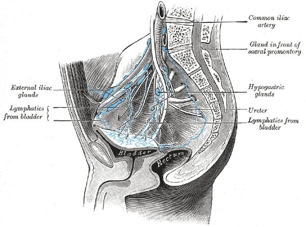
Long description
Lymphatics of the bladder. (Cunéo and Marcille.)

Figure 1: Lymphatics of the bladder (Cunéo and Marcille.).
| Structure | Primary Drainage Destination |
|---|---|
| Testis | Para-aortic nodes |
| Ovary | Para-aortic nodes |
| Cervix | Internal/External iliac nodes |
| Vulva | Superficial inguinal nodes |
Lymphatic Transport
- Lymphatic CapillariesBlind-ended vessels collect interstitial fluid.
- Afferent VesselsTransport lymph into the lymph node cortex.
- Lymph NodeFiltration via subcapsular sinus and medullary cords.
- Efferent VesselsTransport filtered lymph toward larger trunks.
- Venous SystemDumping into the junction of jugular and subclavian veins.
The lymphatic system serves as the essential mechanism for returning interstitial fluid to the bloodstream, a process critical for maintaining fluid balance. Blood pressure drives the leakage of plasma from capillaries into the interstitial space, where it becomes interstitial fluid. In humans, approximately 20 liters of plasma are filtered into the tissues daily[1]. While 17 liters are reabsorbed directly by blood vessels, the remaining three liters are collected by the lymphatic system[1]. This fluid, now termed lymph, enters the system via lymphatic capillaries, also known as terminal lymphatics[1]. These capillaries are located in almost every tissue, interlaced with arterioles and venules, with notable exceptions including the central nervous system, bone marrow, bones, teeth, and the cornea[1].
DefinitionLymphedema
An inappropriate accumulation of protein-rich interstitial fluid due to blocked or damaged lymph vessels, leading to serious medical consequences[1].
The origin of pelvic lymphatic drainage lies within the deep and superficial tissues of the pelvis and lower limbs. Lymphatic capillaries are formed by a single layer of endothelial cells with overlapping edges[1]. When interstitial pressure is low, these flaps remain closed to prevent backflow[1]. As pressure increases, collagen filaments anchor the capillaries to surrounding structures, pulling the endothelial flaps open to allow fluid entry[1]. This mechanism ensures that excess fluid, including immune cells and dietary lipids, is efficiently captured
NoteAnatomical Context
The pelvic girdle attaches the legs to the axial skeleton, and the hip joint is where the pelvic girdle and leg meet[1]. Muscles such as the psoas major and iliacus form the iliopsoas group, while the gluteal muscles (maximus, medius, minimus) are among the largest and most powerful in the body[1]. [2]
The course of pelvic lymph follows the vascular supply. Superficial and deep lymphatics eventually merge to form larger lymphatic trunks[1]. Specifically, the pelvic lymph drains into the lumbar trunks. The thoracic duct begins just beneath the diaphragm in the cisterna chyli, a sac-like chamber that receives lymph from the lower abdomen, pelvis, and lower limbs via the left and right lumbar trunks and the intestinal trunk[1].
The termination of this drainage pathway is the venous system. On the left side of the body, the thoracic duct drains into the left subclavian vein[1]. This ensures that the three liters of excess fluid, along with immune cells and dietary lipids absorbed in the gut, are returned to the bloodstream[1]. The lymphatic system thus completes its cycle, removing interstitial fluid and supporting immune surveillance[1].
| Muscle Group | Primary Action | Nerve Supply |
|---|---|---|
| Iliopsoas (Psoas major, Iliacus) | Flexion of the femur | Femoral nerve, Lumbosacral plexus |
| Gluteus Maximus | Extension and abduction of the femur | Inferior gluteal nerve |
| Gluteus Medius/Minimus | Abduction of the femur | Superior gluteal nerve |
| Lateral Rotators (Piriformis, etc.) | Lateral rotation of the femur | Nerves to quadratus femoris, etc. |
Lymphatic drainage of the pelvis is organised into a series of interconnected node groups that follow the major vascular pathways. These nodes act as biological filters, removing debris and pathogens from the interstitial fluid. Dendritic cells and macrophages within these nodes internalise and destroy pathogens, while the nodes also serve as sites for adaptive immune responses involving T and B cells. [1]
The nodes are grouped according to their anatomical proximity to the major pelvic vessels. The external iliac nodes lie along the external iliac artery and vein, receiving lymph primarily from the lower limb and the deep structures of the anterior abdominal wall. The internal iliac nodes are situated around the internal iliac vessels, draining the pelvic viscera, including the bladder, prostate, and uterus.
| Node Group | Primary Drainage Area |
|---|---|
| External iliac nodes | Lower limb and anterior abdominal wall |
| Internal iliac nodes | Pelvic viscera (bladder, prostate, uterus) |
| Common iliac nodes | Internal and external iliac node groups |
DefinitionLymph node function
Lymph nodes are bean-shaped organs surrounded by a tough connective tissue capsule, divided into internal compartments by trabeculae, which function to filter lymph and facilitate immune surveillance. [1]
The pelvic girdle itself is a rigid, weight-bearing structure formed by the two hip bones, the sacrum, and the coccyx. Unlike the pectoral girdle, which prioritises mobility, the pelvic bones are strongly united to transfer the weight of the upper body to the lower limbs. The hip bone consists of the ilium, ischium, and pubis, which fuse during the late teenage years. When standing, the pelvis is tilted anteriorly, such that the anterior superior iliac spines and the pubic tubercles lie in the same vertical plane. This structural stability is essential for supporting the body’s centre of gravity. [3]
Lymphatic drainage pathway
- Peripheral tissuesLymphatic capillaries collect interstitial fluid
- Regional nodesExternal or Internal iliac nodes filter lymph
- Common iliac nodesConvergence of regional drainage
- Central circulationLymph moves towards the thoracic duct
The lymphatic drainage of the pelvis is organised into distinct regional groups that follow the major vascular pathways. These nodes act as the primary filters for the pelvic viscera, including the bladder, prostate, uterus, and rectum. Understanding these territories is essential for staging pelvic malignancies, as the pattern of spread typically follows the anatomical drainage routes.
Figure 2Gray620
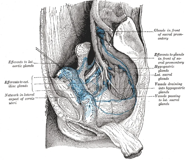
Long description
Lymphatics of the uterus. (Cunéo and Marcille.)

Figure 2: Lymphatics of the uterus (Cunéo and Marcille.).
DefinitionPelvic Lymphatic Drainage
The lymphatic system of the pelvis consists of interconnected node groups that drain the pelvic organs into the common iliac and para-aortic chains.
The external iliac nodes are situated along the external iliac artery and vein, extending from the deep inguinal ring to the bifurcation of the common iliac vessels. These nodes primarily receive lymph from the lower limb, the lower abdominal wall, and the bladder. Medial to these, the internal iliac nodes lie in the fat surrounding the internal iliac artery and its branches. These nodes are the primary drainage site for the pelvic viscera, including the prostate, cervix, and rectum.
The obturator nodes are a specific subset located within the obturator fossa, adjacent to the obturator nerve and vessels. They are often the first site of nodal involvement for tumours arising from the prostate or bladder. The common iliac nodes represent the final common pathway for pelvic lymph before it ascends to the para-aortic nodes. They are positioned along the common iliac vessels and receive efferent vessels from both the internal and external iliac groups.
| Nodal Group | Primary Location | Principal Drainage Area |
|---|---|---|
| External Iliac | Along external iliac vessels | Bladder, lower limb, abdominal wall |
| Internal Iliac | Along internal iliac vessels | Prostate, cervix, rectum, vagina |
| Obturator | Within obturator fossa | Prostate, bladder, pelvic floor |
| Common Iliac | Along common iliac vessels | Efferents from internal/external iliacs |
The drainage pattern is highly predictable, though cross-communication between the sides of the pelvis is common. Lymphatic vessels from the pelvic viscera form a dense plexus within the organ walls before coalescing into larger trunks that accompany the arteries. These trunks ascend along the pelvic sidewalls, passing through the nodal groups described above. Obstruction of these pathways, whether by malignancy or surgical resection, can lead to significant lymphoedema in the lower extremities or pelvic region. Clinically, the internal iliac nodes are considered the most critical relay station for the majority of pelvic organ cancers, making them a primary focus during surgical lymphadenectomy.
Lymphatic Flow
- Pelvic VisceraInitial lymphatic plexus
- Regional NodesInternal/External/Obturator groups
- Common Iliac NodesFinal pelvic relay
- Para-aortic NodesSystemic circulation
The pelvic lymphatic system relies on extensive interconnections to ensure drainage even if primary pathways are obstructed. These connections, known as anastomoses, allow lymph to bypass blockages by flowing through collateral channels. Understanding these routes is critical for predicting the spread of malignancies and managing lymphoedema.
The internal iliac lymph nodes receive drainage from the pelvic viscera, including the bladder, rectum, and reproductive organs. Their efferent vessels do not terminate directly in the common iliac nodes but instead form anastomoses with the external iliac chain. This connection provides an alternative route for lymph from the pelvic floor to reach the abdominal cavity.
Efferent vessels from these nodes curve laterally and superiorly. They connect with the external iliac nodes, which lie along the external iliac vessels.
The obturator nodes are centrally located in the pelvis. They receive lymph from the bladder, prostate, cervix, and upper vagina. Their efferent vessels typically join the external iliac nodes. However, they also anastomose with the internal iliac nodes. This creates a triangular network of drainage between the internal iliac, external iliac, and obturator nodes.
| Structure | Origin | Course | Relations | Termination |
|---|---|---|---|---|
| Obturator Nodes | Pelvic viscera | Along obturator vessels | Between external and internal iliac vessels | External iliac nodes |
| Internal Iliac Nodes | Pelvic walls | Along internal iliac artery | Posterior to internal iliac vein | Common iliac nodes |
| External Iliac Nodes | Lower limb | Along external iliac vessels | Anterior to external iliac artery | Common iliac nodes |
The anastomosis between the obturator and external iliac nodes is particularly significant. It allows lymph from the deep perineal structures to reach the abdominal lymphatic system. This route is often utilized in surgical staging for gynaecological cancers. If the obturator nodes are removed, lymph can still drain via the internal iliac pathway. This redundancy is a key feature of pelvic lymphatic anatomy.
The final common pathway for pelvic lymphatic drainage is the common iliac nodes. Both the internal and external iliac chains converge here. The anastomoses between these chains ensure that lymph from the entire pelvis can reach the common iliac nodes. This convergence point is crucial for systemic drainage.
These trunks drain into the cisterna chyli. The anastomotic network in the pelvis ensures that even if one pathway is blocked, lymph can still reach the common iliac nodes. This is vital for maintaining fluid balance and immune surveillance in the pelvic region.
The nodes are situated lateral to the aorta and inferior vena cava.
The lymphatic drainage of the pelvis is characterised by a complex network of nodes and vessels that follow the major vascular pathways. While the general pattern of drainage is consistent, anatomical variations in the number and precise location of these nodes are common. The pelvic lymph nodes are primarily organised into groups associated with the iliac vessels, which serve as the main conduits for lymph returning from the pelvic viscera and the lower limbs.
DefinitionPelvic Lymphatic System
The pelvic lymphatic system consists of a series of interconnected nodes that drain the pelvic organs, perineum, and lower extremities, ultimately directing lymph towards the lumbar lymphatic trunks.
The iliac lymph nodes are categorised based on their relationship to the iliac arteries and veins. These nodes are the primary filters for the pelvic contents.
- External iliac nodes: These nodes are situated along the external iliac vessels. They receive lymph primarily from the lower limb, the anterior abdominal wall below the umbilicus, and the pelvic viscera, including the bladder and the prostate.
- Internal iliac nodes: Located around the internal iliac artery and its branches, these nodes drain the pelvic viscera, the perineum, and the gluteal region. They are critical in the lymphatic spread of pelvic malignancies.
- Common iliac nodes: These nodes are found along the common iliac vessels. They receive efferent vessels from both the external and internal iliac nodes and act as a final relay before lymph reaches the para-aortic nodes.
| Node Group | Primary Drainage Area |
|---|---|
| External Iliac | Lower limb, bladder, prostate |
| Internal Iliac | Pelvic viscera, perineum, gluteal region |
| Common Iliac | External and internal iliac nodes |
In addition to the iliac groups, specific nodes are located in the deep pelvic spaces. The sacral nodes lie on the concave anterior surface of the sacrum and receive lymph from the rectum and the posterior pelvic wall. The obturator nodes are found in the obturator canal, associated with the obturator nerve and vessels. These nodes are significant because they may be the first site of metastasis for certain pelvic cancers.
Pelvic Lymphatic Drainage Path
- Pelvic VisceraLymph originates in the organs
- Regional NodesInternal/External Iliac nodes
- Common Iliac NodesConvergence of regional drainage
- Lumbar TrunksFinal destination
The drainage pattern is highly integrated. Lymphatic vessels from the pelvic organs often form plexuses before entering the regional nodes. Variations in the number of nodes, which can range from a few to over 20 in specific chains, do not typically alter the fundamental drainage pathway. Understanding these pathways is essential for clinical assessment, particularly when evaluating the spread of disease within the pelvic cavity.
The clinical assessment of the pelvic region relies heavily on identifying superficial bony landmarks that serve as reference points for deeper structures, including lymphatic drainage pathways. When a clinician places their hands on the waist, they can palpate the arching, superior margin of the ilium along the waistline. This curved, superior margin of the ilium is the iliac crest. The rounded, anterior termination of the iliac crest is the anterior superior iliac spine. This important bony landmark can be felt at your anterolateral hip. Inferior to the anterior superior iliac spine is a rounded protuberance called the anterior inferior iliac spine. Both of these iliac spines serve as attachment points for muscles of the thigh. Posteriorly, the iliac crest curves downward to terminate as the posterior superior iliac spine. Muscles and ligaments surround but do not cover this bony landmark, thus sometimes producing a depression seen as a “dimple” located on the lower back. More inferiorly is the posterior inferior iliac spine. [3]
DefinitionIliac Spines
The anterior superior and inferior iliac spines are key attachment points for thigh muscles, while the posterior spines anchor strong ligaments supporting the sacroiliac joint.
The auricular surface of the ilium is located at the inferior end of a large, roughened area. The auricular surface articulates with the auricular surface of the sacrum to form the sacroiliac joint. Both the posterior superior and posterior inferior iliac spines serve as attachment points for the muscles and very strong ligaments that support the sacroiliac joint. Understanding these landmarks is crucial for locating lymph nodes and assessing potential lesions in the pelvic region. [3]
| Landmark | Location | Clinical Significance |
|---|---|---|
| Iliac Crest | Superior margin of ilium | Reference for waistline palpation |
| Anterior Superior Iliac Spine | Anterolateral hip | Attachment for thigh muscles |
| Posterior Superior Iliac Spine | Lower back | Associated with visible depression |
| Auricular Surface | Inferior end of ilium | Forms sacroiliac joint |
The sacroiliac joint is a critical structure for stability and movement. The auricular surface articulates with the auricular surface of the sacrum to form the sacroiliac joint. Both the posterior superior and posterior inferior iliac spines serve as attachment points for the muscles and very strong ligaments that support the sacroiliac joint. These ligaments provide significant support to the joint, ensuring stability during weight-bearing activities. Any injury to these ligaments or the surrounding muscles can lead to pain and dysfunction in the pelvic region.
Clinical pearlSacroiliac Joint Stability
The strong ligaments attached to the posterior iliac spines are essential for maintaining the stability of the sacroiliac joint.
In clinical practice, identifying these landmarks helps in diagnosing conditions related to the pelvic lymphatic system. Similarly, pain in the lower back near the posterior superior iliac spine could suggest issues with the sacroiliac joint or surrounding soft tissues. Accurate identification of these structures is fundamental for effective diagnosis and treatment planning.
Exam trapPalpation Error
Do not confuse the anterior superior iliac spine with the anterior inferior iliac spine; the former is higher and more lateral.
Understanding the anatomical relationships between these bony landmarks and the underlying structures is vital for medical students. The iliac crest, anterior superior iliac spine, posterior superior iliac spine, and auricular surface are all critical for assessing pelvic health. By mastering these landmarks, students can better interpret clinical findings and contribute to accurate diagnoses.
- Trap: Remember that NK cells are part of the innate immune response, whereas B and T cells are part of the adaptive immune response. * Trap: The thymus cortex is densely packed and stains more intensely than the medulla due to high thymocyte density.
DefinitionLymphocyte maturation
Lymphocytes originate in the bone marrow; B cells mature there, while T cells complete their maturation within the thymus gland. [1]
| Organ | Primary Function |
|---|---|
| Lymph nodes | Site of immune responses to antigens in lymph |
| Spleen | Filter of the blood; removes microbes and dying red blood cells |
| Tonsils | Develop immunity to common environmental pathogens [1] |
Lymphocyte development
- Bone MarrowAll lymphocytes originate here
- Question: Maturation site?
Bone Marrow
- B cells
Thymus
- T cells
- Secondary Lymphoid OrgansNaïve lymphocytes mount immune responses here
Students often struggle with the complex, interconnected nature of the pelvic lymphatic system. Avoiding these common errors will improve your anatomical understanding and clinical accuracy.
- Confusing superficial and deep inguinal nodes: Students frequently mistake the superficial inguinal nodes for the primary drainage point of the deep pelvic viscera. The deep inguinal lymph nodes are located in the femoral triangle and serve as a critical relay for the pelvic lymphatic system, whereas superficial nodes primarily drain the lower limb and perineum. [2]
- Ignoring the femoral triangle boundaries: The femoral triangle is formed at the junction between the hip and the leg and includes the femoral nerve, the femoral artery, the femoral vein, and the deep inguinal lymph nodes. Students often forget that the deep inguinal nodes are situated within this specific anatomical space.
- Misunderstanding the role of the iliac region: The ilium is the fan-like, superior region that forms the largest part of the hip bone. Students often fail to associate the lymphatic pathways that track along the iliac vessels with the bony landmarks, such as the arcuate line of the ilium. [3]
- Neglecting the pubic anatomy: The pubis forms the anterior portion of the hip bone. Lymphatic drainage from the lower pelvic structures often follows the path of the superior pubic ramus, which students frequently overlook when tracing the path of lymphatic vessels from the pelvic floor. [3]
- Overlooking the obturator foramen: The large opening in the anteroinferior hip bone between the ischium and pubis is the obturator foramen. This space is a critical landmark for the passage of vessels and lymphatics that drain the deep pelvic structures. [3]
Exam trapThe Femoral Triangle
The deep inguinal lymph nodes are found within the femoral triangle, not the superficial subcutaneous tissue of the thigh.
Knowledge of pelvic lymphatic drainage is essential for clinical practice, particularly in oncology and surgery.
- Bedside examination: During physical examination, palpation of the femoral triangle is used to assess for lymphadenopathy. Because the deep inguinal lymph nodes are located in the femoral triangle, enlargement here can indicate malignancy or infection originating from the pelvic viscera or the lower limb.
- Operating theatre: Surgeons must identify the anatomical boundaries of the pelvic bones, such as the pubic tubercle and the ischiopubic ramus, to navigate the pelvic space safely. Lymph node dissection requires precise knowledge of the vessels accompanying the lymphatics, particularly near the obturator foramen.
- Imaging: Radiologists use the bony landmarks of the pelvis to localise lymph nodes. The arcuate line of the ilium and the pubic symphysis serve as key reference points on CT and MRI scans to delineate the extent of lymphatic chains.
- Examinations: You will be expected to trace the lymphatic drainage from the pelvic organs to the deep inguinal nodes and subsequently to the iliac chains. Understanding that the pectineus muscle is located in the femoral triangle helps you orient yourself to the location of the deep inguinal nodes during anatomical dissection or when interpreting cross-sectional imaging.
| Structure | Anatomical Significance |
|---|---|
| Femoral Triangle | Contains deep inguinal lymph nodes |
| Pubic Tubercle | Superior landmark on the pubic body [3] |
| Obturator Foramen | Space between ischium and pubis |
| Arcuate Line | Inferior margin of the iliac fossa |
Pelvic Lymphatic Assessment
- Identify Femoral TriangleLocate the junction between the hip and the leg.
- Question: Palpate for nodes?
Yes
- Assess deep inguinal nodesCheck for enlargement within the femoral triangle.
No
- Review imagingUse bony landmarks like the pubic symphysis.
- The deep inguinal lymph nodes are situated within the femoral triangle.
- The femoral triangle contains the femoral nerve, artery, vein, and deep inguinal nodes.
- The ilium forms the largest part of the hip bone.
- The pubic body features the pubic tubercle.
- The obturator foramen is located between the ischium and pubis.
- The arcuate line marks the inferior margin of the iliac fossa.
- The pubic symphysis joins the two pubic bodies.
Answer each one in your head first, then open the answer.
Where are the deep inguinal lymph nodes located?
Show answerHide answer
The deep inguinal lymph nodes are located in the femoral triangle.What structures form the femoral triangle?
Show answerHide answer
The femoral triangle is formed at the junction between the hip and the leg and includes the femoral nerve, the femoral artery, the femoral vein, and the deep inguinal lymph nodes.What is the large opening between the ischium and pubis?
Show answerHide answer
The large opening in the anteroinferior hip bone between the ischium and pubis is the obturator foramen.What is the ridge on the superior margin of the superior pubic ramus?
Show answerHide answer
The narrow ridge running along the superior margin of the superior pubic ramus is the pectineal line of the pubis.What is the inferior margin of the iliac fossa?
Show answerHide answer
The inferior margin of this space is formed by the arcuate line of the ilium.
- Betts JG, Young KA, Wise JA, et al. Anatomy of the Lymphatic and Immune Systems. OpenStax, Anatomy and Physiology 2e (CC BY-NC-SA 4.0). Accessed .
- Betts JG, Young KA, Wise JA, et al. Appendicular Muscles of the Pelvic Girdle and Lower Limbs. OpenStax, Anatomy and Physiology 2e (CC BY-NC-SA 4.0). Accessed .
- Betts JG, Young KA, Wise JA, et al. The Pelvic Girdle and Pelvis. OpenStax, Anatomy and Physiology 2e (CC BY-NC-SA 4.0). Accessed .
Figure credits
Educational content for medical students and doctors. It is not a substitute for clinical judgement or local guidelines.