Surface anatomy: palpable landmarks
Surface anatomy palpable landmarks for medical students
Surface anatomy identifies structures that can be seen or felt through the skin. Bony landmarks provide fixed reference points; soft tissue landmarks include tendons, muscles, vessels and nerves that project to the surface. Pulse points occur where arteries lie close to bone or fascia. Injection and procedure sites are chosen to avoid neurovascular bundles while accessing target structures. Palpation technique uses systematic pressure to confirm anatomy before clinical interventions.
DefinitionPalpable landmark
A surface feature that can be reliably identified by touch in the living patient, used to locate deeper structures.
As the brachial artery approaches the coronoid fossa, it bifurcates into the radial and ulnar arteries[1].
| Vein | Origin | Course | Termination |
|---|---|---|---|
| Cephalic | Dorsal venous network of hand | Lateral forearm, along biceps brachii | Axillary vein |
| Basilic | Dorsal venous network of hand | Medial forearm, pierces deep fascia | Brachial veins |
It is palpable in patients without excessive adipose tissue[1].
The areola is typically circular and can vary in size from 25 to 100 mm in diameter. The areolar region is characterized by small, raised areolar glands that secrete lubricating fluid during lactation. [3]
Palpation sequence for vascular access
- Identify bony landmarksClavicle, medial epicondyle, femoral condyles
- Question: Target vessel superficial?
Yes
- Visualize veinCephalic at biceps, saphenous at ankle
No
- Palpate pulseBrachial at elbow, femoral at inguinal ligament
- Confirm with ultrasoundIf anatomy uncertain
Many arteries and veins share the same names, parallel one another throughout the body, and are very similar on the right and left sides[1]. Names of vessels can change with location[1]. Spinal nerves project sensory information to the opposite side of the brain.
Surface anatomy landmarks are grouped by the type of structure that can be felt through the skin. The main categories are bony prominences, arterial pulses, venous landmarks, tendinous and muscular margins, nerve points, and organ borders. Each group follows a consistent regional sequence from head to toe so that examination proceeds in an orderly fashion.
Figure 1Gray1244
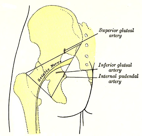
Long description
Left gluteal region, showing surface markings for arteries and sciatic nerve.

Figure 1: Left gluteal region, showing surface markings for arteries and sciatic nerve.
Figure 2Gray1237
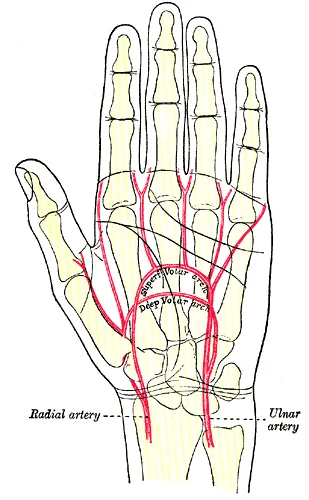
Long description
Palm of left hand, showing position of skin creases and bones, and surface markings for the volar arches.

Figure 2: Palm of left hand, showing position of skin creases and bones, and surface markings for the volar arches.
| Category | Representative landmarks |
|---|---|
| Bony prominences | Sternal angle, medial and lateral epicondyles of humerus and femur, medial and lateral malleoli, anterior superior iliac spine, pubic tubercle |
| Arterial pulses | Carotid, brachial, radial, femoral, popliteal, posterior tibial, dorsalis pedis |
| Venous landmarks | Great saphenous vein at medial malleolus and along medial thigh, dorsal venous arch of foot |
| Tendinous / muscular margins | Biceps brachii tendon, patellar tendon, Achilles tendon, borders of sternocleidomastoid and trapezius |
| Nerve points | Ulnar nerve posterior to medial epicondyle, common fibular nerve at fibular neck |
| Organ borders | Lower costal margin (liver), splenic notch, kidney poles in thin individuals |
The great saphenous vein is a prominent surface vessel located on the medial surface of the leg and thigh that collects blood from the superficial portions of these areas. It runs anterior to the medial malleolus, ascends along the medial leg and thigh, and pierces the cribriform fascia to join the femoral vein. Its superficial course makes it a reliable landmark for venous access and for identifying the saphenofemoral junction. The dorsal venous arch on the dorsum of the foot is also visible and palpable in many individuals [1]
In the scrotum, the raphae is a raised medial thickening on the surface that marks the line of fusion of the labioscrotal folds. The dartos muscle lies beneath the skin and continues internally to form the scrotal septum, dividing the scrotum into two compartments. The cremaster muscles, descending from the internal oblique, cover each testis and can elevate the testes toward the body. [4]
Figure 3Gray1243
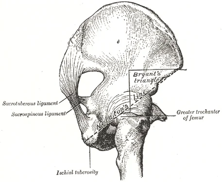
Long description
Nélaton’s line and Bryant’s triangle. Labelled illustration from Wikimedia Commons, used under its open licence.

Figure 3: Nélaton’s line and Bryant’s triangle.
Figure 4Gray1232
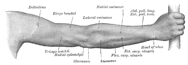
Long description
Back of right upper extremity. Labelled illustration from Wikimedia Commons, used under its open licence.

Figure 4: Back of right upper extremity.
Bony landmarks provide the most consistent reference points because they do not vary with body habitus as much as soft-tissue structures. The sternal angle (angle of Louis) lies at the level of the second costal cartilage and the T4/T5 vertebral level, serving as a key reference for counting ribs and locating the tracheal bifurcation. The anterior superior iliac spine and pubic tubercle define the inguinal ligament, under which the femoral artery, vein, and nerve pass. The medial and lateral epicondyles of the humerus and femur, together with the medial and lateral malleoli, anchor measurements of limb length and joint alignment.
Figure 5Gray1213
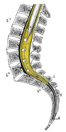
Long description
Sagittal section of vertebral canal to show the lower end of the medulla spinalis and the flum terminale. (Testut.) Li, Lv. First and fifth lumbar vertebra. S?? Second sacral vertebra. 1. Dura mater. 2. Lower part of subarachnoid cavity. 3. Lower extremity of medulla spinalis. 4. Filum terminale int

Figure 5: Sagittal section of vertebral canal to show the lower end of the medulla spinalis and the flum terminale (Testut.) Li, Lv.
Arterial pulses are assessed for rate, rhythm, and character. The femoral pulse is palpated just inferior to the inguinal ligament, midway between the anterior superior iliac spine and the pubic tubercle. The popliteal pulse is felt in the popliteal fossa with the knee flexed. The posterior tibial pulse lies posterior to the medial malleolus, and the dorsalis pedis pulse is on the dorsum of the foot lateral to the extensor hallucis longus tendon.
Figure 6Gray1242
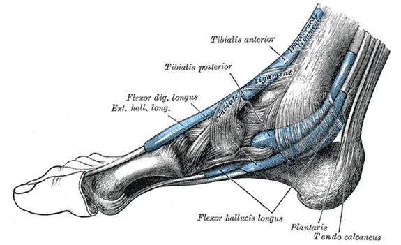
Long description
The mucous sheaths of the tendons around the ankle. Medial aspect.

Figure 6: The mucous sheaths of the tendons around the ankle (medial aspect).
Figure 7Gray1240
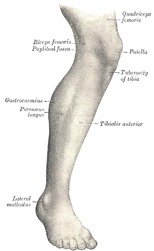
Long description
Lateral aspect of right leg. Labelled illustration from Wikimedia Commons, used under its open licence.

Figure 7: Lateral aspect of right leg.
Figure 8Gray1238
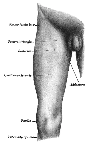
Long description
Front and medial aspect of right thigh. Labelled illustration from Wikimedia Commons, used under its open licence.

Figure 8: Front and medial aspect of right thigh.
Nerve landmarks are important for avoiding injury during procedures. The ulnar nerve is palpable in the groove posterior to the medial epicondyle of the humerus. The common fibular nerve winds around the neck of the fibula, where it can be rolled against the bone.
Organ borders are estimated by percussion and palpation. The liver edge is sought below the right costal margin in the midclavicular line. The spleen is not normally palpable; an enlarged spleen may be felt below the left costal margin. The kidneys are ballotable in the flanks, particularly in thin individuals.
Figure 9Gray1231
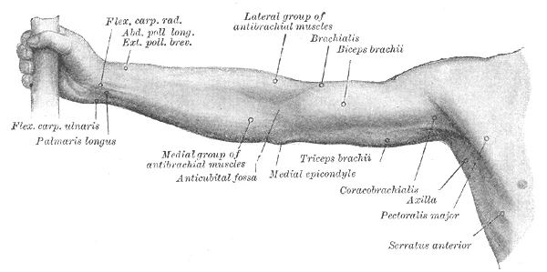
Long description
Front of right upper extremity. Labelled illustration from Wikimedia Commons, used under its open licence.

Figure 9: Front of right upper extremity.
Clinical pearlExamination sequence
Work systematically from superficial to deep and from proximal to distal, using bony landmarks as fixed reference points before seeking vascular or neural structures.
Surface anatomy palpable landmarks include arterial pulses that can be felt through the skin. In the lower limb, the anterior tibial artery lies between the tibia and fibula, supplying the anterior tibial region. Upon reaching the tarsal region, it becomes the dorsalis pedis artery, which branches repeatedly over the tarsal and dorsal foot. The posterior tibial artery runs on the posterior surface of the tibial region, supplying the posterior compartment. [1]
Figure 10Gray1211
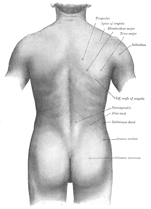
Long description
Surface anatomy of the back. Labelled illustration from Wikimedia Commons, used under its open licence.

Figure 10: Surface anatomy of the back.
DefinitionPalpable arterial landmarks of the lower limb
The dorsalis pedis pulse is felt on the dorsum of the foot lateral to the extensor hallucis longus tendon; the posterior tibial pulse is felt posterior to the medial malleolus.
The tibia lies medially, the fibula laterally. The extensor hallucis longus tendon lies medial to the artery at the ankle. The fibular artery diverges laterally from the posterior tibial artery deep in the calf.
Figure 11Gray1241
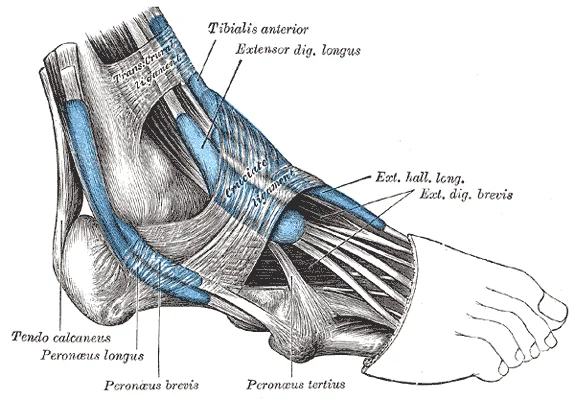
Long description
The mucous sheaths of the tendons around the ankle. Lateral aspect.

Figure 11: The mucous sheaths of the tendons around the ankle (lateral aspect).
The tibia is anterior and medial, the fibula posterior and lateral. The anterior tibial artery sits in the anterior compartment between the tibialis anterior and extensor digitorum longus muscles. The posterior tibial artery lies in the deep posterior compartment between the flexor digitorum longus and tibialis posterior muscles. The interosseous membrane separates anterior and posterior compartments.
| Artery | Surface landmark | Relation |
|---|---|---|
| Anterior tibial / dorsalis pedis | Dorsum of foot, lateral to extensor hallucis longus tendon | Between tibia and fibula proximally; crosses ankle anteriorly |
| Posterior tibial | Posterior to medial malleolus | Deep to flexor retinaculum, between flexor digitorum longus and tibialis posterior |
| Fibular (peroneal) | Not routinely palpated | Runs in lateral compartment along fibula |
Palpable landmarks are essential for clinical examination, allowing clinicians to locate deeper structures without imaging. These landmarks define anatomical planes and guide procedures such as venepuncture, catheterisation, and surgical incisions. Understanding the surface anatomy ensures accuracy and minimises risk to adjacent neurovascular bundles.
The systemic veins return blood to the right atrium, carrying blood that is relatively low in oxygen concentration. While arteries and veins often follow a complementary pattern, the venous circulation exhibits greater variability. Clinically, the superior vena cava drains most of the body superior to the diaphragm. [1]
These vertebral veins arise from the base of the brain and the cervical region of the spinal cord, flowing largely through the intervertebral foramina in the cervical vertebrae. They are the counterparts of the vertebral arteries. [1]
The internal thoracic vein, also known as an internal mammary vein, drains the anterior surface of the chest wall and flows into the brachiocephalic vein. The subclavian vein lies deep to the clavicle, making it less accessible for direct palpation but crucial for central line insertion. [1]
In males, the descent of the testis is a critical developmental process. Cryptorchidism is the clinical term used when one or both of the testes fail to descend into the scrotum prior to birth. The scrotum itself is a palpable landmark for assessing testicular position and size. The spermatic cord, containing the vas deferens and vessels, is palpable superior to the testis. [4]
In females, the breast is a prominent surface landmark. Breast milk is produced by the mammary glands, which are modified sweat glands. The milk exits the breast through the nipple via 15 to 20 lactiferous ducts that open on the surface of the nipple. Breast size differs between individuals and does not affect the amount of milk produced. Palpation of the breast involves checking for lumps in the four quadrants and the axillary tail, as well as assessing the nipple for discharge or retraction. [3]
The surface of the tongue is lined by a stratified squamous epithelium. Raised bumps called papillae contain the structures for gustatory transduction. There are four types of papillae: circumvallate, foliate, filiform, and fungiform. Within the papillae are taste buds containing specialized gustatory receptor cells. These cells release neurotransmitters based on the amount of chemical in the food. Neurotransmitters activate sensory neurons in the facial, glossopharyngeal, and vagus cranial nerves. [2]
This region contains bipolar sensory neurons with dendrites extending into the mucus lining the cavity. The odorant–protein complex binds to a receptor protein within the cell membrane of an olfactory dendrite. These receptors are G protein–coupled, producing a graded membrane potential in the olfactory neurons. While not directly palpable, the nasal cavity is a critical anatomical region for assessing olfactory function. [2]
The carotid pulse is palpated at the anterior border of the sternocleidomastoid muscle where the common carotid artery runs; the right common carotid arises from the brachiocephalic trunk while the left arises directly from the aortic arch. The carotid sinus, a dilatation at the bifurcation of the common carotid into internal and external carotid arteries, houses baroreceptors and chemoreceptors critical for cardiovascular homeostasis; excessive pressure on this sinus during examination can trigger reflex bradycardia or hypotension. The superficial temporal artery, a branch of the external carotid artery, is readily palpable anterior to the tragus of the ear and is a key site for temporal artery biopsy in suspected giant cell arteritis. The femoral pulse is best felt at the mid-inguinal point, halfway between the anterior superior iliac spine and the pubic symphysis, where the femoral artery lies medial to the femoral vein; the femoral vein becomes the external iliac vein as it passes under the inguinal ligament. The brachial artery pulse is located medial to the biceps tendon in the antecubital fossa and is the standard site for blood pressure measurement; it continues the axillary artery distal to the lower border of teres major. The radial artery pulse is palpated at the wrist lateral to the flexor carpi radialis tendon; it is the lateral terminal branch of the brachial artery. The popliteal pulse is found deep in the popliteal fossa with the knee flexed; it represents the continuation of the femoral artery through the adductor hiatus. The dorsalis pedis pulse is palpated on the dorsum of the foot lateral to the extensor hallucis longus tendon; it is the continuation of the anterior tibial artery. During pregnancy, the femoral pulse may be harder to palpate due to increased inguinal ligament tension and soft tissue changes; the carotid and radial pulses remain accessible. In older adults, atherosclerotic stiffening may make peripheral pulses diminished or absent while carotid pulses remain palpable; caution is needed to avoid carotid sinus massage effects. In low-resource settings without Doppler ultrasound, systematic palpation of all major pulses (carotid, brachial, radial, femoral, popliteal, dorsalis pedis, posterior tibial) provides essential vascular assessment; the femoral pulse is particularly valuable as it reflects aortic outflow and is less affected by peripheral vasoconstriction. [1]
Students often confuse the sternal angle with the suprasternal notch when locating the aortic arch. The ascending aorta ends at the sternal angle after a course of approximately 5 cm. The sternal angle marks the level of the second costal cartilage and the aortic arch, not the suprasternal notch.
A frequent error is placing the aortic bifurcation at the wrong vertebral level. The abdominal aorta bifurcates into the common iliac arteries at the level of the fourth lumbar vertebra. This level corresponds to the iliac crests on surface anatomy. [1]
Learners sometimes overlook that the abdominal aorta lies to the left of the vertebral column. The vessel remains to the left of the vertebral column and is embedded in adipose tissue behind the peritoneal cavity. This relationship affects palpation and imaging interpretation. [1]
The clitoris is often missed as a key palpable landmark of the female perineum. The superior, anterior portions of the labia minora come together to encircle the clitoris, an organ that originates from the same cells as the glans penis. It is a consistent midline structure. [3]
Students may assume the hymen is a complete membrane. An intact hymen cannot be used as an indication of virginity; even at birth, this is only a partial membrane, as menstrual fluid and other secretions must be able to exit the body. Its appearance varies widely. [3]
DefinitionSternal angle (angle of Louis)
The manubriosternal joint, palpable as a transverse ridge, marking the level of the second costal cartilage and the aortic arch.
| Vertebral level | Aortic landmark | Surface correlate |
|---|---|---|
| T4/T5 | Sternal angle / aortic arch | Second costal cartilage |
| T12 | Aortic hiatus (diaphragm) | Xiphisternal joint |
| L4 | Aortic bifurcation | Iliac crests |
At the bedside, the sternal angle is used to estimate central venous pressure and to locate the aortic arch for auscultation. The ascending aorta moves in a superior direction for approximately 5 cm and ends at the sternal angle. This landmark also identifies the second intercostal space for needle decompression. [1]
In the operating theatre, the L4 vertebral level guides exposure of the aortic bifurcation during vascular procedures. The abdominal aorta formally ends at approximately the level of vertebra L4, where it bifurcates to form the common iliac arteries. Surgeons use the iliac crests as surface markers for this level. [1]
On imaging, vertebral levels correlate with aortic segments for reporting and intervention planning. The aorta consists of the ascending aorta, the aortic arch, and the descending aorta, which passes through the diaphragm. The transition from thoracic to abdominal aorta occurs at the aortic hiatus (T12).
In examinations, students must identify the sternal angle, xiphisternal joint, and iliac crests as surface projections of the aortic arch, diaphragmatic hiatus, and bifurcation respectively. The aorta bifurcates at the level of the fourth lumbar vertebra into the two common iliac arteries. These landmarks are tested in OSCE stations for cardiovascular and abdominal examination.
Surface landmark to aortic segment
- Palpate suprasternal notchMove fingers down 5 cm to sternal angle
- Identify sternal angleMarks aortic arch, second costal cartilage
- Question: Need diaphragmatic level?
Yes
- Locate xiphisternal jointT12, aortic hiatus
No
- Proceed to iliac crestsL4, aortic bifurcation
- The sternal angle (angle of Louis) is the principal surface landmark for the aortic arch and second rib.
- The abdominal aorta bifurcates at L4, corresponding to the iliac crests.
- The aortic hiatus at T12 aligns with the xiphisternal joint.
- The clitoris is a consistent midline palpable landmark in the female perineum.
- The hymen is a partial membrane from birth and not a marker of virginity.
- The abdominal aorta lies left of the vertebral column in the retroperitoneum.
Answer each one in your head first, then open the answer.
At which vertebral level does the abdominal aorta bifurcate?
Show answerHide answer
L4What surface landmark corresponds to the aortic arch?
Show answerHide answer
Sternal angle (angle of Louis)Where is the aortic hiatus located relative to the sternum?
Show answerHide answer
At the xiphisternal joint (T12)Which structure do the labia minora encircle to form the prepuce of the clitoris?
Show answerHide answer
The clitoris (glans clitoris)Is the hymen a complete membrane at birth?
Show answerHide answer
No, it is a partial membrane to allow menstrual flow
- Betts JG, Young KA, Wise JA, et al. Circulatory Pathways. OpenStax, Anatomy and Physiology 2e (CC BY-NC-SA 4.0). Accessed .
- Betts JG, Young KA, Wise JA, et al. Sensory Perception. OpenStax, Anatomy and Physiology 2e (CC BY-NC-SA 4.0). Accessed .
- Betts JG, Young KA, Wise JA, et al. Anatomy and Physiology of the Ovarian Reproductive System. OpenStax, Anatomy and Physiology 2e (CC BY-NC-SA 4.0). Accessed .
- Betts JG, Young KA, Wise JA, et al. Anatomy and Physiology of the Testicular Reproductive System. OpenStax, Anatomy and Physiology 2e (CC BY-NC-SA 4.0). Accessed .
Figure credits
- Figure 1: Henry Vandyke Carter, via Wikimedia Commons, Public domain, Public domain.
- Figure 2: Henry Vandyke Carter, via Wikimedia Commons, Public domain, Public domain.
- Figure 3: Henry Vandyke Carter, via Wikimedia Commons, Public domain, Public domain.
- Figure 4: Henry Vandyke Carter, via Wikimedia Commons, Public domain, Public domain.
- Figure 5: Henry Vandyke Carter, via Wikimedia Commons, Public domain, Public domain.
- Figure 6: Henry Vandyke Carter, via Wikimedia Commons, Public domain, Public domain.
- Figure 7: Henry Vandyke Carter, via Wikimedia Commons, Public domain, Public domain.
- Figure 8: Henry Vandyke Carter, via Wikimedia Commons, Public domain, Public domain.
- Figure 9: Henry Vandyke Carter, via Wikimedia Commons, Public domain, Public domain.
- Figure 10: Henry Vandyke Carter, via Wikimedia Commons, Public domain, Public domain.
- Figure 11: Henry Vandyke Carter, via Wikimedia Commons, Public domain, Public domain.
Educational content for medical students and doctors. It is not a substitute for clinical judgement or local guidelines.