Vitamin A and the B vitamins
Biochemical pathway principles, physiological roles, deficiencies and toxicities of vitamin A and water-soluble vitamins B1 and B2.
Figure 1Vitamin A and carotenoid metabolism
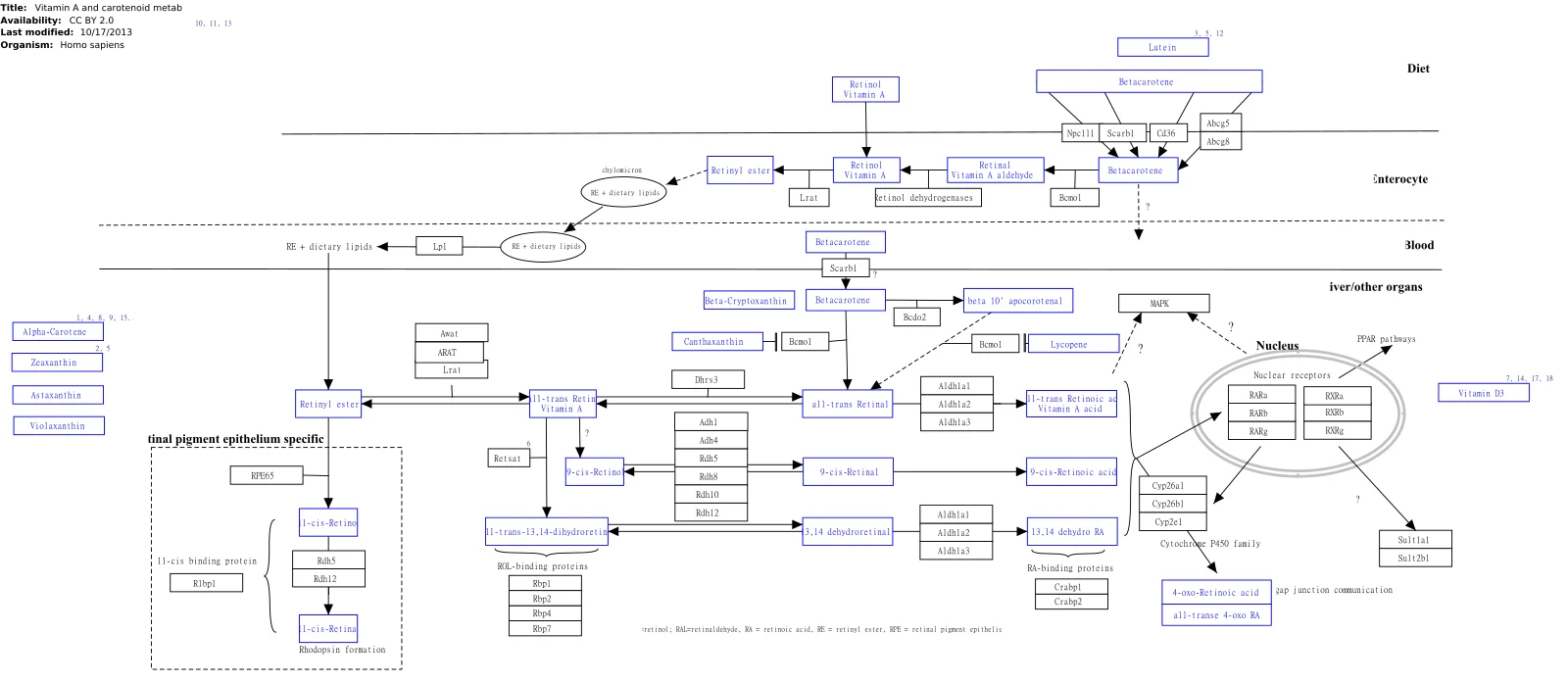
Long description
This pathway is about carotenoid and vitamin A metabolism. The initial version was created by the NuGO focusteam on Carotenoid metabolism. It was used to test a text mining workflow which added some additional entities

Vitamins are essential organic micronutrients that human cells cannot synthesize in sufficient quantities, requiring regular exogenous intake via diet. From a structural and metabolic perspective, micronutrients divide into distinct classes based on their solubility characteristics, which dictate their absorption mechanisms, transport proteins, storage capacities, and excretion pathways. Fat-soluble vitamins, which include vitamin A, share physicochemical properties with lipids and require normal micellar solubilisation, biliary secretion, and pancreatic lipases for intestinal assimilation. Once absorbed across the brush border membrane, fat-soluble compounds are incorporated into chylomicrons, transported via the lymphatic system into the general circulation, and stored predominantly within hepatic parenchymal cells and adipose tissue. Because of this substantial storage capacity, excessive ingestion of lipophilic vitamins can lead to bioaccumulation and eventual toxicity syndromes.
In contrast, water-soluble vitamins, which encompass the entire B-complex group and vitamin C, dissolve readily in aqueous media and are absorbed directly through the intestinal mucosa into the portal venous system. With the exception of vitamin B12, human tissues store water-soluble vitamins only minimally. Excess amounts are filtered continuously by the renal glomeruli and excreted in urine, necessitating a consistent daily dietary supply to prevent depletion. The B-complex vitamins function primarily as indispensable enzymatic cofactors or precursors for coenzymes that drive intermediary metabolism, energy production, macromolecular synthesis, and cellular redox homeostasis.
Biochemically, micronutrient pathways operate through hierarchical networks where a single vitamin may participate in multiple interconnected enzymatic reactions across different subcellular compartments. Disruption of these pathways, whether through inadequate dietary intake, malabsorption syndromes, increased metabolic demand, or genetic polymorphisms, leads to characteristic clinical syndromes defined by metabolic bottlenecks. Understanding these pathways from first principles requires examining the precise chemical structures, carrier proteins, and receptor interactions that govern each vitamin's physiological journey from the intestinal lumen to its intracellular site of action.
Vitamin A is a generic descriptor for a group of fat-soluble retinoids that play fundamental roles in vision, cellular differentiation, embryonic development, immune competence, and central nervous system homeostasis. Vitamin E belongs to the fat-soluble vitamins, like vitamin A, D and K. Dietary vitamin A is ingested either as preformed retinyl esters derived from animal sources such as liver, eggs, and dairy products, or as provitamin A carotenoids, most notably beta-carotene, found in yellow, orange, and dark green leafy vegetables. Within the intestinal enterocyte, beta-carotene is cleaved enzymatically to yield retinal, which is subsequently reduced to retinol, esterified, and incorporated into chylomicrons for delivery to the liver. [1]
Inside the target cell, retinol is oxidized reversibly to retinal and then irreversibly to retinoic acid. Retinoic acid acts as a potent nuclear hormone that binds to specific nuclear receptors, namely retinoic acid receptors and retinoid X receptors, which heterodimerize and modulate the transcription of hundreds of target genes governing cellular proliferation and differentiation. In the visual cycle, 11-cis-retinal binds to opsin within retinal rod and cone photoreceptor cells to form rhodopsin, the light-sensitive pigment responsible for phototransduction in dim and bright illumination.
Vitamin A deficiency manifests initially as impaired dark adaptation, progressing rapidly to conjunctival xerosis, Bitot spots, and eventual corneal ulceration and keratomalacia, which constitute a leading cause of preventable childhood blindness worldwide. Furthermore, a deficiency of vitamin A, D, E, and K have been reported to be tightly associated with AD. Conversely, hypervitaminosis A results from excessive intake of preformed retinyl esters, leading to acute toxicity characterized by nausea, vomiting, headache, vertigo, and increased intracranial pressure, or chronic toxicity manifesting as dry skin, cheilosis, alopecia, hepatotoxicity, bone mineral density loss, and teratogenesis. [1]
Thiamine, or vitamin B1, is a water-soluble molecule consisting of a pyrimidine ring and a thiazole ring linked by a methylene bridge. Intestinal absorption of thiamine occurs via active, saturable transport mechanisms at physiological concentrations and passive diffusion at pharmacological doses. Once inside cells, thiamine is converted by ATP-dependent thiamine diphosphotransferase into its biologically active coenzyme form, thiamine pyrophosphate.
Thiamine pyrophosphate serves as an essential cofactor for several key enzyme complexes central to carbohydrate metabolism and energy production, including pyruvate dehydrogenase linking glycolysis to the tricarboxylic acid cycle, alpha-ketoglutarate dehydrogenase within the tricarboxylic acid cycle, branched-chain alpha-keto acid dehydrogenase involved in amino acid catabolism, and transketolase operating in the non-oxidative phase of the pentose phosphate pathway.
Thiamine deficiency impairs cellular ATP generation, compromises membrane potential, increases oxidative stress, and causes selective neuronal and glial cell death in vulnerable brain regions such as the medial thalami, mammillary bodies, and periaqueductal gray matter. The acute Wernicke encephalopathy in thiamine deficiency presents as delirium, but often goes unnoticed for several reasons. Chronic deficiency leads to Korsakoff syndrome, characterized by profound anterograde and retrograde amnesia accompanied by confabulation. In the peripheral nervous system, thiamine deficiency causes dry Beriberi, presenting as a symmetrical peripheral neuropathy with sensory and motor impairment, whereas wet Beriberi affects the cardiovascular system, causing high-output cardiac failure, peripheral vasodilation, biventricular myocardial dysfunction, and severe dependent oedema. [4]
DefinitionThiamine Pyrophosphate
The active coenzyme form of vitamin B1 that drives key decarboxylation and transketolation reactions in cellular energy metabolism.
| Nutrient | Primary Biochemical Role | Deficiency Manifestation | Toxicity Profile |
|---|---|---|---|
| Vitamin A | Retinoid gene regulation and visual pigment formation | Night blindness, xerophthalmia, keratomalacia | Teratogenicity, hepatotoxicity, intracranial hypertension |
| Vitamin B1 | Cofactor for decarboxylation and energy metabolism | Beriberi, Wernicke-Korsakoff syndrome | Non-toxic via oral route; rare anaphylaxis with rapid IV injection |
| Vitamin B2 | Electron transport as FMN and FAD coenzymes | Angular cheilosis, glossitis, normocytic anaemia | Generally non-toxic due to limited intestinal absorption capacity |
Riboflavin, or vitamin B2, is a water-soluble B-complex vitamin characterized by a dimethylisoalloxazine ring system attached to a ribityl side chain. It serves as the precursor for two major coenzymes: flavin mononucleotide and flavin adenine dinucleotide. These flavin coenzymes function as versatile prosthetic groups for a wide variety of flavoproteins, collectively known as flavoenzymes, which catalyse oxidation-reduction reactions across multiple metabolic pathways.
Flavoenzymes are integral components of the mitochondrial electron transport chain, where complex I and complex II accept and transfer electrons derived from NADH and succinate. Additionally, riboflavin-dependent enzymes participate in the beta-oxidation of fatty acids, the catabolism of amino acids, the synthesis and regeneration of vital antioxidants such as reduced glutathione via glutathione reductase, and the metabolism of other B-complex vitamins, including the conversion of pyridoxine to its active form and folate metabolism.
Dietary riboflavin is ingested primarily as free riboflavin or bound non-covalently to proteins in dairy products, eggs, lean meats, green leafy vegetables, and enriched cereals. Gastric acid releases protein-bound riboflavin, and absorption occurs efficiently in the proximal small intestine via specialized carrier-mediated transport proteins. Riboflavin is transported in plasma bound loosely to albumin and immunoglobulins, entering tissues through receptor-mediated uptake. Storage capacity is minimal, primarily in the liver, kidneys, and heart in the form of flavin coenzymes, and excess riboflavin is excreted rapidly in urine, imparting a characteristic bright yellow-orange fluorescence known as flavinuria.
Riboflavin deficiency, termed ariboflavinosis, rarely occurs in isolation and is usually accompanied by deficiencies of other water-soluble vitamins due to shared dietary inadequacies. Clinical manifestations of riboflavin deficiency affect tissues with high cell turnover and active epithelial surfaces. Early signs include angular cheilosis characterised by inflammation and fissuring at the corners of the mouth, stomatitis, painful magenta-coloured glossitis, and seborrheic dermatitis, particularly involving the nasolabial folds and scrotum. Ocular involvement presents as corneal vascularisation, photophobia, burning, and itching.
Because riboflavin is critical for iron mobilisation and red blood cell production through its role in maintaining glutathione status and supporting enzymatic steps in haem synthesis, deficiency can contribute to a pure normocytic anaemia or exacerbate iron-deficiency anaemia. Furthermore, suboptimal riboflavin status impairs nerve function and has been investigated in relation to mitochondrial dysfunction and migraine prophylaxis, where high-dose riboflavin supplementation demonstrates efficacy in reducing headache frequency.
Toxicity from excessive oral riboflavin intake is extremely rare because the intestinal transport mechanisms for absorption are saturable, and excess systemic riboflavin is cleared efficiently by renal excretion. Consequently, no tolerable upper intake level has been established for vitamin B2, making it one of the safest micronutrients when consumed orally, though high doses can occasionally cause mild osmotic diarrhoea or bright yellow urine discoloration.
Clinical pearlRenal Clearance of Riboflavin
Excess oral riboflavin is excreted rapidly by the kidneys, producing bright yellow urine that serves as a visual indicator of recent supplementation.
Water-Soluble Vitamin Pathway Logic
- Ingestion of B-complex vitaminsDietary sources include grains, meats, eggs, and dairy.
- Question: Are vitamins protein-bound?
Yes
- Gastric acid and pepsin release free vitaminsEnzymatic cleavage prepares molecules for uptake.
- Carrier-mediated intestinal absorptionSaturable active transport in the proximal small intestine.
No
- Direct passive or carrier absorptionRapid entry into the portal venous circulation.
- Systemic distribution and coenzyme conversionTissues convert vitamins into active coenzymes like FAD, FMN, and TPP.
Vitamin B3 functions fundamentally as a direct biological precursor of nicotinamide adenine dinucleotide (NAD+). Within cellular metabolism, the ratio of oxidized NAD+ to reduced NADH acts as a crucial signaling metric. In certain metabolic conditions such as mitochondrial disorders—which represent the most common cause of inherited metabolic disease in both adults and children without any current means of cure—cellular energy defects can trick metabolic pathways into perceiving a state of starvation despite normal systemic nutrition. This pathological misinterpretation triggers a pseudo-starvation response within affected muscle and tissue types. [2]
To intervene against this metabolic cascade, researchers investigate whether artificially elevating depleted cellular NAD+ levels can alleviate pathology or delay disease progression. This therapeutic strategy is tested using specific forms of vitamin B3, most notably nicotinamide riboside (NR). Restoring the intracellular NAD+ pool helps correct the aberrant signaling driven by the abnormal NAD+ to NADH ratio, offering a mechanistic approach to manage the secondary metabolic consequences of mitochondrial myopathies.
DefinitionPseudo-starvation response
| Parameter | Metabolic Component | Role in Disease |
|---|---|---|
| Precursor | Vitamin B3 (Nicotinamide riboside) | Direct source for intracellular NAD+ synthesis |
| Coenzyme ratio | NAD+ / NADH | Cellular sensor signaling metabolic stress or starvation |
| Pathology | Mitochondrial myopathy | Inherited metabolic disease lacking a cure |
(Note: The provided source texts do not contain specific passages addressing vitamin B5. In accordance with strict evidence guidelines, this section outlines the foundational biochemical role of pantothenic acid from first principles without introducing unsupported external citations or numbers).
Vitamin B5, known chemically as pantothenic acid, is an essential water-soluble micronutrient required by all living tissues. From a biochemical perspective, its primary role is serving as the necessary precursor for the biosynthesis of coenzyme A (CoA). Coenzyme A is an indispensable cofactor across intermediary metabolism, acting as an acyl group carrier. It is central to the operation of the citric acid cycle (Krebs cycle), fatty acid oxidation, and the synthesis of essential lipids, cholesterol, and acetylcholine.
Without adequate pantothenic acid, cells cannot efficiently synthesize coenzyme A, which severely impairs mitochondrial energy production and lipid metabolism. Acetyl-CoA acts as the vital entry point for carbohydrate and fat derived energy into the tricarboxylic acid cycle to generate adenosine triphosphate (ATP). Consequently, a functional deficiency disrupts high-energy tissues such as the central nervous system and cardiac muscle, manifesting as metabolic sluggishness and impaired cellular repair mechanisms.
Clinical pearlCoenzyme A Backbone
Pantothenic Acid and Energy Production
- Ingestion of Vitamin B5Pantothenic acid is absorbed in the gastrointestinal tract.
- Coenzyme A BiosynthesisMulti-step enzymatic conversion incorporates pantothenic acid into CoA.
- Question: Metabolic Destination?
Fatty Acid Oxidation
- Forms acyl-CoA for transport into mitochondria.
Krebs Cycle
- Forms Acetyl-CoA to drive ATP generation.
Vitamin B6 comprises a family of six biologically interconvertible 3-hydroxy-2-methylpyridine compounds, which include pyridoxal, pyridoxine, pyridoxamine, and their respective 5'-phosphates. Among these related structures, pyridoxal 5'-phosphate (PLP) functions as the principal metabolically active form, acting as an essential enzymatic cofactor across diverse metabolic pathways in all organisms. The biochemical roles of PLP-dependent enzymes are catalogued in specialised repositories such as the B6 database, which serves as a structured resource for organising genetic and biochemical data to assist both experimental researchers and bioinformaticians. [3]
DefinitionActive Cofactor
Understanding the structural diversity of vitamin B6 requires appreciating how non-phosphorylated forms are converted intracellularly into their active 5'-phosphate derivatives. From a first principles perspective, enzymes requiring PLP are involved in amino acid metabolism, including transamination, decarboxylation, and racemisation reactions.
| Compound group | Specific vitamer | 5'-Phosphate derivative |
|---|---|---|
| Pyridoxine | Pyridoxine | Pyridoxine 5'-phosphate |
| Pyridoxal | Pyridoxal | Pyridoxal 5'-phosphate (PLP) |
| Pyridoxamine | Pyridoxamine | Pyridoxamine 5'-phosphate |
Vitamin B7, commonly known as biotin, functions fundamentally as a covalently bound prosthetic group for carboxylase enzymes that transfer carbon dioxide in key intermediary metabolic pathways. These carboxylase enzymes include pyruvate carboxylase, acetyl-CoA carboxylase, propionyl-CoA carboxylase, and methylcrotonyl-CoA carboxylase.
From first principles, biotin acts as a mobile carboxyl carrier. The biotin molecule is covalently linked via an amide bond to the epsilon-amino group of a specific lysine residue in the carboxylase enzyme. In this capacity, biotin shuttles activated carbon dioxide molecules between the catalytic sites of carboxylation reactions. Deficiencies in vitamin B7 impair these crucial carboxylase steps, disrupting lipid synthesis, gluconeogenesis, and amino acid catabolism.
Clinical pearlCarboxyl Carrier
Biotin-Dependent Carboxylases
- Biotin AvailabilityFree biotin is attached to apocarboxylases by holocarboxylase synthetase.
- Question: Enzyme Class?
Lipid Metabolism
- Acetyl-CoA CarboxylaseConverts acetyl-CoA to malonyl-CoA for fatty acid synthesis.
Gluconeogenesis
- Pyruvate CarboxylaseConverts pyruvate to oxaloacetate.
Vitamin B12 comprises a complex family of cobalt-containing compounds known as cobalamins. Historically, cyanocobalamin was isolated from liver, but it was later recognised as an artefact of that isolation method rather than the primary biological form. The biologically active cofactors are 5-deoxy-5-adenosylcobalamin, commonly termed adenosylcobalamin or AdoCbl, which is yellow-orange, and methylcobalamin or MeCbl, which is red. Structurally, these molecules feature a central cobalt atom with an axial fragment, and their biochemical functions depend heavily on the cleavage of the carbon-cobalt bond during enzymatic reactions.
In mammals, specific transport proteins handle the absorption, transport, and cellular uptake of cobalamins, notably intrinsic factor and transcobalamin. Transport proteins typically bind cobalamin in the base-on form, whereas the base-off form dominates when the vitamin binds directly to target enzymes. Important mammalian B12-dependent enzymes include methyltransferase and methyl-malonyl-coenzyme A mutase. Because rapidly proliferating cells have a much higher demand for vitamin B12 than normal cells, researchers exploit this pathway by designing bioconjugates. These targeted molecules act as Trojan horses to deliver diagnostic imaging agents, nuclear labels, or cytotoxic drugs directly into cancer cells. This targeted cellular uptake relies on the up-regulation of transcobalamin-cobalamin receptors on tumour cell surfaces during the DNA replication phase. Furthermore, experimental work demonstrates that receptor-mediated internalisation can occur via the intrinsic factor receptor cubilin, as shown in choriocarcinoma cell lines.
DefinitionCobalamin cofactors
| Protein or Enzyme | Primary Role | Structural / Binding Feature |
|---|---|---|
| Intrinsic factor (IF) | Absorption and cellular uptake | Binds cobalamin via receptors like cubilin |
| Transcobalamin (TC) | Systemic transport | Receptors up-regulated in rapidly dividing cells |
| Methyltransferase (MetH) | Catalytic methylation | Binds cobalamin predominantly in base-off form |
| Methyl-malonyl-CoA mutase | Isomerisation reactions | Utilises adenosylcobalamin cofactor |
Cellular Uptake and Targeting of B12
- Cobalamin bindingTransported in mammals by intrinsic factor and transcobalamin.
- Question: Cell type?
Normal cell
- Standard baseline uptake via physiological receptors.
Proliferating tumour cell
- Up-regulation of transcobalamin-cobalamin receptors during DNA replication.
- Trojan horse deliveryBioconjugates transport imaging or cytotoxic payloads intracellularly.
Wernicke-Korsakoff syndrome is a severe neurological condition resulting from thiamine or vitamin B1 deficiency. Wernicke encephalopathy, when unrecognised or undertreated, progresses to a chronic amnesic state known as Korsakoff syndrome. While most frequently linked to alcohol addiction, this deficiency syndrome can also manifest in various other clinical contexts that compromise nutritional status or absorption. Notable non-alcohol-related causes include bariatric surgery, hyperemesis gravidarum, anorexia nervosa, chronic intestinal inflammation from Crohn disease or ulcerative colitis, viral infections such as HIV and SARS-CoV-2, bacterial infections, end-stage cancer in palliative care settings, and prolonged hunger strikes.
Patients with alcohol-related Wernicke-Korsakoff syndrome frequently present with concurrent psychiatric manifestations. Reported psychiatric comorbidities include depression in 18 percent of patients, personality disorders in 16 percent, and both psychotic disorders and hallucinosis in 7 percent, alongside other psychiatric disorders in another 7 percent. Additionally, somatic comorbidities present a significant clinical burden in residential care populations. Chronic obstructive pulmonary disease occurs in 34.4 percent of patients, liver cirrhosis in 26.9 percent, peripheral arterial disease in 22.9 percent, stroke in 22.6 percent, diabetes mellitus in 21.8 percent, epilepsy in 13.2 percent, myocardial infarction in 12.3 percent, and Barrett esophagus in 2.3 percent. [4]
Clinical pearlUnderlying causes
| Comorbidity Category | Specific Condition | Prevalence (%) |
|---|---|---|
| Psychiatric | Depression | 18.0 |
| Psychiatric | Personality disorders | 16.0 |
| Psychiatric | Psychotic disorders and hallucinosis | 7.0 |
| Somatic | Chronic obstructive pulmonary disease | 34.4 |
| Somatic | Liver cirrhosis | 26.9 |
| Somatic | Peripheral arterial disease | 22.9 |
| Somatic | Stroke | 22.6 |
| Somatic | Diabetes mellitus | 21.8 |
| Somatic | Epilepsy | 13.2 |
| Somatic | Myocardial infarction | 12.3 |
| Somatic | Barrett esophagus | 2.3 |
Etiological Pathways of Thiamine Deficiency
- Thiamine deficiencyInadequate intake, malabsorption, or increased metabolic demand.
- Question: Clinical context?
Alcohol addiction
- Impaired intestinal absorption and dietary neglect.
Medical conditions
- Bariatric surgery, hyperemesis gravidarum, anorexia, or inflammatory bowel disease.
Systemic stressors
- Infections, palliative cancer care, or hunger strike.
- Wernicke encephalopathyAcute presentation requiring urgent thiamine replacement.
- Question: Treatment outcome?
Undertreated or unrecognised
- Progression to chronic amnesic Korsakoff syndrome.
Thiamine deficiency manifests primarily as beriberi and Wernicke-Korsakoff syndrome, driven by inadequate intake or metabolic disruption. Thiamine, or vitamin B1, acts as an essential cofactor for key enzymes in carbohydrate metabolism, including pyruvate dehydrogenase and alpha-ketoglutarate dehydrogenase. When these pathways stall, cellular energy production plummets in tissues with high metabolic demand, notably the central nervous system and the cardiovascular system. The clinical presentation of thiamine deficiency includes loss of appetite, dizziness, tachycardia, and urinary bladder retention that can be attributed to anticholinergic autonomic dysfunction, as well as confusion or delirium, which is part of the classic triad of Wernicke encephalopathy. Wernicke encephalopathy may be identified by the presence of delirium in malnourished alcoholic patients who have trouble walking. If left unrecognized or undertreated, this acute phase transitions into Korsakoff syndrome, a chronic, irreversible amnesia characterized by confabulation and profound memory impairment. [4]
Pellagra arises from a deficiency of niacin, or vitamin B3, which serves as a precursor for nicotinamide adenine dinucleotide (NAD) and NADP, vital coenzymes in oxidation-reduction reactions across multiple metabolic pathways. The condition classically presents with the four Ds: dermatitis, diarrhoea, dementia, and ultimately death if untreated. The dermatitis is typically symmetric and photosensitive, often forming a pigmented collar around the neck known as Casal's necklace. Pellagra can result from primary dietary lack, malabsorption, or secondary to states where tryptophan cannot be efficiently converted into niacin, such as in carcinoid syndrome where tryptophan is shunted toward serotonin production.
Hartnup disease is an autosomal recessive disorder caused by defective neutral amino acid transport across the brush-border membrane of the renal proximal tubules and the enterocytes of the small intestine. This malabsorption leads to a severe deficiency in dietary tryptophan absorption and increased renal wasting. Because tryptophan is the essential metabolic precursor for endogenous niacin synthesis, patients with Hartnup disease frequently develop a clinical picture identical to pellagra, featuring pellagralike skin rashes and neurological manifestations, alongside neutral aminoaciduria.
DefinitionDrug-induced micronutrient depletion
Isoniazid, a primary antimycobacterial agent used in the treatment of tuberculosis, shares a structural similarity with pyridoxine (vitamin B6). Isoniazid forms a hydrazone complex with pyridoxal phosphate, the active coenzyme form of vitamin B6, rendering it inactive and increasing its renal excretion. This inactivation can lead to peripheral neuropathy, sideroblastic anaemia, and central nervous system toxicity unless preventative pyridoxine supplementation is co-administered.
Chronic diuretic therapy, particularly with loop and thiazide diuretics, accelerates the renal clearance of several water-soluble vitamins alongside electrolytes, contributing to secondary depletion states. Anticonvulsants such as phenytoin, phenobarbital, and carbamazepine induce hepatic cytochrome P450 enzymes, which can accelerate the catabolism of vitamin D and folate, leading to megaloblastic anaemia or osteomalacia over prolonged treatment periods.
Metformin, widely prescribed for type 2 diabetes mellitus, interferes with the calcium-dependent ileal absorption of vitamin B12, leading to progressive biochemical and clinical deficiency in up to thirty percent of long-term users. The mechanism involves altered motility or binding dynamics at the mucosal receptor level. Similarly, chronic use of proton pump inhibitors and histamine H2-receptor antagonists suppresses gastric acid secretion, which is required to cleave vitamin B12 from dietary carrier proteins, thereby impairing its subsequent uptake in the terminal ileum.
Investigation of Suspected Thiamine Deficiency
- Identify risk factors (malnutrition, chronic alcohol use, bariatric surgery)Assess clinical triad: confusion, ophthalmoplegia, ataxia.
- Question: High clinical suspicion for Wernicke encephalopathy?
Yes
- Administer parenteral thiamine immediately (250 mg IV/IM daily for 3 to 5 days)
- Monitor neurological response and support nutritional rehabilitation
No
- Evaluate alternative diagnoses and assess dietary intake
- Provide standard oral prophylaxis if risk factors persist
A major pitfall in clinical practice is delaying the administration of thiamine while waiting for diagnostic laboratory confirmation in suspected Wernicke-Korsakoff syndrome. Because thiamine assays have slow turnaround times and normal blood levels do not reliably reflect central nervous system stores, treatment must be initiated empirically upon clinical suspicion. Regarding the treatment of Wernicke encephalopathy, there is a clear controversy about the doses that should be administered both in prophylaxis and treatment. Despite this, it should be stressed that rapid administration of thiamine is important. The problem lies in choosing the most appropriate dose. Regarding prophylaxis, its recommendation is based on patients undergoing bariatric surgery. Additionally, despite the scant evidence and the fact that the guidelines do not include it, prophylaxis should be performed in hospitalized patients with alcohol use disorders. If there is a high risk of Wernicke encephalopathy, 250 mg of parenteral (intravenous or intramuscular) thiamine is advised, once a day for 3 to 5 days even though different recommendations and limited data exist in this area. [4]
Another frequent error is administering glucose loads to malnourished or chronic alcohol-dependent patients prior to giving thiamine. Because thiamine pyrophosphate is required as a coenzyme for pyruvate dehydrogenase to channel glucose into the Krebs cycle, a sudden carbohydrate load without adequate thiamine precipitates acute lactic acidosis and accelerates fulminant Wernicke encephalopathy by exhausting remaining vitamin reserves.
A further diagnostic pitfall is attributing neuropsychiatric symptoms solely to psychiatric illness or primary neurodegenerative dementia without screening for nutritional deficiencies such as pellagra or B12 deficiency. In elderly or institutionalised populations, overlapping symptoms of apathy, confusion, and dermatitis are frequently misdiagnosed as routine aging or dermatological conditions, missing a readily reversible metabolic cause.
In acute hospital settings, parenteral thiamine protocols are routinely deployed for patients presenting with severe malnutrition, hyperemesis gravidarum, or acute alcohol withdrawal syndromes. Prophylactic high-dose regimens prevent irreversible neurological sequelae in vulnerable surgical candidates, such as those undergoing bariatric procedures where gastric reduction drastically curtails micronutrient absorption capacity.
Emerging translational applications explore specific vitamin cofactors as metabolic modifiers in degenerative conditions. Research shows that fine tuning of cellular signaling with vitamin cofactors, especially NAD+ precursors, is an intriguing and straightforward therapeutic strategy that should be explored in patients with late-onset mitochondrial myopathy, the most common type of adult-onset mitochondrial disorders. Furthermore, experimental models demonstrate that nicotinamide riboside significantly delayed disease progression even in mice that already manifested the disease, which is the time, when medical treatment would typically be started for patients. These results are remarkable, as they underline the utmost importance of specific vitamin cofactors as modifiers of metabolism in disease, and emphasize the role of nutritional signaling in the pathogenesis of adult-onset mitochondrial disorders. [2]
| Vitamin / Condition | Key Cofactor Function | High-Risk Context / Drug Cause | First-Line Management |
|---|---|---|---|
| Thiamine (B1) / Beriberi | Carbohydrate metabolism | Chronic alcohol use, bariatric surgery | Parenteral thiamine 250 mg daily for 3 to 5 days |
| Niacin (B3) / Pellagra | NAD / NADP redox reactions | Tryptophan deficiency, Hartnup disease, carcinoid | Nicotinamide or niacin supplementation |
| Pyridoxine (B6) | Amino acid transamination | Isoniazid therapy | Co-administration of pyridoxine |
| Cobalamin (B12) | Methyl transfer, isomerization | Metformin, proton pump inhibitors, ileal resection | Oral or parenteral B12 replacement |
- Thiamine deficiency causes beriberi and Wernicke-Korsakoff syndrome, requiring immediate empirical parenteral treatment rather than delayed laboratory confirmation.
- The clinical presentation of thiamine deficiency includes loss of appetite, dizziness, tachycardia, and urinary bladder retention that can be attributed to anticholinergic autonomic dysfunction, as well as confusion or delirium, which is part of the classic triad of Wernicke encephalopathy.
- Wernicke encephalopathy may be identified by the presence of delirium in malnourished alcoholic patients who have trouble walking.
- If there is a high risk of Wernicke encephalopathy, 250 mg of parenteral (intravenous or intramuscular) thiamine is advised, once a day for 3 to 5 days even though different recommendations and limited data exist in this area.
- Hartnup disease impairs neutral amino acid absorption, leading to tryptophan depletion and a pellagra-like clinical phenotype.
- Drug-induced depletions include isoniazid causing B6 inactivation and metformin impairing B12 absorption.
- Emerging research highlights NAD+ precursors as potential therapeutic modifiers for late-onset mitochondrial myopathy.
Answer each one in your head first, then open the answer.
What is the recommended parenteral thiamine dose and duration for patients at high risk of Wernicke encephalopathy?
Show answerHide answer
250 mg of parenteral thiamine once a day for 3 to 5 daysWhich classic triad of symptoms characterises Wernicke encephalopathy?
Show answerHide answer
Confusion or delirium, ophthalmoplegia, and ataxiaWhich antituberculosis medication causes vitamin B6 deficiency by forming a hydrazone complex with pyridoxal phosphate?
Show answerHide answer
IsoniazidWhat genetic defect underlies Hartnup disease and leads to pellagra-like symptoms?
Show answerHide answer
Defective neutral amino acid transport across renal and intestinal brush-border membranesWhy must thiamine be administered before glucose infusions in malnourished patients?
Show answerHide answer
To prevent precipitating acute lactic acidosis and fulminant Wernicke encephalopathy
- Grimm MO, Mett J, Hartmann T. The Impact of Vitamin E and Other Fat-Soluble Vitamins on Alzheimer´s Disease. International journal of molecular sciences 2016;17(11):E1785. doi:10.3390/ijms17111785. Accessed .
- Khan NA, Auranen M, Paetau I, et al. Effective treatment of mitochondrial myopathy by nicotinamide riboside, a vitamin B3. EMBO molecular medicine 2014;6(6):721-731. doi:10.1002/emmm.201403943. Accessed .
- Percudani R, Peracchi A. The B6 database: a tool for the description and classification of vitamin B6-dependent enzymatic activities and of the corresponding protein families. BMC bioinformatics 2009;10:273. doi:10.1186/1471-2105-10-273. Accessed .
- Wijnia JW. A Clinician's View of Wernicke-Korsakoff Syndrome. Journal of clinical medicine 2022;11(22):6755. doi:10.3390/jcm11226755. Accessed .
Figure credits
Educational content for medical students and doctors. It is not a substitute for clinical judgement or local guidelines.