Hidradenitis suppurativa, cysts and dermatofibroma
HS, epidermal cysts, eyelid lesions, dermatofibroma, ichthyosis vulgaris and ageing skin: recognition, staging and management.
- Hidradenitis suppurativa (HS) is a chronic, recurrent, inflammatory skin disease of the terminal hair follicle. It produces painful nodules, abscesses, draining tunnels and rope-like scars in apocrine-gland-rich areas such as the axillae, groin, buttocks and under the breasts.
- It is classified as an inflammatory form, graded with the International Hidradenitis Suppurativa Severity Score (IHS4), and a predominantly non-inflammatory form, graded with Hurley stages I to III. Surgery is chosen by Hurley stage; drugs are chosen by IHS4 severity.
- First-line drug therapy for mild disease is topical clindamycin 1% twice daily for up to 12 weeks or oral tetracyclines for 3 months. For moderate to severe disease, approved biologics are adalimumab (from age 12), secukinumab and bimekizumab (adults). [1][3]
- Epidermal inclusion cysts are very common, slow-growing, keratin-filled nodules with a central punctum; they are male-predominant and rare before puberty, and small uncomplicated cysts can be left alone.
- Chalazion, hordeolum and xanthelasma are common eyelid lesions: a chalazion is a sterile lipogranuloma of a meibomian gland, a hordeolum is an acute bacterial infection of a lid gland, and xanthelasma is a cholesterol-rich yellow plaque that may signal dyslipidaemia.
- Dermatofibroma is a benign firm skin nodule up to 1 cm, usually on the limbs, that often shows a central dimple when pinched. Ichthyosis vulgaris and intrinsic/extrinsic skin ageing complete this group of common dermatology topics. [6]
By the end of this page you should be able to:
- Define HS and classify a patient by inflammatory severity (IHS4) and Hurley stage.
- Choose and dose first-line and biologic drug therapy for HS from the EHSF 2024 and AAD 2026 guidance, and explain when surgery is added.
- Explain the pathogenesis and the dimple, punctum, and foam-cell mechanisms that make these lesions recognisable.
- Describe the diagnosis and management of epidermal inclusion cysts, chalazion, hordeolum, xanthelasma and dermatofibroma.
- Recognise ichthyosis vulgaris and age-related skin change, and how they differ from the more inflammatory conditions.
- Use the approach flowchart and check-yourself questions to work through a clinical presentation.
This page collects six conditions that a student meets in the same part of the dermatology curriculum: hidradenitis suppurativa, epidermal inclusion cysts, eyelid lesions (chalazion, hordeolum, xanthelasma), dermatofibroma, ichthyosis vulgaris and age-related skin change. They share little in mechanism, but they are compared constantly at the bedside: is this an inflammatory nodule, a cyst, a tumour-like lump, or a change of skin with age? The page therefore leads with HS, the only one of the group that dominates a patient's life, then covers the others in the order a student needs them.
Hidradenitis suppurativa has been under-recognised for years: diagnosis is often delayed by years to a decade. Missed diagnosis, shame about the sites involved and self-management of "boils" all contribute. That delay matters because the destructive scarring of HS is partly irreversible. The EHSF S2k guideline (2024, part 2) and the joint AAD-HSF guideline (published 20 Oct 2026) both pair anti-inflammatory drug therapy with surgery once tunnels have formed.
DefinitionHidradenitis suppurativa (HS), or acne inversa
A chronic, recurrent, inflammatory skin disease of the terminal hair follicle, characterised by painful deep-seated nodules, abscesses, draining tunnels and scars in apocrine gland-bearing areas: axillae, groin, perianal area, buttocks, under the breasts and, in men who shave, the beard area. It is not an infection of the sweat gland, despite the name.
Classification is the backbone of treatment. The EHSF S2k guideline classifies HS into two forms by how much inflammation is present: an inflammatory form, graded by the International Hidradenitis Suppurativa Severity Score (IHS4), and a predominantly non-inflammatory form, graded by Hurley stage[1]. IHS4 is calculated as nodules (times 1) plus abscesses (times 2) plus draining tunnels (times 4). A total of 3 or fewer is mild, 4 to 10 moderate, and 11 or more severe. The Hurley stage describes how much fixed tissue damage there is: Hurley stages describe fixed structural damage (below); it is a static system and does not measure disease activity or treatment response[2].
| Stage | Features |
|---|---|
| I | Single or multiple abscesses without cicatrization (scarring) or sinus tracts |
| II | Single or multiple recurrent abscesses, widely separated lesions, with cicatrization and sinus tracts |
| III | Diffuse or near-diffuse involvement across a whole area with multiple interconnected sinus tracts and abscesses |
The HiSCR (Hidradenitis Suppurativa Clinical Response) defines a responder as achieving at least a 50% reduction in abscess and inflammatory nodule count without an increase in abscesses or draining tunnels relative to baseline[1].
DefinitionEpidermal inclusion (epidermoid) cyst
A benign intradermal or subcutaneous cavity lined by stratified squamous epithelium with a granular layer, filled with lamellated keratin. The old name "sebaceous cyst" is wrong: the wall has no sebaceous glands. Common synonyms are infundibular, epidermal, epidermoid and epidermoid inclusion cyst.
They are slow-growing, dome-shaped nodules that often tether to the skin through a central punctum (the black keratin plug). Telangiectasia may overlie large lesions. Most are 1 to 4 cm, but giant lesions occur.
- Chalazion: a chronic, sterile lipogranuloma of a meibomian gland of the tarsal plate. Deep chalazia come from a tarsal meibomian gland; superficial ones from a gland of Zeis (a "collar-stud" chalazion). It is usually painless and slow-growing.
- Hordeolum (stye): an acute, painful bacterial infection of an eyelid gland. An external hordeolum (stye) involves a gland of Zeis or an eyelash follicle and points at the lid margin; an internal hordeolum involves a meibomian gland and points on the conjunctival side.
- Xanthelasma palpebrarum: the commonest cutaneous xanthoma - symmetrical, soft to firm, yellowish plaques at the medial canthus, more often on the upper lid. Histology shows perivascular foam cells (histiocytes with lipid vacuoles) in the superficial reticular dermis. It has no premalignant potential but may mark dyslipidaemia.
DefinitionDermatofibroma (fibrous histiocytoma)
A common, almost always benign, usually asymptomatic cutaneous nodule: firm, hyperkeratotic, red-brown, up to 1 cm in diameter, on the limbs in most patients[6]. It often shows the characteristic central dimple sign when pinched or pressed laterally. Histology is a non-encapsulated dermal proliferation of spindle-shaped fibrocytes with overlying epidermal hyperplasia, acanthosis and basal hyperpigmentation, sometimes with a collarette.
Common variants (hemosiderotic, epithelioid, aneurysmal, cellular, atrophic, lipidized, palisading) can mimic malignant lesions and may need biopsy. Dermatofibrosarcoma protuberans is the key mimic: locally aggressive, high recurrence after wide excision, with no focal classical dermatofibroma areas at the periphery.
DefinitionIchthyosis vulgaris
An inherited disorder of keratinization caused by loss-of-function mutations in the filaggrin gene (FLG), presenting with xerosis, fine scaling, keratosis pilaris and palmar/plantar hyperlinearity (exaggerated dermatoglyphics), with a strong association with atopic dermatitis, asthma and allergic rhinitis.
Inheritance is autosomal semi-dominant: people with two FLG mutations are more severely affected than those with one.
With age the skin thins (epidermis especially in women) through combined intrinsic and extrinsic (UV, environment) processes, epidermal lipid content falls by as much as 65%[8], moisturising factor falls, and repair slows. The result is xerosis and asteatotic (eczema craquele) and seborrhoeic patterns, senile purpura and fragility, photoageing with solar elastosis, and a rising incidence of basal and squamous cell carcinoma and melanoma. These are physiological changes, not an inflammatory dermatosis, although they can be mistaken for one.
HS affects roughly 0.7 to 1.2% of European and US populations, with a global estimate of 0.00033 to 4.1% depending on method[2]. Onset is most often between 20 and 40 years; HS is rare before puberty. Women are affected more often in most series, and the condition is more frequent in people of African descent and in smokers. Onset is uncommon in older adults but does occur.
HS is often diagnosed only years after symptoms start: missed diagnosis, shame about the sites involved and self-management of 'boils' all contribute.
Epidermal inclusion cysts are very common: they occur at any age, are rare before puberty, are more common in males, and most arise in the third and fourth decades[5]. Multiple or early-onset cysts raise suspicion of Gardner syndrome or another genodermatosis.
Xanthelasma palpebrarum has a prevalence of roughly 1.1% in women and 0.3% in men, with a variable incidence of 0.56 to 1.5% in Western populations and peak onset between 30 and 50 years[9].
Ichthyosis vulgaris is common: FLG mutations are found in roughly 7.7% of Europeans and 3.0% of Asians, and are uncommon in darker-skinned populations. Prevalence of manifest IV falls with high ambient humidity[7].
Dermatofibroma and age-related change are not reported with firm worldwide figures; both increase with adult age.
HS starts in the terminal hair follicle. Infundibular keratosis and epithelial hyperplasia occlude the follicle, secretions accumulate, the follicle ruptures and its contents spill into the dermis, triggering an intense innate immune response (interleukin-1, TNF-alpha, and, in chronic lesions, interferon-gamma and IL-17 pathways). Bacteria are secondary colonisers of the resulting tracts; HS is not primarily an infection[2]. Genetic susceptibility is well documented: gamma-secretase complex mutations (NCSTN, PSEN1, PSENEN) have been linked to familial HS[2].
Risk factors and associations:
- Smoking: the strongest modifiable association.
- Obesity: mechanical friction, moisture and adipose-driven inflammation.
- Hormonal influence: female predominance, onset after puberty, flares premenstrually, in pregnancy or at menopause.
- Comorbidities: metabolic syndrome, type 2 diabetes, hypertension, dyslipidaemia, axial spondyloarthritis, inflammatory bowel disease, depression, anxiety and (in women) polycystic ovary syndrome. Screen for these[4].
Epidermal inclusion cysts arise from implantation of epidermis (trauma, surgery, inflammation) or follicular infundibular occlusion (acne, comedones). Multiple or early cysts raise suspicion of Gardner syndrome or other genodermatoses.
Chalazion risks include blepharitis, rosacea, demodicosis, smoking, dry eye, low vitamin A and gastrointestinal inflammation.
Xanthelasma associates with (but does not prove) type II or IV hyperlipidaemia, hypothyroidism, diabetes and drugs such as glucocorticoids, cyclosporine, estrogens, retinoids and antiepileptics. Up to half of patients have a normal lipid profile, so absence of dyslipidaemia does not exclude xanthelasma. [9]
Ichthyosis vulgaris is caused by FLG mutation (chromosome 1q21). Age-related change is driven by intrinsic cellular senescence plus extrinsic UV, smoking and cumulative damage.
HS: follicular occlusion -> retention -> rupture -> dermal inflammation -> abscess -> sinus tract -> scarring. The abscess is deep, round and painful; rupture into the dermis releases keratin and bacteria, driving chronic inflammation and tunnelling. Systemic features (fatigue, sometimes anaemia or fever in florid disease) reflect the chronic inflammatory load.
Epidermal inclusion cyst: squamous epithelium proliferates in the dermis and produces concentric lamellated keratin. Rupture spills keratin into the dermis, causing an acute foreign-body granulomatous reaction with erythema, swelling and pain that mimics infection.
Chalazion: meibomian lipid leaks through an obstructed duct and incites a chronic granulomatous (lipogranulomatous) reaction with multinucleated giant cells.
Hordeolum: acute pyogenic infection, usually Staphylococcus species, of a Zeis gland (external) or meibomian gland (internal).
Xanthelasma: dermal perivascular foam cells with esterified cholesterol; a marker, not a disease.
Dermatofibroma: reactive dermal proliferation of fibrocytes/histiocytes after minor trauma or insect bite, with overlying epidermal hyperplasia and pigment.
Ichthyosis vulgaris: filaggrin deficiency -> defective cornification and barrier, reduced natural moisturising factor, fine scale worst on extensor surfaces and lower legs, spare of flexures.
Ageing skin: keratinocyte proliferation and collagen/elastin content fall, dermal-epidermal junction flattens, lipids and water fall, appendage function declines; repair capacity and immune surveillance decline.
Typical sites: axillae, groin, perianal, buttocks, inframammary folds, natal cleft. Ask about painful "boils", drainage, odour, staining of underwear, fever, weight loss, family history, smoking, BMI, menstrual and breast surgery history, and quality of life (DLQI or pain score).
Lesion vocabulary matters and is tested: double-ended comedones (blackheads in unusual sites) and "tombstone" scars are early clues; papules and pustules are superficial; nodules and abscesses are deep and painful; tunnels (sinus tracts) draining pus or blood; bridges and rope-like scars. HS is staged on examination after gentle palpation and eversion of tracts.
Secondary: axillary or inguinal lymph nodes may be reactive. Examine all four quadrants even if only one is symptomatic, and document sites and counts. Consider anogenital involvement always; it is common and embarrassing for patients.
Firm, mobile, dome-shaped intradermal nodule with normal overlying skin and a punctum. Contents express as cheesy malodourous keratin. Common on face, neck, scalp (but pilar/trichilemmal cysts lack a punctum there), trunk and scrotum. Sudden enlargement with redness and pain suggests rupture/foreign body reaction rather than infection.
- Chalazion: painless, firm, round tarsal nodule; single or multiple; upper lid more than lower because there are more meibomian glands. Overlying skin normal unless secondarily infected. Vision can blur if large enough to indent the cornea.
- Hordeolum: acute tender, erythematous, often pustular swelling at the lid margin (external) or a tender deep nodule (internal). Usually unilateral.
- Xanthelasma: flat to slightly raised yellow plaques at the medial upper lids, bilateral in over half. Look for corneal arcus and signs of systemic disease.
Firm, hyperkeratotic brown-red nodule on a limb, usually 1 cm or less, often after an insect bite. The dimple sign: when pressed or pinched laterally the lesion indents centrally. Asymptomatic or mildly itchy. Multiple eruptive dermatofibromas occur in immunosuppression (HIV, systemic lupus) and should prompt testing.
Onset in infancy or childhood: fine white-to-grey polygonal scale on extensor arms and legs, worse in winter and dry climates, with unaffected flexures; keratosis pilaris on the upper arms and thighs; palmar hyperlinearity; atopic dermatitis is common among people with ichthyosis vulgaris[7].
Thinning, fragility (skin tears), xerosis, asteatotic eczema on the shins, senile purpura, telangiectasia, seborrhoeic changes, lentigines, actinic keratoses and skin cancers. Assessment should include sun exposure history and skin checks.
| Condition | Key distinguishing features |
|---|---|
| HS | Deep painful nodules/abscesses, tunnels, scarring in apocrine sites; double-ended comedones; delayed diagnosis |
| Epidermal inclusion cyst | Mobile intradermal nodule with central punctum; cheesy keratin; male predominance |
| Chalazion | Chronic non-tender tarsal nodule; upper lid; meibomian gland origin |
| Hordeolum | Acute tender red pustular lid margin or deep nodule |
| Xanthelasma | Yellow medial eyelid plaques; lipid-laden foam cells |
| Dermatofibroma | Firm red-brown limb nodule 1 cm or less, central dimple on pressure |
| Furuncle/carbuncle | Acute, staphylococcal, single lesion with pustular head; responds to incision |
| Sebaceous carcinoma | Recurrent or atypical chalazion in older patients; biopsy essential |
| Dermatofibrosarcoma protuberans | Slow-growing plaque/nodule, locally aggressive, high recurrence |
| Atopic dermatitis/ichthyosis | Flexural or generalised eczema; filaggrin-related barrier defect |
Figure 1Hidradenitis suppurativa (stage II) in axilla
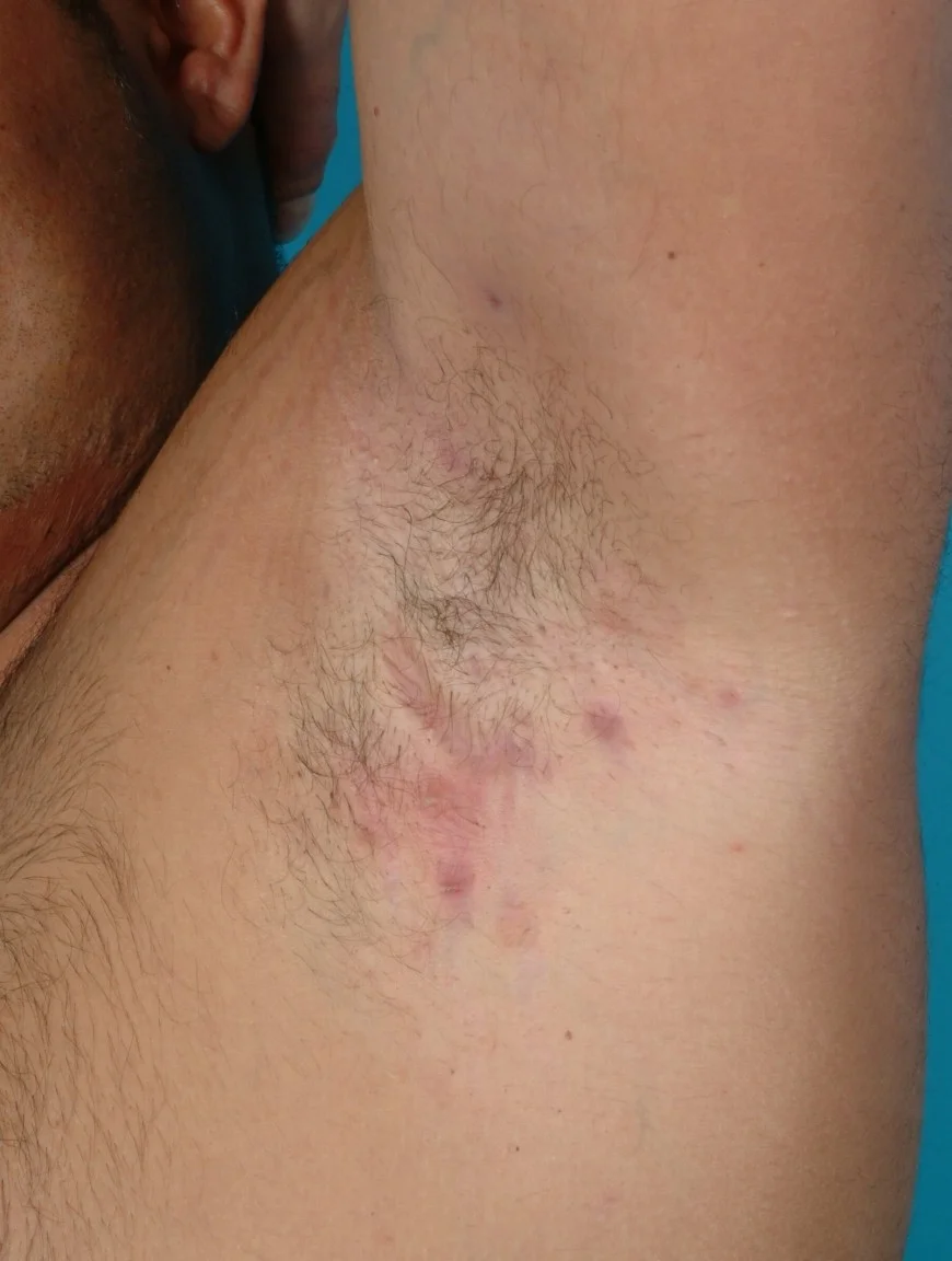
Long description
A clinical photograph titled 'Hidradenitis suppurativa (Hurley's Staging II) in the left axilla'.

Figure 2Pigmented and tenderness dermatofibroma
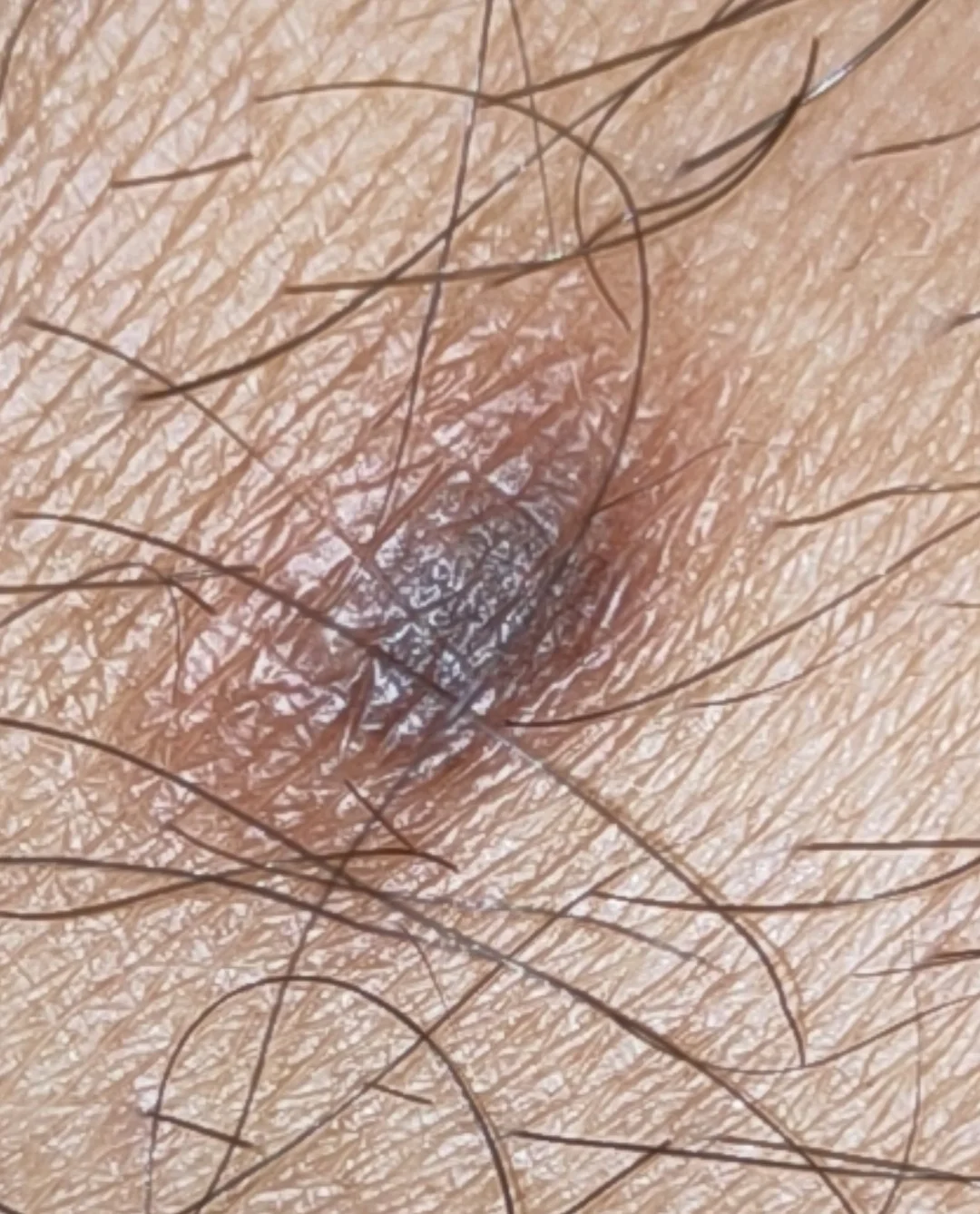
Long description
A pigmented and tender dermatofibroma on the leg of a 34-year-old man.

Figure 3Inflamed epidermal inclusion cyst
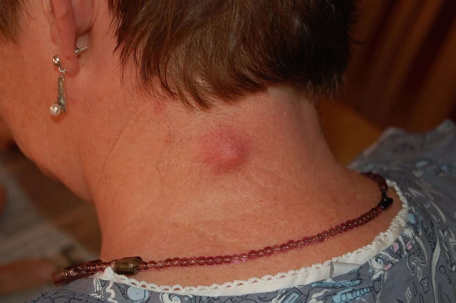
Long description
An inflamed epidermoid cyst (aka epidermal inclusion cyst or sebaceous cyst) on the neck.

Diagnosis of HS is clinical. There is no laboratory test for a typical lesion. The EHSF and BAD guidance suggest baseline recording and screening:
- Record site, lesion count (nodules, abscesses, tunnels) and Hurley stage; calculate IHS4 at baseline and at follow-up.
- Pain score and DLQI (or Skindex).
- Screen for cardiovascular and metabolic risk: BMI, blood pressure, lipids, HbA1c; depression and anxiety (NICE CKS also lists mental-health and pain support as part of primary-care management).
- Swab draining lesions if atypical; blood cultures only if systemic infection is suspected.
- Ultrasound (local) can detect early tunnels not visible clinically and refine Hurley stage.
- Punch biopsy only when a lesion is atypical or to exclude malignancy; skin biopsy for recurrent chalazion in the elderly.
- Bioclinical: no routine tests for cysts, dermatofibroma or xanthelasma. Fasting lipids and thyroid function for new xanthelasma, particularly in the young.
HS diagnosis rests on three clinical criteria: (1) nodules, (2) in typical sites, (3) chronicity or recurrence. Hurley stage describes fixed damage (I/II/III); IHS4 grades inflammatory activity (mild 3 or fewer, moderate 4 to 10, severe 11 or more points). Refined Hurley staging addresses the limits of the original three-stage system for dynamic assessment[2]. Table above gives thresholds. [1]
For ichthyosis vulgaris, diagnosis is clinical with family history; FLG genotyping is mainly a research and specialist-dermatogenetics tool. Histology shows reduced or absent F-type keratohyaline granules and reduced filaggrin.
Approach to a patient with a painful nodule or skin lump in the differential of HS and cysts
- Presentation with a painful or persistent skin lesionCharacterise site, depth, tunnel, abscess and scar
- Question: Inflammatory deep nodule or abscess in an apocrine site (axilla, groin, buttocks, inframammary)?
Yes
- Hidradenitis suppurativaRecord Hurley stage, IHS4, sites, lesion counts and quality of life
- Mild (IHS4 3 or fewer, Hurley I or mild II)Topical clindamycin 1% twice daily up to 12 weeks, or oral tetracycline for 3 months; plus general measures
- Moderate to severe (IHS4 4 or more)Clindamycin plus rifampicin 2 x 300 mg/day orally 10 to 12 weeks; reassess at 12 weeks
- Non-response or fixed tunnelsStep to adalimumab, secukinumab or bimekizumab, and add deroofing or wide excision under cover of medical therapy
No
- Question: Central punctum or expresses keratin?
Yes
- Epidermal inclusion cystObserve if small; excise completely with intact wall if symptomatic or recurrent
No
- Question: Eyelid lesion?
Chronic tarsal nodule
- ChalazionWarm compresses, lid hygiene; steroid or incision and curettage if persistent; biopsy atypical or recurrent
Acute tender pustule
- HordeolumWarm compresses 7 to 10 days; topical antibiotics; incision if pointing
Yellow plaque
- XanthelasmaFasting lipids, thyroid and glucose; cosmetic removal optional
- Question: Firm limb nodule 1 cm or less with central dimple on pressure?
Yes
- DermatofibromaReassure; excise completely if symptomatic, cosmetically troublesome or atypical
No
- Other differential (furuncle, malignancy, ichthyosis, age change)Biopsy evolving, atypical, pigmented or recurrent lesions
All HS patients should receive general advice: stop smoking, weight management, antiseptic washes (chlorhexidine 4% or octenidine), avoid tight clothing and shaving of affected areas, pain relief (NSAIDs first line; acetaminophen; topical lidocaine; neuropathic agents for chronic pain), wound care with absorbent atraumatic dressings, and a patient information leaflet. Screen for and treat depression, anxiety, sexual dysfunction and pain burden. Offer psychosocial support.
For an acute flare: oral antibiotics (often the same agents used for maintenance), incision and drainage of a pointing abscess, intralesional triamcinolone 10 to 40 mg/mL for an inflamed nodule or tunnel, and short courses of systemic corticosteroids (0.5 to 0.7 mg/kg/day prednisolone equivalent for flares, rapidly tapered; or 10 mg/day as adjunct in refractory disease)[1].
The EHSF treatment ladder by inflammatory severity[1], mirrored by AAD/HSF 2026 and BAD 2018[3][4]:
-
Mild (IHS4 3 or fewer, Hurley I or mild II): topical clindamycin 1% gel/lotion/cream twice daily in a flare for up to 12 weeks (may be prolonged if indicated); or oral tetracyclines (doxycycline 100 mg twice daily or equivalent, such as lymecycline 300 mg/day, or subantimicrobial modified-release doxycycline 40 mg/day) for 3 months. Tetracyclines should not be given to pregnant women or children under 9 because of tooth discoloration[1]. Oral clindamycin 2 times 300 mg/day for 12 weeks or intravenous clindamycin 3 times 600 mg/day over 5 days are alternatives. Topical resorcinol 15% twice daily in flares and once daily for maintenance up to 16 weeks is useful for localised mild disease without tunnels, and can give rapid relief. [1]
-
Moderate to severe (IHS4 4 or more): the oral combination clindamycin and rifampicin, each 2 times 300 mg/day orally for 10 to 12 weeks, is equally effective to tetracyclines in most patients. Rifampicin strongly induces cytochrome P450 and reduces clindamycin plasma levels; the combination may also reduce oral contraceptive efficacy, so counsel on contraception. Dapsone 50 to 200 mg/day (minimum 3 months) for mild-to-moderate disease where first-line agents fail (not teratogenic but avoid in breastfeeding). Ertapenem 1 g/day intravenously for 6 weeks for severe disease or down-staging before surgery. Colchicine, acitretin for men and non-fertile women, and other agents are specialist options. [1]
-
Biologics (moderate to severe, inadequate response to conventional systemic therapy):
- Adalimumab (TNF-alpha inhibitor): EMA- and FDA-approved for active moderate to severe HS in patients 12 years and older. Dosing: adults, 160 mg on day 1, 80 mg on day 15, then 40 mg weekly or 80 mg every 2 weeks; for 12 years and older and 30 kg or more, 80 mg day 1 then 40 mg every 2 weeks from day 8, escalating to 40 mg weekly or 80 mg every 2 weeks if response is inadequate. Re-introduction after a break is 40 mg weekly or 80 mg every 2 weeks. Antibiotics may be continued. Adalimumab continuation for at least 2 years maintains response with an acceptable safety profile. The AAD/HSF 2026 guideline strongly recommends adalimumab among others[3]. [1]
- Secukinumab (IL-17A inhibitor): EMA- and FDA-approved for adult moderate to severe HS with inadequate response to conventional therapy. Dosing 300 mg at weeks 0, 1, 2, 3 and 4, then monthly maintenance; increase maintenance to 300 mg every 2 weeks if response is inadequate. [1]
- Bimekizumab (IL-17A/F inhibitor): EMA-approved for adults with moderate to severe HS with inadequate response to conventional therapy. Dosing 320 mg subcutaneously every 2 weeks for 16 weeks, then every 4 weeks. [1]
- NICE recommends adalimumab (technology appraisal TA392, 2016) and secukinumab (TA935) for moderate to severe HS after conventional systemic therapy has failed; secukinumab only if adalimumab is unsuitable, ineffective or lost effect. NICE CKS supports primary-care recognition, baseline assessment and referral.
- Other (off-label/specialist): infliximab, ustekinumab, upadacitinib (JAK inhibitor), spironolactone for women (85% lesion reduction and 55% complete clearing reported in small studies; teratogenic - avoid in pregnancy)[3], intravenous ertapenem, hormonal and metabolic measures.
Response should be assessed at about 12 weeks (IHS4 or HiSCR plus DLQI/pain) and therapy modified if there is no significant reduction in inflammatory activity or quality of life[1].
Exam trapAntibiotics are not treating an infection
In HS, antibiotics work as anti-inflammatory and biofilm-breaking drugs at the doses above, often for 12 weeks or more. Cultures are usually sterile or show secondary colonisers. Prolonged courses are standard practice, not misuse, but review at 12 weeks and step up therapy if there is no response.
Surgery is for the predominantly non-inflammatory form and is chosen by Hurley stage[1]:
- Hurley stage I/early II: incision and drainage of an acute abscess is a temporising measure only; deroofing (unroofing) of tunnels under local anaesthesia is an effective office procedure; CO2 laser evaporation for focal lesions.
- Hurley stage II/III: wide surgical excision of irreversibly damaged tissue, with healing by secondary intention, skin grafting or flap reconstruction; CO2 laser excision (low recurrence, 1.1% over 1 to 19 years in one series). [1]
- Recurrence rates from the EHSF table: deroofing 17% (median 3 years), CO2 laser evaporation 29% (1 year), wide excision with secondary intention 12 to 19% in meta-analysis and 24% at 5 years in one large study. [1]
Medical therapy is not stopped for surgery; continuing adalimumab through the perioperative period improves outcomes. In the SHARPS trial, adding adalimumab 40 mg weekly before (12 weeks), during (2 weeks) and after (10 weeks) radical surgery achieved HiSCR-type benefit in 48% versus 34% with surgery plus placebo (p=0.049)[1]. A retrospective series combining infliximab with surgery (deroofing/incisions) left 37% disease-free at 50 months versus 13% with biologic alone[1].
Small uncomplicated cysts need no treatment. When removed, the intact cyst wall must be excised to prevent recurrence; initial incision and drainage alone commonly recurs. Local anaesthesia with epinephrine is preferred. Electrocautery and laser ablation are options but may leave small scars. Ruptured cysts produce a foreign-body granulomatous reaction and should be treated with anti-inflammatory measures and, if infected, appropriate antibiotics. Counsel that these cysts are benign; rare malignant transformation (most often to squamous cell carcinoma, then basal cell carcinoma) is documented but exceptional[5].
- Chalazion: self-limiting in many; conservative treatment with warm compresses, lid massage, lid hygiene and, if needed, topical antibiotic-steroid drops. Intralesional diluted triamcinolone or 5-FU is effective; incision and curettage is standard for persistent or large lesions (inner-conjunctival approach, no visible scar). Systemic tetracyclines (doxycycline) are used for severe or recurrent disease and associated meibomian gland dysfunction. Refer any atypical, recurrent (especially in older patients) or pigmented chalazion for biopsy to exclude sebaceous carcinoma[10].
- Hordeolum: conservative first - warm compresses about 8 or 9 days, two or three times daily, about 8 minutes each, eyelid scrubbing, topical antibiotic drops/ointment. Oral antibiotics if extensive or recurrent; incision and drainage if pointing and persistent. Topical antibiotics are preferred first-line; most specialists give 5 to 14 days of non-surgical care before procedures[10].
- Xanthelasma: treat the underlying lipid disorder (lifestyle, statins, thyroid and diabetes control) first; efficacy of cholesterol-lowering on lesions is variable and slow. Cosmetic removal by surgical excision (with or without blepharoplasty), cryotherapy, trichloroacetic acid peels, radiofrequency or laser. Choice follows size and site; recurrence is common, particularly with argon laser and when hyperlipidaemia persists. No premalignant potential[9].
No treatment is needed for the typical asymptomatic lesion; reassurance is often safer than excision, because the scar may be worse than the nodule. Excise completely when symptomatic, cosmetically bothersome or atypical. Cryosurgery or shave removal are alternatives but recur more often. Consider biopsy for rapid growth, large size, lymphadenopathy or immunosuppression. Multiple eruptive dermatofibromas should trigger screening for HIV and lupus[6].
No cure; aim is barrier repair and scale removal:
- Emollients with urea, lactic acid or salicylic acid; bath oils; avoidance of drying soaps.
- Topical retinoids for thick scale (specialist use, avoid in pregnancy).
- Advise sun protection and skincare; monitor for atopic dermatitis, asthma, allergic rhinitis and nickel/irritant sensitivity; check vitamin D.
- Screen relatives; counsel on semidominant inheritance.
- Emollients, soap substitutes and regular lipid-rich moisturisers; avoid over-washing.
- Treat asteatotic eczema, seborrhoeic dermatitis, pruritus and xerosis proactively.
- Full skin examination with dermoscopy; treat actinic keratoses (cryotherapy, 5-fluorouracil, photodynamic therapy) and remove suspicious lesions; vitamin D and nutrition; emollients reduce skin tears.
- Repair skin tears with paper tape/strip closure rather than sutures when appropriate.
- HS is mostly diagnosed clinically; teach pattern recognition (deep tender nodules, tunnels, apocrine sites, double-ended comedones). Where ultrasound is unavailable, rely on examination.
- Oral doxycycline or tetracycline and topical clindamycin are cheap and widely available; combine with antiseptics and weight/smoking advice.
- Biologics and many surgical options may not be available; refer complex cases early to a surgical or dermatology centre. Incision and drainage plus antibiotics prevents some acute morbidity.
- Cysts can be excised with local anaesthesia; dermatofibroma needs no treatment unless symptomatic.
- Xanthelasma can be observed; treatment of lipids takes precedence.
HS: scarring with contracture and sinus tracts, lymphoedema, fistulae to urinary or genital tract in perianal disease, anaemia of chronic disease, squamous cell carcinoma in long-standing perianal tunnels (rare but reported), sepsis, pain, depression, sexual dysfunction, infertility from scarring, impaired mobility.
HS treatment complications: tetracycline tooth staining (avoid under 9 and in pregnancy), rifampicin drug interactions (including reduced efficacy of oral contraceptives), biologic infection risk including TB reactivation (screen before starting), corticosteroid effects.
Cysts: recurrence after incomplete excision, rupture and foreign-body granuloma, secondary infection, (rare) malignant change. Dermatofibroma: cosmetic, (rare) local recurrence after incomplete removal, (rare) metastasising variants. Xanthelasma: recurrence after removal while lipid disorder persists; ophthalmic complications are not usually caused by the plaque itself. Ageing: skin tears, pressure injury, infection, melanoma and non-melanoma skin cancer. Ichthyosis: fissures, secondary infection, barrier-related atopic disease.
HS is chronic and relapsing; there is no cure. Early, escalating treatment slows progression and preserves quality of life; surgical removal of fixed tunnels is the only durable remedy for that tissue. Severity often lessens after menopause in women. Mental-health and pain support are part of routine care.
Cysts, dermatofibroma, xanthelasma and age-related change have an excellent or indolent prognosis; xanthelasma and cysts recur if the underlying trigger persists. Ichthyosis vulgaris improves with age and humidity and is managed, not cured.
HS: no prophylaxis; reduce smoking and obesity, avoid friction and trauma to apocrine sites, treat early. NICE CKS and BAD guidance both frame HS as a disease to recognise early in primary care. Screening for cardiovascular/metabolic risk, depression and pain at baseline and annually. Biologic patients need infection, TB and hepatitis screening per local protocol, and pregnancy counselling.
Xanthelasma: screen lipids, thyroid and glucose; cardiovascular risk assessment. Dermatofibroma: none. Ichthyosis: genetic counselling; barrier and atopy prevention in children. Ageing: sun protection, skin self-examination, regular dermoscopic review in high-risk patients.
- Pregnancy: tetracyclines contraindicated (tooth and bone effects); many biologics avoided (adalimumab is generally stopped in the third trimester; secukinumab/bimekizumab data are limited; infliximab may be used in selected cases). Prednisolone can be used short term with the usual cautions. Spironolactone is contraindicated. HS often worsens in pregnancy due to hormonal change. Surgery is deferred where possible. Intralesional steroids are used with caution. NICE and BAD guidance support multidisciplinary care.
- Children: HS is rare before puberty; when present, consider familial forms and refer. Tetracyclines are contraindicated under 9; adalimumab is licensed from age 12. Dermatofibroma is rare in children. Ichthyosis vulgaris is diagnosed in infancy/childhood. Eyelid lesions (chalazion, hordeolum) are common in children; 81% of specialists extend conservative management and delay surgery in paediatric cases[10].
- Older adults: HS occurs but is less common; HS-like nodules in the elderly can be hidradenocarcinoma, so biopsy persistent, unilateral or suspicious lesions. Ageing skin changes dominate dermatology in older adults; cancer surveillance is central.
- Most of this group is diagnosed clinically; teaching pattern recognition (deep tender nodules with tunnels, punctum-bearing cyst, dimple sign) replaces tests that are unavailable.
- Tetracyclines, topical clindamycin, antiseptics, emollients and incision and drainage are low-cost and widely available.
- Refer complex HS (Hurley stage III, large cysts, suspected malignancy) rather than attempting biologic therapy locally.
- Acne vulgaris
- Rosacea and perioral dermatitis
- Psoriasis ( inverse psoriasis differential)
- Skin cancer (basal cell, squamous cell, melanoma)
- Atopic dermatitis and skin barrier
- Wound healing and surgical site infection
- Drug eruptions and hypersensitivity
- Neurofibromatosis and genodermatoses
- HS is a chronic follicular inflammatory disease: follicular occlusion, rupture and dermal inflammation, not a sweat-gland infection. Stage and treat by IHS4 (inflammatory) and Hurley (structural) severity.
- First-line therapy for mild HS: topical clindamycin 1% twice daily (up to 12 weeks) or oral tetracyclines for 3 months. For moderate to severe HS, approved biologics are adalimumab (from age 12), secukinumab and bimekizumab (adults).
- Adalimumab dosing: adults 160 mg day 1, 80 mg day 15, then 40 mg weekly (or 80 mg every 2 weeks); 12 years and older: 80 mg day 1 then 40 mg every 2 weeks, escalating as needed. Assess at 12 weeks and escalate if no response.
- Surgery is chosen by Hurley stage; combining adalimumab with radical surgery improved outcomes (48% vs 34%) in the SHARPS trial.
- Epidermal inclusion cysts are benign keratin-filled nodules, male-predominant, rare before puberty, with a central punctum; remove with intact wall to prevent recurrence.
- Chalazion is a sterile lipogranuloma of a meibomian gland; hordeolum is an acute bacterial lid-gland infection; both usually resolve with warm compresses, lid hygiene and time.
- Xanthelasma is a cholesterol-rich yellow eyelid plaque with 1.1% prevalence in women and 0.3% in men; it can mark dyslipidaemia but is harmless and has no premalignant potential.
- Dermatofibroma is a firm red-brown nodule of 1 cm or less on a limb, often with a central dimple on pressure; usually needs no treatment. [6]
- Ichthyosis vulgaris is autosomal semi-dominant FLG-related xerosis and scaling, 7.7% of Europeans carrying FLG mutations, strongly associated with atopy.
- Age-related skin thinning reduces lipids by up to 65% and raises risks from asteatotic eczema to skin cancer; management is emollients, sun protection and surveillance.
- NICE, BAD and EHSF guidance all stress early recognition and referral of HS in primary care.
Answer each one in your head first, then open the answer.
A 24-year-old smoker with BMI 33 presents with recurrent tender nodules and a draining sinus in both axillae and the groin, with rope-like scars. What is the diagnosis and what two severity measures do you use?
Show answerHide answer
Hidradenitis suppurativa. Grade inflammatory severity with IHS4 (mild 3 or fewer, moderate 4 to 10, severe 11 or more points, from nodules, abscesses and draining tunnels) and structural damage with Hurley stage (I/II/III). Both guide drug and surgical choice.
A patient has mild HS with a few inflammatory nodules and no tunnels. Which two medical options are appropriate first-line, and for how long?
Show answerHide answer
Topical clindamycin 1% twice daily for up to 12 weeks, or an oral tetracycline (for example doxycycline 100 mg twice daily) for 3 months. Tetracyclines are avoided in pregnancy and in children under 9.
The same patient has not responded after 12 weeks. She is 26, moderate to severe, otherwise well. Outline the next steps in order.
Show answerHide answer
Escalate to clindamycin plus rifampicin 2 times 300 mg/day orally for 10 to 12 weeks (or continue tetracycline); reassess at 12 weeks by IHS4 and quality of life. If still inadequate, an EMA/FDA-approved biologic: adalimumab 160 mg day 1, 80 mg day 15, then 40 mg weekly (first choice in many algorithms), else secukinumab 300 mg at weeks 0 to 4 then monthly, or bimekizumab 320 mg every 2 weeks for 16 weeks then every 4 weeks. Add deroofing or wide excision for fixed tunnels (Hurley II/III), and a surgical opinion early.
A firm 8 mm red-brown nodule on a shin indents in the centre when you pinch it laterally. What is it and what do you do?
Show answerHide answer
Dermatofibroma. Reassure; no treatment needed unless symptomatic or cosmetically bothersome, then complete surgical excision (cryotherapy/shave recur more). Biopsy if large, fast-growing, or in an immunosuppressed patient.
A child has dry, fine, grey polygonal scale on the extensor legs, keratosis pilaris on the arms and marked palmar lines. The mother has eczema. Most likely diagnosis and inheritance?
Show answerHide answer
Ichthyosis vulgaris (FLG mutation), autosomal semi-dominant. Heterozygotes are milder; homozygotes more severe. Strongly associated with atopic dermatitis.
A painless yellow plaque sits at the inner corner of each upper eyelid in a 42-year-old woman. What is it, what labs do you order, and why is she reassured?
Show answerHide answer
Xanthelasma palpebrarum. Order fasting lipids, thyroid function and glucose because it can associate with dyslipidaemia, hypothyroidism and diabetes, even though up to half of patients have normal lipids. Reassure: no premalignant potential, no threat to vision.
An epidermal inclusion cyst has been incised and drained twice and keeps refilling. Why, and what is the definitive step?
Show answerHide answer
Drainage alone leaves the cyst wall in place, so recurrence is expected. Definitive treatment is complete surgical excision of the intact cyst wall under local anaesthesia.
- Zouboulis CC, Bechara FG, Benhadou F, et al. European S2k guidelines for hidradenitis suppurativa/acne inversa part 2: Treatment. J Eur Acad Dermatol Venereol 2025;39:899-941. doi:10.1111/jdv.20472. Accessed .
- Pandey A. Essentials of hidradenitis suppurativa: a comprehensive review of diagnostic and treatment perspectives. Arch Med Surg (PMC11374290). Accessed .
- American Academy of Dermatology. Hidradenitis suppurativa: diagnosis and treatment. AAD patient guidance (citing the joint AAD-HSF guideline, published online 20 Oct 2026). Accessed .
- Ingram JR, Collier F, Brown D, et al. British Association of Dermatologists guidelines for the management of hidradenitis suppurativa (acne inversa) 2018 (summary). HS Foundation summary of Br J Dermatol 2019;180:1009-1017. doi:10.1111/bjd.17537. Accessed .
- PMC6732711 (StatPearls-cited review). Overview of epidermoid (epidermal inclusion) cyst. Int J Dermatol educational review (open access). Accessed .
- PMC12333637 (Dove Medical Press). Dermatofibroma: Reappraisal and Updated Review. Clin Cosmet Investig Dermatol 2025. Accessed .
- Thyssen JP, et al. Ichthyosis vulgaris: the filaggrin mutation disease. Br J Dermatol 2013;168:1155-1166. doi:10.1111/bjd.12219. Accessed .
- Farage MA, et al. Characteristics of the aging skin. Adv Wound Care 2013;2:5-11. doi:10.1089/wound.2011.0356. Accessed .
- Nair PA, Singhal R. Xanthelasma palpebrarum - a brief review. Clin Cosmet Investig Dermatol 2018;11:1-5. doi:10.2147/CCID.S130116. Accessed .
- Shin HJ, et al. Management practice for hordeolum and chalazion: survey of KSOPRS members. Korean J Ophthalmol 2025;39:222-240. doi:10.3341/kjo.2025.0024. Accessed .
Figure credits
Educational content for medical students and doctors. It is not a substitute for clinical judgement or local guidelines.