Basal cell carcinoma, squamous cell carcinoma and Merkel
How BCC, SCC and Merkel cell carcinoma present, are risk-stratified and excised, and when radiotherapy and systemic therapy are used.
- Keratinocyte cancers (basal cell carcinoma and squamous cell carcinoma) are the common skin cancers and are usually cured locally.
- Risk tracks cumulative ultraviolet light, fair skin, age, and the intensity of immunosuppression.
- Basal cell carcinoma grows locally with very low metastatic potential; squamous cell carcinoma can metastasise, especially when high risk.
- Excision with measured margins, Mohs micrographic surgery for selected high-risk or anatomically sensitive tumours, radiotherapy and topical or field treatments all have defined roles.
- Merkel cell carcinoma is a rare neuroendocrine skin cancer with early nodal spread and a 48 to 63% five-year survival across all stages.
- Cutaneous lymphoma is a separate group, dominated by mycosis fungoides, managed by disease stage rather than by simple excision.
By the end of this page you should be able to:
- Recognise the clinical types of basal cell carcinoma and cutaneous squamous cell carcinoma and list their high-risk features.
- Explain why immunosuppression raises the risk of skin cancer and how management changes in transplant recipients.
- Choose an excision margin for a given cutaneous squamous cell carcinoma using the published guideline thresholds.
- Describe how Mohs micrographic surgery differs from standard excision and when it is indicated.
- State the radiotherapy doses used for Merkel cell carcinoma and the systemic options for advanced disease.
- Recognise Merkel cell carcinoma clinically and distinguish it from keratinocyte cancers and cutaneous lymphoma.
- Outline how cutaneous T-cell lymphoma is staged and why its management differs.
This topic groups three skin cancers: basal cell carcinoma (BCC), a slow-growing tumour of basaloid cells that almost never metastasises; cutaneous squamous cell carcinoma (cSCC), an invasive cancer of keratinocytes that can spread to lymph nodes; and Merkel cell carcinoma (MCC), a rare aggressive neuroendocrine cancer of the skin. Despite different cell origins they share sun-exposed sites, ultraviolet light as a driver and decisions about excision, margin control, radiotherapy and systemic therapy. A short overview of cutaneous lymphoma is included because it is the other major skin cancer group a dermatology clinic sees. [3]
Keratinocyte carcinomas (BCC and cSCC) are tumours of the epidermis or its adnexae. They are collectively the most common human cancers. Non-melanoma skin cancer (NMSC) includes both; in one recent global analysis the age-standardised incidence of all NMSC rose from 45.0 to 74.1 per 100 000 people between 1990 and 2021.[6] BCC accounts for about 75% of NMSC and is the most common human cancer overall, whereas cSCC accounts for up to 25% of NMSC but is the leading cause of NMSC death.[6][5]
MCC is not a keratinocyte cancer: it is a neuroendocrine tumour of the skin, characterised by small round blue cells and expression of cytokeratin 20 with a paranuclear dot pattern.
Cutaneous lymphomas are clonal proliferations of skin-homing lymphocytes. The commonest is mycosis fungoides (MF), usually an indolent disease of patches and plaques; Sézary syndrome is the leukemic, erythrodermic, aggressive counterpart.
CSCC makes up roughly 20 to 25% of skin tumours.[3] Exact counts are uncertain because many registries group cSCC with mucosal or metastatic forms and because some in-situ lesions are never counted. England's 2013 to 2015 data gave first registered cSCC rates of 77.3 per 100 000 person-years for men and 34.1 for women, and 2.4% of men and 1.1% of women with cSCC developed metastases within 36 months.[3] In New South Wales, where overall NMSC rates are very high, cSCC occurred at about 856 cases per 100 000 from 2016 to 2019.[3] Over 90% of primary cSCC patients have a favourable prognosis, but immunosuppressed and socially disadvantaged patients are over-represented among the advanced cases.[3]
Merkel cell carcinoma is rare everywhere, and global rates are hard to calculate. The RARECARE database gave a European crude incidence of 0.13 per 100 000 in 1995 to 2002.[4] SEER rates in the United States rose from 0.22 per 100 000 in 1986 to 0.79 per 100 000 in 2011, with mortality rising from 0.03 to 0.43 per 100 000 over the same period.[4] Highest rates are in Australia, where the age-adjusted rate in Queensland was 1.6 per 100 000.[4] MCC appears at a median age of 76 years, affects Caucasian men about eight times more often than people of colour and about twice as often as women.[4] Ten percent of MCC patients are immunosuppressed (organ transplant recipients, haematological malignancy or HIV infection).[4] In Europe and North America roughly 80% of MCC is driven by Merkel cell polyomavirus and 20% by ultraviolet damage; in Australia this is reversed.[4]
The dominant carcinogen for both BCC and cSCC is ultraviolet radiation (UVR), both solar and artificial (psoralen plus UVA, indoor tanning). Chronic cumulative exposure explains the rise with age and the higher rates at lower latitudes. In most cSCC the tumour arises on chronically sun-exposed skin of the head and neck and the dorsa of the hands and forearms, and is more common in outdoor workers. [3]
Genetic skin type matters: fair (Fitzpatrick I to II) skin is more susceptible, and this acts synergistically with exposure. Genodermatoses (xeroderma pigmentosum, oculocutaneous albinism, epidermodysplasia verruciformis) and chronic inflammation such as burns, scars, chronic ulcers and epidermolysis bullosa all raise cSCC risk. Human papillomavirus plays a part, particularly in immunosuppressed patients and in digital, anal and genital squamous carcinoma.
Immunosuppression and skin cancer is a major theme. Immunosuppression impairs immune surveillance against tumour cells and against human papillomavirus. Solid organ and haematopoietic stem-cell transplant recipients, patients on systemic immunosuppression for autoimmune disease, patients with haematological malignancy and those with advanced solid cancer are all at increased risk. Iatrogenic immunosuppression increases the risk of cSCC by a factor of 40 to 250 compared with the general population, depending on the graft, skin phototype, ultraviolet exposure and the degree and duration of immunosuppression.[6] cSCC, not BCC, is the dominant tumour in transplant recipients: it is more frequent, more numerous and more aggressive in that population, and is the leading cause of NMSC death. The same immunosuppression also raises MCC risk: 10% of MCC patients are immunosuppressed.[4]
Other chemical and treatment-related triggers matter: arsenic exposure, chronic PUVA, voriconazole and BRAF inhibitors (which can accelerate the growth of ultraviolet-damaged keratinocytes and promote SCC) raise risk.
Prevention remains inconsistent. Sunscreen SPF 30 to 50 is a sensible part of photoprotection for exposed people but the evidence for cSCC prevention is of very low quality; in the Nambour trial regular sunscreen reduced the number of cSCC per person but not the proportion of people developing one.[3] Chemoprevention with oral retinoids or nicotinamide has a small but real effect on new lesion counts in high-risk patients, and is usually reserved for immunosuppressed patients or those with numerous lesions.
| Factor | Effect |
|---|---|
| Cumulative ultraviolet exposure | Main driver of both BCC and cSCC; explains age-related and latitude-related incidence |
| Fair skin phototype | Synergistic with ultraviolet damage |
| Intensity of immunosuppression | 40 to 250 times cSCC risk in transplant recipients; raises BCC and MCC risk |
| Genodermatoses (xeroderma pigmentosum, albinism) | Markedly raised risk, often in young patients |
| Chronic inflammation (scars, ulcers, epidermolysis bullosa) | Predisposes to cSCC |
| Human papillomavirus | Promotes cSCC, especially digital, anal and genital |
| Age, male sex | Raised incidence of cSCC and MCC |
BCC grows as lobules or strands of basaloid cells with peripheral palisading. Nodular BCC is the commonest facial type; superficial BCC lies on the trunk; morphoeic (sclerosing) and micronodular or infiltrative types have ill-defined margins and invade deeply; basosquamous carcinoma has mixed features and a worse local course.
Morphological growth pattern matters more than size. Well-defined tumours (nodular, superficial) are lower risk; infiltrative, micronodular, morphoeic and basosquamous growth patterns are high risk and are the classic indications for Mohs surgery. They spread along tissue planes with "roots" invisible clinically, which is why incomplete excision and recurrence are common. Clinically, BCC presents as a pearly or waxy nodule with fine telangiectasia, an ulcer with a raised rolled edge, or a slowly enlarging pink plaque.
cSCC ranges from actinic keratosis and Bowen disease (squamous cell carcinoma in situ) to invasive tumour. Invasive cSCC is usually a firm scaly nodule, plaque or keratotic horn on sun-exposed skin. It may be verrucous, poorly differentiated, spindle-cell or desmoplastic, and can arise in old scars, chronic ulcers or fields of radiotherapy (Marjolin ulcer).
Staging is by AJCC8 or the Brigham and Women's Hospital system. The latter is more specific for predicting nodal metastasis and disease-specific death, because AJCC8 leaves a broad T2/T3 group containing about 23% of head and neck cSCC patients, whereas the Brigham system identifies the same poor outcomes within only about 9% of cases.[6]
MCC classically presents as a painless, firm, rapidly growing, dome-shaped red-violaceous or pink nodule or plaque, 1 to 2 cm in size, on sun-exposed skin of older patients. Sites: head and neck (45%), upper limb (24%), lower limb (10%), trunk or other (under 10%); in 11% no primary is found. Ulceration occurs in advanced lesions. It grows fast, spreads in transit through skin and to first-echelon nodes (26% at presentation) and then to distant sites such as bone, liver and brain (8% at presentation).[4] It is easily mistaken for BCC, cSCC, amelanotic melanoma, adnexal tumours, primary cutaneous B-cell lymphoma or pyogenic granuloma. Dermatoscopy may show polymorphous vessels or milky-red areas.
- BCC vs cSCC vs MCC vs melanoma: dermoscopy, clinical morphology, and biopsy with immunohistochemistry. MCC needs H and E with an immunopanel: CK20 paranuclear dot, chromogranin A, synaptophysin, CD56, neurofilament and negative melanoma (S100, SOX10, Melan-A), CK7 and TTF1 markers in most cases.
- BCC vs intradermal naevus or sebaceous hyperplasia: pearly nodules lack the uniform yellow globules of sebaceous lesions and dermoscopy helps.
- cSCC vs keratoacanthoma vs actinic keratosis: biopsy when invasive change (induration, ulceration, tenderness, rapid growth, diameter over 1 cm) is suspected.
- MCC vs primary cutaneous B-cell lymphoma, adnexal tumours, small-cell lung cancer metastasis: tissue diagnosis is essential.
- cSCC in transplant recipients vs warty viral lesions, porokeratosis, seborrhoeic keratosis: biopsy suspicious or changing lesions.
A mass seen in the skin of an immunosuppressed patient should be biopsied rather than frozen or "watched".
Figure 1BCC Nodular type
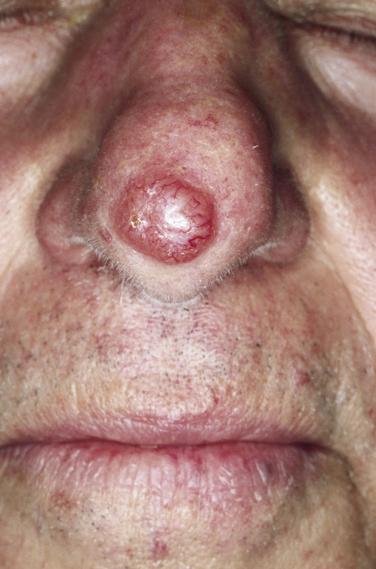
Long description
A colour clinical photograph of a basal cell carcinoma described as a red, waxy nodule on the tip of the nose with visible telangiectasias over the surface.

Figure 2Advanced squamous cell carcinoma scalp
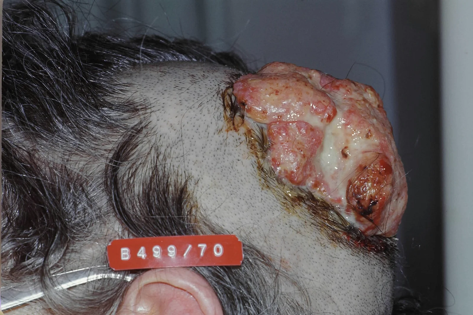
Long description
Clinical photograph described as a neglected squamous cell carcinoma of the scalp (dated 1992), used in the English Wikipedia article on cutaneous squamous-cell carcinoma.

Biopsy is the diagnostic step for all three. A punch or shave biopsy suffices for BCC; incisional or excisional biopsy with adequate tissue is needed for cSCC and MCC. For MCC an excisional biopsy with the histology report should record tumour size, involvement of fascia, muscle, cartilage or bone, surgical margins, tumour depth, lymphovascular invasion, intratumoural lymphocyte infiltration, immunohistochemical profile, polyomavirus status and mitotic rate.
For cSCC, histopathology defines grade, depth, Clark level, perineural and lymphovascular invasion, margin status, desmoplasia and high-risk size. The histological thresholds that raise risk include thickness over 2 mm and depth to Clark level IV or V for Mohs indications, poor differentiation, perineural or lymphovascular invasion. High-risk cSCC markers include a vertical thickness over 6 mm with high-risk features or invasion of subcutaneous tissue in the source guidelines.
For MCC, staging studies depend on presentation. Ultrasound of regional nodes for clinical stage I and II disease, CT of chest, abdomen, pelvis and head and neck for head and neck primaries; if available, whole-body 18F-FDG PET-CT is preferred over contrast-enhanced CT alone, because PET-CT upstages about 16.8% of patients versus about 6.9% with CT alone.[4] Sentinel lymph node biopsy with an immunopanel is the most reliable way to find subclinical nodal disease: it is positive in roughly 20% of T1 and 40 to 50% of T2 MCCs, and is false-negative in about 17.1% of immunocompromised patients or head, neck or midline trunk tumours.[4]
Histology in MCC: small blue round cell tumour with a vesicular nucleus and scant cytoplasm, high mitotic and apoptotic rates, infiltration of dermis and subcutis with epidermis usually spared, intra-lymphatic emboli possible. CK20 positive in about 90% (paranuclear dot), CK8/18/19, chromogranin A, synaptophysin, CD56, neuron-specific enolase. CK20-negative cases (under 10%) have high mutational burden and are virus-negative.[4] MCC is usually negative for TTF1, S100, Melan-A, CK7 and vimentin.
MCC uses UICC TNM eighth edition (developed from 9 387 patients in the National Cancer Database). Staging and risk assessment shape management. The stage groups are: no metastases stage I-II with a 64% five-year survival, regional node involvement stage III with 51%, unknown primary with 68%, distant metastases stage IV with 17 to 29%. Unfavourable prognostic factors are nodal or distant metastasis, primary tumour diameter over 2 cm, extension beyond dermis, head or neck site, age over 75 years, male sex and comorbidity, especially immunosuppression. Favourable histological markers are intratumoural CD8+ T cells and viral large T antigen expression; unfavourable are positive margins, high mitotic rate, infiltrative growth, lymphovascular invasion and p63 expression. Median age at diagnosis is 76 years. PD-L1 expression is frequent but is not a prognostic factor.[4]
cSCC staging by AJCC8 or Brigham and Women's Hospital system as noted; BCC is not formally staged because metastasis is rare.
Recognition to treatment of a suspected skin cancer
- Suspicious skin lesionPersistent, growing, bleeding, ulcerated or non-healing lesion on sun-exposed or immunosuppressed skin
- Clinical assessmentSite, size, borders, depth, growth rate, risk factors, immunosuppression
- Question: Does the lesion raise suspicion of squamous cell carcinoma?
Yes
- Suspected cancer pathway referralBiopsy to confirm type, grade, thickness and stage
No - basal cell carcinoma suspected
- Non-urgent referral or excisionTypical low-risk facial BCC can be excised by a trained clinician
- Biopsy if the diagnosis or risk is uncertainMohs indicated only for high-risk or anatomically sensitive tumours
- Multidisciplinary treatment planExcision with measured margins, Mohs surgery, radiotherapy, topical or systemic therapy
The recommendations in this page are based on the following current guidance and reviews, each named with the edition actually used:
- NICE NG12, "Suspected cancer: recognition and referral", published 23 June 2015 and last updated 15 April 2026 (skin cancers section 1.7).[1]
- British Association of Dermatologists (BAD) guideline for basal cell carcinoma in adults, 2021 (Nasr I, et al.; Br J Dermatol 2021;185:899-920).[2]
- Italian Association of Medical Oncology (AIOM) cutaneous squamous cell carcinoma guideline, a GRADE-based guideline published in ESMO Open in 2024.[3]
- ESMO-EURACAN Merkel-cell carcinoma guideline, approved February 2024.[4]
- NCCN Clinical Practice Guidelines in Oncology for basal cell skin cancer, squamous cell skin cancer, Merkel cell carcinoma and cutaneous lymphomas, current versions available through the NCCN website.[8]
- European guidelines quoted in the AIOM guideline: EDF, EADO and EORTC; and the German Dermatology Society recommendations.[3]
The NICE NG12 guideline on suspected cancer (last updated 15 April 2026) organises recognition rules for primary care.[1] For skin cancers:
- Consider a suspected cancer pathway referral if a skin lesion raises the suspicion of squamous cell carcinoma.
- Consider a non-urgent referral if a lesion raises the suspicion of basal cell carcinoma (typical features: ulcer with raised rolled edge, prominent fine vessels around a lesion, nodule that is pearly or waxy).
- A suspected cancer pathway referral for basal cell carcinoma is considered only if there is particular concern that delay may significantly affect the outcome, because of factors such as lesion site or size.
- Basal cell carcinoma excision should follow the NICE skin tumours guidance on who should excise.[1]
A persistent skin lesion that bleeds or does not heal, is rapidly growing, ulcerates, or shows depth/induration in a sun-exposed or immunosuppressed patient should be biopsied promptly, not observed.
United Kingdom (NICE)
In the UK, the 2-week-wait suspected cancer pathway applies to squamous cell carcinoma and to basal cell carcinoma only where delay would be harmful because of site or size. Non-urgent referral is appropriate for a straightforward facial basal cell carcinoma in primary care.
Complete surgical excision with clear pathological margins is the backbone of treatment for both BCC and cSCC. Desmoplastic, infiltrative or high-risk cSCC needs wider margins than low-risk nodular lesions.
The Italian Association of Medical Oncology cSCC guideline (a GRADE-based guideline published in ESMO Open in 2024, endorsed by ESMO) summarises two long-standing and conflicting margin conventions:
- NCCN recommendations based on the Brodland and Zitelli prospective excision study of 1992: well-defined, low-risk cSCC under 2 cm can be excised with a 4 mm margin and complete excision is achieved in over 95% of cases; low-risk lesions over 2 cm need a 6 mm margin.[3]
- For high-risk locations (scalp, ears, eyelids, nose, lips) or high-risk characteristics (histological grade 2 or higher, subcutaneous invasion), lesions under 1 cm, 1 to 1.9 cm and over 2 cm need free margins of 4 mm, 6 mm and 9 mm respectively.[3]
- The European EDF, EADO and EORTC guidelines recommend a minimum 5 mm margin for low-risk tumours (vertical thickness under 6 mm with no other risk factors), and 6 to 10 mm for high-risk tumours.[3]
- The German Dermatology Society recommends a free margin of at least 6 mm for cSCC over 2 cm in diameter, thicker than 6 mm, or with other high-risk prognostic features, to achieve a 95% complete response rate at 5 years.[3]
Practical conclusions that the Italian guideline supports: margins of 4 mm or more should be considered first-line for low-risk cSCC and margins of 6 mm or more for high-risk cSCC (conditional, expert-opinion strength, because there are no comparative trials fixing the minimum safe margin). Excisions with larger free margins trade better oncological control for worse scarring, pain and bleeding, and are not automatically better. [3]
For MCC the ESMO-EURACAN 2024 guideline recommends wide local excision with an adequate margin where feasible, because a resection margin over 2 cm has been associated with improved survival in one publication, while other data do not show additional benefit beyond 1 cm. If an adequate margin is not technically achievable, a narrower margin with adjuvant radiotherapy is also acceptable.[4]
Excisional surgical therapy is highly effective for cSCC, with healing rates near 95%. Mohs surgery is not standard first-line for all SCC; it is reserved for recurrent or high-risk cases (see below). [3]
Mohs micrographic surgery (MMS) is a specialised technique: the tumour is excised with a narrow rim, divided, colour-coded, frozen and examined horizontally so that the whole peripheral and deep margin is checked in real time. Further tissue is removed only where tumour persists, in increments of roughly 1 to 2 mm, until clearance is histologically confirmed.[6] It therefore combines the highest cure rate with the greatest tissue preservation, which is why it is used on the face, around the eyes, nose, lips, ears and genitalia and in cosmetically or functionally important sites. It is also used in older patients needing local anaesthetic, with lidocaine remaining first-line and epinephrine added for haemostasis.[6]
Indications rest on national appropriate-use criteria:
- NCCN and AAD/ACMS/ASDSA/ASMS criteria (USA): area H of the face (central face, eyelids, nose, lips, ears, chin, temples, preauricular sites, plus hands, feet, ankles, nail units, nipple/areola complex and genitalia) is considered high risk at any size; area M (cheeks, forehead, scalp, neck, jawline, pretibial surface) is high risk for most subtypes except superficial BCC under 6 mm in a healthy patient; area L (trunk and extremities excluding special sites) is high risk for recurrent or aggressive or nodular lesions, primary aggressive BCC over 0.5 cm, or nodular BCC over 2 cm in healthy and over 1 cm in immunocompromised patients.[8]
- European guidelines (EADO) recommend MMS for high-risk BCC and cSCC: recurrent tumours, aggressive subtypes (infiltrative, morpheaform, micronodular, basosquamous), perineural invasion, tumours in critical anatomical sites and poorly defined margins. In Europe, outside Germany, Switzerland and the Netherlands, MMS is offered in fewer centres because of cost and infrastructure.
- High-risk SCC indications: MMS for area H at any size; area M tumours of 1 cm or more; area L tumours of 2 cm or more; poorly defined borders; prior radiotherapy or chronic inflammatory process; rapidly growing tumour; neurologic symptoms; high-risk histologic subtype; poorly differentiated; depth 2 mm or more or Clark level IV or V; perineural or lymphovascular involvement; recurrence.
MMS has better cure rates than standard excision. A Dutch randomised trial of facial BCC showed 2.5% versus 4.1% recurrence for primary BCC treated with MMS versus standard excision over 60 months, and 2.4% versus 12.1% for recurrent BCC. A 10-year update found 4.4% versus 12.2% recurrence in primary BCC and 3.9% versus 13.5% in recurrent BCC.[6] A systematic review found an overall recurrence rate of 3.1% after MMS versus 5.3% after standard excision, and a BCC subgroup incidence rate ratio for recurrence of 0.37. For cSCC, one cohort of 579 patients with 672 tumours found 3% recurrence after MMS versus 8% after standard excision, and high-risk cSCC treated with MMS had a 2.9% local recurrence rate.[6] The Italian cSCC guideline found a 2.7% local recurrence rate among 1 045 high-risk or recurrent cSCC treated in MMS literature, and a 12.4% distant metastasis rate in 460 patients.[3][6]
Perioperative MMS care follows common-sense rules that the Mohs literature supports. Oral anticoagulants and antiplatelets are usually continued because thromboembolic risk outweighs bleeding risk; only if much larger excision with flaps or grafts is needed is warfarin stopped 5 days beforehand (INR under 1.5) or direct oral anticoagulants stopped 1 to 2 days beforehand. Routine prophylactic antibiotics are not recommended for most patients, but preoperative single-dose prophylaxis can be considered in immunosuppression, diabetes, radiotherapy, lower-limb reconstruction, flap or graft repair or a facility-based setting. Angiotensin risks and choice of anaesthetic (lidocaine with epinephrine) are well described.
MMS is not the default for every low-risk tumour. For low-risk BCC or cSCC in a non-critical site standard excision is equally good and MMS may be overtreatment. For frail or older patients in whom surgery is risky, electrochemotherapy with bleomycin is an alternative tissue-sparing option that has shown efficacy in selected studies.
For in-situ disease and field cancerization (actinic keratosis with multiple or ill-defined cSCC in situ, especially Bowen disease) destructive or topical treatments are used, and the Italian guideline systematically searched this evidence:
- Photodynamic therapy with aminolaevulinic acid achieved the highest complete clinical response rate (93%), followed by cryotherapy (86%) and topical 5-fluorouracil (83%) in a multicentre study of 225 lesions.[3]
- A 12-month study showed higher complete response for photodynamic therapy (88% versus 67%) with lower recurrence (6.8% versus 27.3%) than 5-fluorouracil.[3]
- In a retrospective 8-year study of 263 lesions, recurrence after photodynamic therapy was 18% versus 0.4% with surgery and 5% with cryotherapy (larger and more infiltrated lesions were treated with photodynamic therapy).[3]
- Topical agents in use for field cancerization include imiquimod 5% or 3.75%, diclofenac gel 3%, 5-fluorouracil and ingenol mebutate.[3]
- Topical and destructive options work by clearing the field of cancerization but give no histological margin analysis. If there is any clinical doubt that a lesion is invasive rather than in situ, biopsy or excision is needed.
Topical 5-fluorouracil, imiquimod and photodynamic therapy are not substitutes for surgery for invasive cSCC.
Radiotherapy has definitive, adjuvant and palliative roles:
- For patients unsuitable for surgery (comorbidity, refusal, inoperable tumour) definitive radiotherapy provides high in-field control: in MCC a systematic review showed almost 90% in-field control, with 11.7% cumulative post-radiotherapy in-field recurrence.[4]
- Postoperative radiotherapy is indicated for high-risk cSCC with extensive perineural invasion, unresectable or persistently positive margins, and substantial nodal disease, decided case by case with multidisciplinary review.
- The Italian cSCC guideline reports a relapse-free survival of 80.6% at 10 years in 180 high-risk cSCC patients treated with radiotherapy, and recurrence rates around 8.1% in radiotherapy series (these are observational and are not a reason to prefer radiotherapy over surgery when both are possible).[3]
- For MCC, adjuvant radiotherapy to the tumour bed at 50 to 60 Gy is recommended for tumours of 1 cm or more, or with negative prognostic factors.[4] 50 to 60 Gy is a commonly recommended range to attain in-field control, and an Australian dose-response series reported no in-field relapse in macroscopic MCC at doses over 56 Gy. Definitive radiotherapy for inoperable MCC is typically prescribed at European consensus and NCCN recommended doses of 60 to 66 Gy for grossly positive margins or clinically evident lymphadenopathy.[4][8]
- In patient subgroups unsuitable for surgery, radiotherapy is combined with systemic therapy only after multidisciplinary review; routine combination with checkpoint inhibitors is not yet supported by phase III evidence.
For locally advanced or metastatic cSCC not amenable to curative surgery or radiotherapy, PD-1 inhibitors are standard first-line. Cemiplimab showed an objective response rate around 47% in pivotal data and durable responses.[5] pembrolizumab is also effective; cosibelimab is another approved option. In real-world cohorts head-and-neck primaries show higher response rates (most recent 2026 data), but this is retrospective and not a validated selection criterion. Neoadjuvant cemiplimab produces high pathological response rates but its exact role in the surgical pathway is still being defined.
For high-risk cSCC after surgery and radiotherapy, the C-POST trial showed that adjuvant cemiplimab reduces disease recurrence (hazard ratio 0.32; estimated 24-month disease-free survival 87.1% versus 64.1%), with grade 3 or higher adverse events in 23.9% versus 14.2% and discontinuation in 9.8% versus 1.5%.[5] The US Food and Drug Administration approved adjuvant cemiplimab for high-risk cSCC in October 2025. Overall survival was not significantly different at first analysis, partly because trial design allowed cemiplimab at relapse. Solid-organ transplant recipients were excluded from the pivotal and adjuvant cemiplimab trials, and checkpoint inhibitors in transplant recipients must be balanced against allograft rejection, with most published prospective experience in kidney recipients. Active surveillance, surgery, radiotherapy, or systemic agents each need individualised multidisciplinary decision-making.
For BCC, most tumours are cured by surgery (including Mohs for high-risk or facial lesions) or radiotherapy. Vismodegib and sonidegib (Hedgehog pathway inhibitors) are the principal first systemic option when surgery or radiotherapy cannot control local spread; their objective response in locally advanced BCC is in the range of roughly 30 to 60%.[5] Cemiplimab after failure, contraindication or intolerance of Hedgehog inhibitors is an established second-line option. Cumulative adverse effects of Hedgehog inhibitors (muscle spasm, dysgeusia, alopecia, appetite and weight loss) drive treatment interruption and discontinuation, and are clinically important in deciding whether to continue, interrupt or switch.
MCC treatment is multidisciplinary from the outset.
Surgical treatment is wide local excision with an adequate margin.[4] Mohs surgery has similar reported recurrence in stage I disease (local recurrence 6.8% versus 8.5% in meta-analyses) and may be considered in selected cases.[4]
Sentinel lymph node biopsy during local therapy detects micrometastases in about one third of clinically node-negative patients. [4]
Radiotherapy is routine in most pathways:
- Adjuvant RT to the tumour bed, 50 to 60 Gy, for tumours of 1 cm or more or with negative prognostic features (stage IB or higher).[4]
- For stage I and II it is recommended, while in very low risk (IA, under 1 cm, no unfavourable factors) observation may be appropriate in expert centres.
- In elderly or frail patients, or where excision is impossible, definitive radiotherapy is curative with in-field control in most patients and a reasonable alternative.
- RT indication in less clear situations is the subject of the guideline debate; a NCCN-guideline-based study of 2 330 stage I-II patients showed that among 1 858 with an indication for adjuvant RT only 57% received it, and those who did had better 5-year survival (76% versus 68%). Conversely, 43% of 472 without an indication received RT without benefit (79% versus 75% survival).[4]
Systemic therapy: PD-(L)1 inhibitors (avelumab, pembrolizumab, nivolumab, retifanlimab) have high and often durable response rates (avelumab has high response rates after prior systemic therapy and first-line; pembrolizumab 58%;[4] combinations being tested). Chemotherapy is reserved for contraindications to immunotherapy. Neoadjuvant and adjuvant immunotherapy are under active investigation but are not yet standard. Management of checkpoint-inhibitor resistance remains an unmet need.
- Immunosuppressed patients: dermatological surveillance and clinical follow-up are strongly favoured in solid organ transplant recipients; intensive surveillance is conditional expert opinion for other immunosuppressed patients. Risk of cSCC rises 40 to 250 times. Management includes sun protection, field treatment of actinic damage, conversion or reduction of immunosuppression only under transplant team supervision, consideration of mTOR (mammalian target of rapamycin) conversion for chemoprevention, and Mohs for high-risk lesions. Anti-PD-1 therapy in transplant recipients is a high-risk decision requiring individual multidisciplinary assessment because of allograft rejection.
- Older adults: MCC and cSCC peak in later life. In MCC, severe comorbidity drives deviations from standard treatment and chemotherapy-related mortality is 4 to 8%. Age alone is not a barrier to cancer therapy; decisions should rest on comorbidity, frailty, immune status and patient goals. Electrochemotherapy is useful for older or frail patients with BCC who are poor surgical candidates.
- Children: BCC and cSCC are uncommon in children; when they occur, look for genodermatoses (Gorlin syndrome, xeroderma pigmentosum) and immunodeficiency. MCC is not a paediatric disease. Cutaneous lymphoma (CTCL) can occur in children and needs specialist management.
- Pregnancy: skin cancers in pregnancy are managed with surgery as usual for localised disease, avoiding radiotherapy and systemic therapy where possible and deferring aggressive treatment after delivery. No keratinocyte cancer in pregnancy is an emergency, but lymphoma and MCC carry very different risk profiles and need specialist review. (Evidence for keratinocyte cancer specifically during pregnancy is limited; decisions rest on oncology and obstetric consensus.)
Cutaneous lymphomas are a separate cancer group. The commonest is mycosis fungoides (MF), usually indolent, presenting with patches and plaques; Sézary syndrome is a distinct, aggressive clinicopathological entity with erythroderma, blood involvement and frequent nodal disease. Advanced MF and Sézary syndrome are aggressive lymphomas with median survivals of 1 to 5 years, depending on clinical and biological risk factors.[7]
Diagnosis requires clinicopathological correlation, immunophenotyping, T-cell receptor clonality testing and repeated biopsies; in the TNMB (tumour-node-metastasis-blood) system, T1 means patches or plaques on less than 10% of body surface area, T2 on 10% or more, T3 means tumour, T4 erythroderma on 80% or more, N and M (visceral) reflect nodal and visceral disease, and blood classification is B0 (fewer than 250 aberrant CD4+ T cells per microlitre), B1 (250 to fewer than 1000) and B2 (1000 or more aberrant cells with a demonstrable clone).[7]
Skin-directed treatment is central in early disease: topical corticosteroids, narrow-band ultraviolet B (NB-UVB) for patch-stage disease, PUVA (psoralen plus UVA) for thicker plaques, topical chlormethine (randomised trial data support it), local radiotherapy for solitary or refractory lesions and low-dose (10 to 12 Gy) total skin electron beam therapy for extensive or refractory cutaneous disease.[7] Systemic options for advanced, relapsed or refractory disease include bexarotene (a response rate of about 45% at 300 mg per m2 per day in a multinational open-label study, with improvements in pruritus and quality of life), interferon, extracorporeal photopheresis (particularly relevant for erythrodermic disease and Sézary syndrome with blood involvement), low-dose methotrexate, gemcitabine, pegylated liposomal doxorubicin, histone deacetylase inhibitors (vorinostat, romidepsin), brentuximab vedotin for CD30-positive CTCL (superior to physician's choice in the randomised ALCANZA trial), mogamulizumab (median progression-free survival 7.7 versus 3.1 months in the randomised MAVORIC trial, over vorinostat, in previously treated MF and Sézary syndrome), pembrolizumab in a multicentre phase II study, and allogeneic haematopoietic stem cell transplantation for selected fit patients. Guidelines that address this disease group include ESMO, NCCN and BAD/UK Cutaneous Lymphoma Group guidance.[7]
The usual distinction here is between lymphoma (managed by stage with skin-directed and systemic therapy, potentially curative transplant in selected patients) and keratinocyte carcinoma (usually cured by local treatment).
- Local tissue destruction by untreated BCC, cosmetic and functional damage (nasal ala, eyelid, ear, periorbital region), poor healing after wide excision, recurrence after incomplete excision and perineural invasion in infiltrative SCC.
- Metastasis from cSCC (regional nodal then distant) with substantial mortality in high-risk cohorts; post-transplant cSCC has worse prognosis.
- In MCC, local recurrence and in-transit metastasis are common; five-year survival 48 to 63% overall (64% without metastases, 51% with nodal involvement, 17 to 29% with distant disease), 68% with unknown primary.[4] The prognosis worsens with tumour diameter over 2 cm, extension beyond dermis, head or neck site, age over 75 years, male sex and immunosuppression.
- Complications of MMS: bleeding rarely serious, infection uncommon, scarring. Anticoagulation is generally continued; prophylactic antibiotics are usually unnecessary.
- Complications of radiotherapy: radiodermatitis, late fibrosis and field cancerization.
- Complications of systemic therapy: PD-1 immune-related adverse events (including allograft rejection) and Hedgehog inhibitor toxicity.
- Cutaneous lymphoma: advanced MF and Sézary syndrome carry median survivals of 1 to 5 years depending on risk; Sézary syndrome is aggressive with poor long-term control.[7]
- Primary prevention: sun avoidance, protective clothing and hats, shade, and sunscreen SPF 30 to 50 applied correctly; PPE for occupational exposure. Sunscreen reduces the number of new cSCC but has not clearly reduced the number of people affected.
- Secondary prevention: clinical skin examination for high-risk groups, especially transplant recipients and patients with many actinic keratoses; treat actinic keratosis and field disease with cryotherapy, topical therapy or photodynamic therapy.
- Chemoprevention: oral retinoids or nicotinamide in high-risk, immunosuppressed patients or those with many NMSCs; low-level evidence, with toxicity (hepatitis, dyslipidaemia) limiting long-term use.
- Early detection of MCC is difficult because it mimics other tumours; new, rapidly growing, red-violaceous lesions in older patients need prompt biopsy.
- There is no national screening programme for NMSC; surveillance is risk based.
See above for immunosuppressed, older, children and pregnancy. In low-resource settings, after clinical recognition:
- Biopsy and standard histopathology are the diagnostic tools to protect.
- If excision with 4 to 6 mm margins is technically achievable, simple robust excision with histology is superior to topical or destructive improvisation for invasive cSCC.[3]
- If radiotherapy is unavailable, excision becomes even more important; consider Mohs-equivalent tissue conservation only where laboratory support exists.
- For advanced disease, palliative radiotherapy and oral retinoids may be the realistic systemic options where modern immunotherapy is not funded.
- In all settings, refer lesions suspicious of MCC or cutaneous lymphoma to a centre with dermatopathology, and send every excised specimen to histology.
NoteLow-resource and global health note
The Mohs infrastructure (frozen-section laboratory, trained surgeon) exists in a minority of centres worldwide. Where it is unavailable, careful standard excision with adequate margins, rigorous pathological sectioning, and radiotherapy where indicated are the standard of care. Evidence-based practice in low-resource settings is also to treat actinic damage aggressively and to maintain immunosuppressed patients under dermatological care.
- BCC is the most common skin cancer, grows slowly and almost never metastasises; metastatic or unresectable disease is the reason we use Hedgehog inhibitors or PD-1 blockade.
- cSCC is the leading cause of non-melanoma skin cancer death; it invades and metastasises when high-risk features are present.
- Immunosuppression multiplies cSCC risk by 40 to 250 times, and BCC, MCC and aggressive cSCC are all more frequent and more aggressive in transplant recipients.
- Most NMSCs are cured by complete excision; for low-risk cSCC a 4 mm margin is first-line, for high-risk 6 mm, with 4 mm, 6 mm and 9 mm for high-risk site lesions sized under 1 cm, 1 to 1.9 cm and over 2 cm.[3]
- European guidelines favour 5 mm for low-risk and 6 to 10 mm for high-risk cSCC, and German guidelines require at least 6 mm for large or aggressive tumours; no comparative trial fixes the "correct" margin. [3]
- Mohs micrographic surgery gives the highest cure (about 3.1% overall recurrence versus 5.3% for standard excision) and best tissue conservation for high-risk or anatomically sensitive BCC and cSCC.[6]
- Topical and destructive treatments manage in-situ lesions and field cancerization only; they do not replace excision for invasive cSCC.
- Radiotherapy has definitive (inoperable), adjuvant (high-risk, positive or uncertain margins, nodal disease) and palliative roles; for MCC the tumour-bed dose is 50 to 60 Gy.
- Merkel cell carcinoma presents as a rapidly growing red-violaceous nodule in older sun-exposed skin; stage predicts five-year survival from 64% (no metastases) to 17 to 29% (distant)[4], and wide local excision (1 to 2 cm) plus radiotherapy with antitumour systemic immunotherapy forms the management backbone.
- Cutaneous lymphoma, especially mycosis fungoides and Sézary syndrome, behaves as a stage-directed chronic lymphoma with skin-directed and systemic therapies, not as an excised keratinocyte cancer.
Answer each one in your head first, then open the answer.
A 72-year-old, fair-skinned retired farmer has a 1.2 cm, well-defined, pearlescent nodule with telangiectasia on the left cheek. What is the likely diagnosis, what excision approach is appropriate and what margin would you take if it was a squamous cell carcinoma of the same size and site?
Show answerHide answer
Basal cell carcinoma: a pearly nodule with fine vessels on sun-exposed skin of an older man is typical. It is low risk for recurrence on the cheek and can be treated with standard excision or, if tissue conservation is paramount, Mohs micrographic surgery. If it were a well-defined low-risk squamous cell carcinoma under 2 cm, a 4 mm free margin is the usual first-line excision margin; the cheek is an area M site, so Mohs would be reserved for tumours above the site/size thresholds or for high-risk histology.
Which of the following push a basal cell carcinoma from low-risk to high-risk for Mohs micrographic surgery?
Show answerHide answer
Poorly defined borders, area H location (eyelid, nose, lip, ear, central face), prior radiotherapy to the field, onset under 40 years, genetic syndromes such as Gorlin syndrome or xeroderma pigmentosum, aggressive subtypes (morpheaform, infiltrative, micronodular, basosquamous) and perineural invasion. Immunosuppression also upgrades risk (nodular BCC over 1 cm in an immunocompromised patient on area L becomes a high-risk Mohs indication).
A solid organ transplant recipient has developed her fourth squamous cell carcinoma in two years, plus multiple actinic keratoses. What changes in management does this case need?
Show answerHide answer
Recognise the 40 to 250-fold rise in cSCC risk in transplant patients; treat the field and the in-situ disease (topical or photodynamic therapy) as well as excising invasive tumours; move to a risk-stratified dermatological surveillance schedule; consider conversion or reduction of immunosuppression or mTOR-based chemoprevention only with the transplant team; excise high-risk or recurrent lesions by Mohs; biopsy rather than freeze any new suspicious lesion; and avoid PD-1 inhibitors unless multidisciplinary consent to the risk of allograft rejection is obtained.
A 78-year-old man presents with a painless rapidly growing 1.5 cm red-violaceous dome-shaped nodule on the scalp. What is the diagnosis, and what must the histology report include?
Show answerHide answer
Merkel cell carcinoma. The report must include tumour size, involvement of fascia, muscle, cartilage or bone, surgical margins, tumour depth, lymphovascular invasion, intratumoural lymphocyte infiltration, immunohistochemical profile, polyomavirus status and mitotic rate. Staging includes sentinel lymph node biopsy and cross-sectional imaging, and treatment is based on wide local excision with an adequate margin, usually with adjuvant radiotherapy at 50 to 60 Gy.
A patient is diagnosed with erythrodermic cutaneous T-cell lymphoma with a high blood burden of malignant T cells. Under the TNMB system, what blood compartment does he have, how does this disease differ from mycosis fungoides, and what systemic options should be considered?
Show answerHide answer
Blood classification is B2 when 1000 cells or more per microlitre are aberrant and clonally related. Sézary syndrome is a distinct aggressive entity with erythroderma, blood involvement and nodal disease, not merely late mycosis fungoides. Management follows a stage-directed plan: skin-directed (narrow-band ultraviolet B or PUVA, topical corticosteroids, chlormethine), extracorporeal photopheresis, low-dose methotrexate, bexarotene, interferon, brentuximab vedotin for CD30-positive disease and mogamulizumab for CCR4-positive or blood-involved disease, with allogeneic transplant in selected fit patients.
A 65-year-old has an infiltrative basal cell carcinoma with ill-defined margins at the nasal ala, and takes warfarin. How would you plan excision and perioperative care?
Show answerHide answer
Excision by Mohs micrographic surgery (infiltrative tumour with ill-defined borders in area H, nasal ala), because it allows real-time margin assessment and tissue conservation. Anticoagulants should usually be continued: warfarin would be stopped only if the procedure involved much larger excision, flap or graft, in which case it is stopped 5 days in advance to reach an INR under 1.5. Prophylactic antibiotics are reserved for higher-risk circumstances, such as immunosuppression, lower-limb reconstruction, large grafts or facility-based reconstruction.
Skin examination and dermoscopy; actinic keratosis and field cancerization; skin biopsy technique; dermatological surgery and reconstruction; radiotherapy for skin tumours; primary cutaneous lymphomas more broadly; inherited epidermolysis bullosa and sun-sensitive genodermatoses.
- National Institute for Health and Care Excellence. Suspected cancer: recognition and referral. NICE guideline NG12 (published 23 June 2015; last updated 15 April 2026). Accessed .
- Nasr I, McGrath EJ, Harwood CA, et al.; British Association of Dermatologists' Clinical Standards Unit. British Association of Dermatologists guidelines for the management of adults with basal cell carcinoma 2021. British Journal of Dermatology 2021;185(5):899-920. doi:10.1111/bjd.20524. Accessed .
- Queirolo P, Cinquini M, Argenziano G, et al. Guidelines for the diagnosis and treatment of cutaneous squamous cell carcinoma: a GRADE approach for evidence evaluation and recommendations by the Italian Association of Medical Oncology. ESMO Open 2024;9(5):103005. doi:10.1016/j.esmoop.2024.103005. Accessed .
- Lugowska I, Becker JC, Ascierto PA, et al.; ESMO Guidelines Committee. Merkel-cell carcinoma: ESMO-EURACAN Clinical Practice Guideline for diagnosis, treatment and follow-up. ESMO Open 2024;9(5):102977. doi:10.1016/j.esmoop.2024.102977. Accessed .
- Kowalczyk D, Jaworek J, Ciborek A, et al. Contemporary Multimodal Management of Cutaneous Malignancies: Surgery, Radiotherapy, Systemic Therapy, Molecular Targeting and Immunotherapy. Cancers (Basel) 2026;18(18):3029. doi:10.3390/cancers18183029. Accessed .
- Paradisi A, Brunetti F, Jeha GM, Tolkachjov SN. Current and Future Perspectives in Mohs Micrographic Surgery for Non-Melanoma Skin Cancers: A Narrative Review. Journal of Clinical Medicine 2026;15(12):4754. doi:10.3390/jcm15124754. Accessed .
- Heumann P, Mehler S, Steinmann SM, et al. Cutaneous T Cell Lymphoma: Targeting the Survival Network in Mycosis Fungoides and Sézary Syndrome. Cancers (Basel) 2026;18(18):2956. doi:10.3390/cancers18182956. Accessed .
- National Comprehensive Cancer Network. NCCN Clinical Practice Guidelines in Oncology. NCCN guidelines portal, current versions for basal cell skin cancer, squamous cell skin cancer, Merkel cell carcinoma and cutaneous lymphomas. Accessed .
Figure credits
Educational content for medical students and doctors. It is not a substitute for clinical judgement or local guidelines.