Melanoma
Clinical melanoma primer: ABCDE and ugly duckling, dermoscopy, subtypes, AJCC 8 staging from Breslow thickness, margins and sentinel node, adjuvant.
- Melanoma is a malignant tumour of melanocytes, the pigment-producing cells present mainly in the skin. Overexposure to ultraviolet radiation is the best-established modifiable risk factor.
- A mole that changes, is asymmetric, has an irregular border or several colours, or is evolving deserves urgent assessment, especially if it looks different from a person's other moles (the "ugly duckling" sign).
- For most patients the first treatment is complete excision with a 1 cm margin; thicker melanomas need a 2 cm margin and may need removal of the draining lymph nodes. [1]
- Surgery cures most early melanomas. Once it has reached the lymph nodes, adjuvant anti-PD-1 therapy (pembrolizumab or nivolumab) for up to 1 year reduces relapse, and targeted therapy is an option when a BRAF V600E mutation is present. [4][6]
- There is no single blood test or cure-all: diagnosis rests on the biopsy, staging rests on Breslow thickness and nodal status, and every plan should be made by a specialist skin cancer team.
By the end of this page you should be able to:
- Name the risk factors for cutaneous melanoma and how ultraviolet light and melanocytic biology interact to cause it.
- Perform a disciplined skin check using the ABCDE criteria and the ugly duckling sign, and decide when dermoscopy is needed.
- Explain the clinical and histological subtypes and why they look the way they do.
- Stage a melanoma from the pathology report using the AJCC 8th edition, based on Breslow thickness and nodal or distant spread. [3]
- Choose appropriate surgical margins and know when sentinel lymph node biopsy is indicated.
- Explain the place of adjuvant anti-PD-1 and BRAF-targeted therapy, with typical adult doses and durations.
- Outline an evidence-based follow-up plan for each stage.
- Recognise regional guideline differences and state where uncertainty remains.
DefinitionMelanoma
Melanoma is a malignant neoplasm arising from melanocytes, the neural-crest-derived pigment cells found in skin, mucous membranes and the uveal tract. This page covers cutaneous (skin) melanoma, by far the commonest form.
Melanocytic lesions form a spectrum. A benign naevus is a harmless collection of melanocytes. Dysplastic or atypical naevi are larger, more irregular lesions that are not malignant but are more likely to become so and can signal an at-risk person. Melanoma is malignant: it invades the dermis, and once it has invaded it can spread via lymphatics and blood. Melanoma in situ is confined to the epidermis and is Stage 0; it is the last pre-invasive step and is cured by complete removal.
Subtypes matter because they link site, appearance and prognosis:
| Subtype | Typical presentation | Typical site and patient | Behavioural point |
|---|---|---|---|
| Superficial spreading melanoma | Flat or slightly raised patch with uneven colour | Trunk and limbs; younger adults | Most common; shows horizontal growth for some time |
| Nodular melanoma | Rapidly growing dome-shaped dark nodule | Any skin; can occur on normal skin | Often no horizontal phase; may lack ABCDE colour signs early |
| Lentigo maligna melanoma | Large, flat, slowly enlarging patch | Sun-damaged skin of face, neck, forearms | In situ phase may last years; older adults |
| Acral lentiginous melanoma | Dark streak or patch on palmar or plantar skin | Palms, soles, under nails | Not linked to sun exposure; more common in people with darker skin |
| Desmoplastic / spindle cell melanoma | Firm indurated plaque or nodule | Head and neck | Can resemble a scar; immunohistochemistry is needed for diagnosis |
Each melanocyte carries melanin in melanosomes and transfers it to keratinocytes. In melanoma the tumour retains pigment variably, which explains the mottled brown, black, red, grey or white appearance. Amelanotic melanomas lack pigment altogether and are easy to miss. Melanomas on skin of colour are clinically and culturally important because the common textbook pictures are of light skin.
Cutaneous melanoma incidence has risen in fair-skinned populations for several decades, and worldwide it remains a leading cancer in those groups. Risk is concentrated in people of European ancestry in high-ambient-ultraviolet regions such as Australia, New Zealand, the Nordic countries and the United States, and is much lower but rising in populations with darker skin. Mortality is lower than for many other cancers because most melanomas are found while thin and curable by surgery; advanced, thick, ulcerated and stage III-IV disease carries the highest mortality. Registry figures vary by country and year and are sensitive to how thoroughly a country looks for thin melanomas, so exact incidence and mortality numbers are not quoted here. [1]
What matters for clinical practice is that melanoma can occur in any adult, that it is becoming more common, and that the mortality is driven by advanced disease, which is why early recognition saves lives.
The 2025 ESMO guideline (which supersedes the 2019 edition), NICE NG14, the AJCC 8th edition staging system and product labels provide the evidence base for this page.
Two ideas explain the risk profile. First, melanoma is driven by ultraviolet (UV) light-induced mutational damage, especially intermittent, intense sunburn in childhood and youth. Second, risk is determined by host factors: skin phototype, ability to tan, hair and eye colour, and number of naevi. Chronic cumulative sun exposure is more tightly linked to non-melanoma skin cancer and to lentigo maligna; intermittent, burning exposure is the pattern more strongly linked to superficial spreading and nodular melanoma.
Established risk factors:
- Sun exposure and sunburn: intermittent intense exposure and blistering sunburns, especially in childhood, are associated with melanoma; solarium (tanning bed) use further increases risk, including at a young age.
- Fair skin phenotype: skin types that burn easily and tan poorly, red or blond hair, blue or green eyes, freckling.
- High naevus counts and atypical naevi: a large number of common naevi and any dysplastic or large atypical naevi.
- Personal history of melanoma: raises the risk of a second primary melanoma markedly.
- Family history: a first-degree relative with melanoma raises risk; rare high-penetrance melanoma genes exist, and familial cases cluster in a minority of patients.
- Immune suppression: solid-organ transplant recipients, chronic immunosuppression, and a history of childhood cancer treated with radiation or chemotherapy.
- Genodermatoses and unusual naevi: giant congenital melanocytic naevi, xeroderma pigmentosum, and dysplastic naevus syndrome.
- UV-independent (acral) melanoma is not linked to sunlight; it arises on glabrous skin of the palms, soles and under nails and is relatively more common in people with darker skin.
- Age: risk rises with age, although melanoma is one of the most common cancers of young adults.
Most melanomas occur in people with no family history. People of colour are less likely to develop melanoma but, when they do, are more likely to present with thick, advanced disease, partly because lesions arise on acral sites and partly because of diagnostic delay.
Every melanoma can be understood as the same sequence:
- Acquired melanocytic DNA damage. UVB light produces cyclobutane pyrimidine dimers; if repair fails, mutation follows. The dominant signalling pathway in sun-exposed skin is the MAPK pathway, controlled by activating mutations in BRAF, NRAS and, in acral, mucosal or chronically sun-damaged melanomas, KIT.
- Benign naevus phase (often skipped). An activating BRAF mutation on its own drives a harmless melanocytic naevus; further genetic events permit progression.
- Radial growth phase. Single and small clusters of malignant melanocytes spread laterally through the epidermis and superficial dermis. At this stage there is no capacity for metastasis.
- Vertical growth phase. A nodule breaches the basement membrane into the deep dermis. Mitoses appear, vessels are recruited, and the biological behaviour shifts from local to metastasising. Breslow thickness, the vertical measurement from the granular layer to the deepest tumour cell in millimetres, is the single best predictor of outcome and is the T value.
- Metastasis. Lymphatic spread reaches the sentinel nodes (stage III), or haematogenous spread reaches liver, lung, bone, brain and skin (stage IV).
- Immune evasion and checkpoint control. Tumour cells can evade T-cell attack through PD-L1 expression and CTLA-4 signalling; modern immunotherapy releases that brake.
Microscopically the diagnosis rests on:
- asymmetry, poor circumscription and lack of maturation of melanocytes;
- nuclear pleomorphism, prominent nucleoli and mitotic (dividing) figures;
- pagetoid spread (single melanocytes moving upwards through the epidermis);
- dermal invasion measured as Breslow thickness to the nearest 0.1 mm; [2][3]
- presence or absence of ulceration (loss of epidermal surface overlying tumour), which worsens prognosis;
- mitotic count per mm2 (now reported, no longer a staging criterion in AJCC 8);
- lymphovascular and perineural invasion, microsatellites and regression;
- for melanoma in situ (Stage 0), no dermal invasion.
Immunohistochemistry (S100, SOX10, HMB-45, Melan-A) helps in poorly pigmented or desmoplastic lesions. Molecular testing for BRAF, and if needed RAS or KIT, is arranged when systemic treatment might be offered[1].
Patients notice a new lesion or a change in an old one. Ask, and record, using the ABCDE memory aid:
- A - Asymmetry: one half unlike the other.
- B - Border irregularity: notched, scalloped, blurred.
- C - Colour variation: several shades, or red, white or blue-black.
- D - Diameter: any increase in width, or a diameter out of proportion to that person's other moles.
- E - Evolving: any change in size, shape, colour, elevation, or new symptoms such as bleeding, itching or crusting.
The single most powerful addition is the ugly duckling sign: a lesion that looks different from the patient's other moles warrants assessment even if it does not meet ABCDE. A person may have twenty identical small round moles and one irregular black-brown patch; the latter is the ugly duckling. Nodular melanoma in particular may violate ABCDE at first because it is symmetric and uniform in colour while growing rapidly and becoming raised. Lesions on the scalp, back, soles and under nails are easily missed by the patient.
- Full skin examination, including hair, nails, oral and anogenital mucosa, with good lighting and magnification. Nail-unit melanoma may present as a longitudinal brownish-black streak (Hutchinson sign, pigment spreading into the proximal nail fold, is a warning sign).
- Dermoscopy by a trained clinician is recommended and increases diagnostic accuracy for pigmented lesions[2]. A dermatoscope reveals patterns (pigment networks, globules, streaks, blue-white veil, vascular patterns) invisible to the naked eye. [1]
- Photographic follow-up with review of baseline dermoscopic photographs at three months is used for clinically atypical lesions that do not need immediate excision, in line with NICE NG14[1].
- Regional lymph nodes are palpated in the draining basins; enlarged or hard nodes convert the stage.
- Perform a whole-body skin check and compare bilateral areas.
| Lesion | Why it resembles melanoma | Separating feature |
|---|---|---|
| Benign or dysplastic naevus | Asymmetry, variegated colour | Stable, symmetric pigment network on dermoscopy; may be multiple and similar |
| Seborrhoeic keratosis | Dark, irregular lump | Waxy, "stuck on" surface, milia-like cysts and comedo-like openings on dermoscopy |
| Pigmented basal cell carcinoma | Dark patch or nodule | Pearly margin, telangiectasia, blue-grey ovoid nests |
| Pigmented squamous (Bowen) disease | Flat scaly pigmented plaque | Scales, irregular glomerular vessels |
| Haemangioma / angiokeratoma | Dark red-blue papule | Red-blue lacunae on dermoscopy; sometimes bleeds |
| Pyogenic granuloma | Red-black papule, often on a stalk | Rapid growth; no melanin pigment network |
| Atypical Spitz naevus | Symmetric red-pink or pigmented nodule in a child or young adult | Histology is essential; discuss in the specialist skin cancer multidisciplinary team |
| Subungual haematoma | Dark nail discoloration | History of trauma; grows out with the nail |
| Melanotic macule, freckle, lentigo | Flat brown patch | Uniform pigmentation, no change over time |
Figure 1Photograph of lentigo maligna melanoma
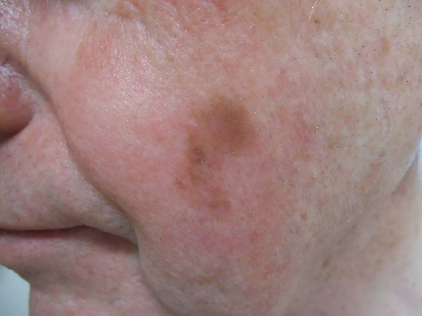
Long description
A lentigo maligna melanoma presenting as a 2 x 1 cm brown patch on the patient's left cheek.

Figure 2Melanoma (1)
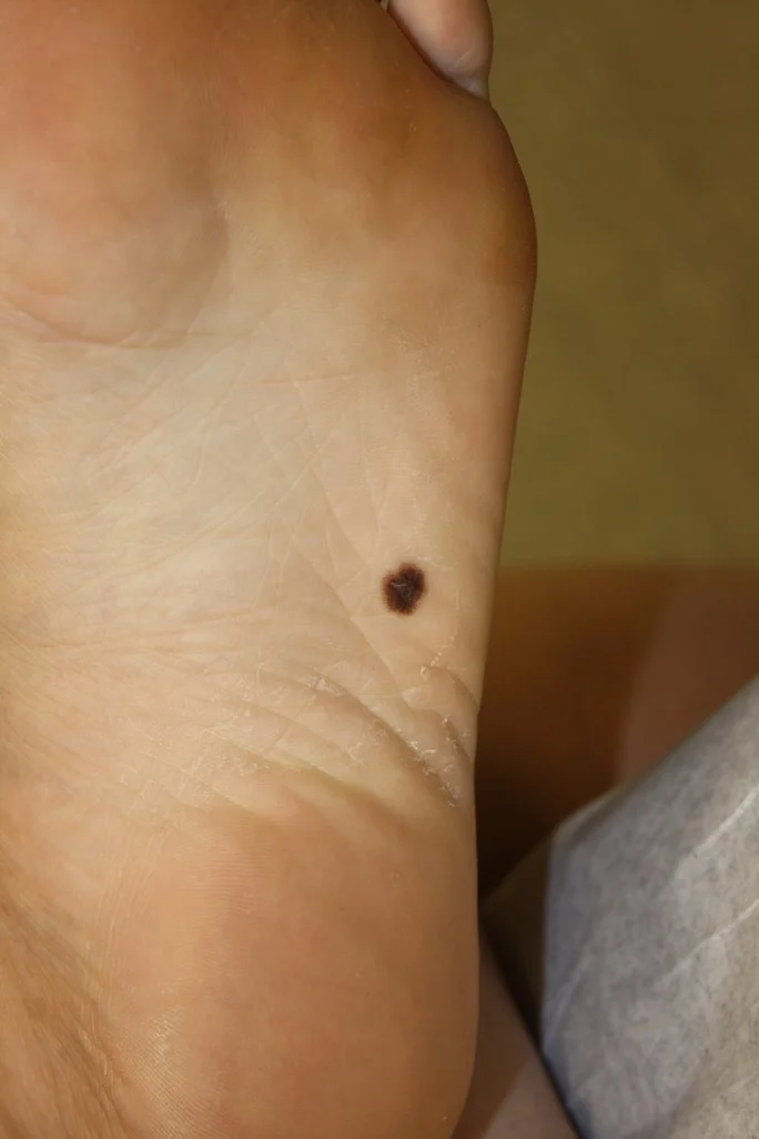
Long description
An asymmetrical, brown lesion on the skin on the bottom of the foot, described as melanoma with characteristic asymmetry, border irregularity, color variation and large diameter; filed under Acral lentiginous melanoma.

Figure 3Photography of nodular melanoma
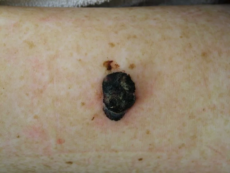
Long description
A clinical photograph captioned 'Photography of nodular melanoma', categorized under Nodular melanoma on Wikimedia Commons and sourced from DermNetNZ via StatPearls Publishing LLC.

Figure 4Melanoma border
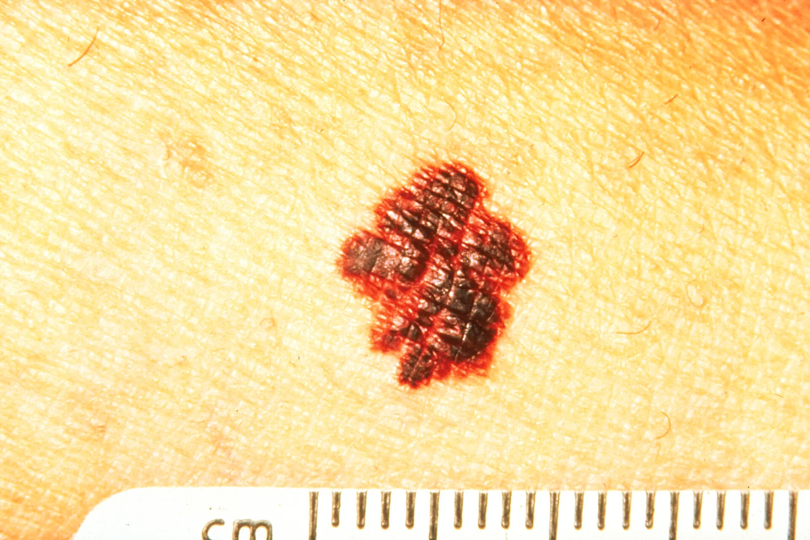
Long description
A clinical photograph of melanoma whose border is uneven, ragged, or notched, presented as part of the ABCD features for melanoma detection.

The diagnostic sample should be full-thickness and include the thickest (deepest) part of the lesion:
- an excisional biopsy with a narrow margin of normal skin is preferred, because it gives the pathologist the whole lesion and a clean surgical base;
- a deep shave or punch biopsy may be used when the lesion is too large to excise safely without reconstruction; a superficial shave risks missing a thin invasive component;
- incisional biopsy is reserved for very large facial or acral lesions where excising the whole lesion is impractical;
- do not ablate, curette or shave-flatten a strongly pigmented lesion without a specimen.
- There is no blood test for melanoma. Melanoma antigen tests are research tools.
- Lactate dehydrogenase (LDH) is raised in many advanced cases and is part of M staging in AJCC 8.
- Staging imaging is not used for thin, node-negative disease. Whole-body and brain contrast-enhanced CT is considered for stage IIB and offered for stage IIC to IV melanoma before adjuvant therapy, with MRI brain or whole-body MRI in children and young adults (birth to 24 years), pregnant women and in brain-metastasis follow-up, according to NICE NG14[1].
- BRAF V600 testing is mandatory for stage IIB to IV resected melanoma and should be considered in stage IIA in the 2025 ESMO guideline[2]; NICE NG14 does not offer BRAF analysis in stage IA or IB, considers it in IIA and tests in IIB to IV[1]. The differences are real and the local skin cancer multidisciplinary team's pathway should be followed.
- Testing is done on melanoma tissue (preferably nodal or metastatic if available) so that targeted therapy is an option.
- NRAS, KIT and NTRK testing is optional or guided by acral, mucosal or sun-damaged sites; gene-expression profiling tests are research tools and are not routine practice[2].
Staging at diagnosis uses the 8th edition of the AJCC melanoma staging system, implemented in 2018[3]. The T number is the Breslow thickness to the nearest 0.1 mm and whether the epidermis is ulcerated.
| T category | Breslow thickness | Ulceration |
|---|---|---|
| Tis | In situ | None |
| T1a | Less than 0.8 mm | Absent |
| T1b | 0.8 to 1.0 mm (with or without ulceration); or less than 0.8 mm with ulceration | Present if less than 0.8 mm [3] |
| T2a / T2b | Over 1.0 to 2.0 mm | Absent / present |
| T3a / T3b | Over 2.0 to 4.0 mm | Absent / present |
| T4a / T4b | Over 4.00 mm | Absent / present |
N category is clinico-pathological nodal status: clinically occult nodal metastasis found by sentinel node biopsy (microscopic, N1a/2a/3a), clinically apparent nodal metastasis (macroscopic, N1b/2b/3b), and in-transit or satellite disease (N1c/2c/3c), with higher numbers for more nodes. M category is site of distant disease (M1a skin/soft tissue/nodes, M1b lung, M1c other viscera, M1d brain), subdivided by LDH normal (0) or elevated (1). The order of prognosis broadly follows stage: pathologically staged AJCC 8 stage IIIA has a 5-year melanoma-specific survival of about 93% and stage IIID about 32%[3].
Points that commonly cause confusion:
- Mitotic rate is no longer a T criterion. It remains important prognostically and must be reported[3]; the older 7th-edition rule that one or more mitoses per mm2 upstaged a thin melanoma no longer applies.
- Pathological stage IA (pT1bN0M0) has better 5-year survival than clinical stage IB (cT1bN0M0), because the first group had a negative sentinel node on histology, so AJCC 8 moved it to IA. [3]
- Stage categories up to IIC stay node-negative. Stage III is any T with nodal disease and stage IV is distant disease.
- T1b lesions have a sentinel-node positivity of 5-12%, compared with under 5% for T1a[3].
SLNB is a staging test, not a treatment, but it changes risk and therefore adjuvant decisions. A sentinel node is the first draining lymph node in the basin, mapped by lymphoscintigraphy and blue dye, and removed in theatre.
- Do not offer SLNB in stage IA (Breslow less than 0.8 mm without ulceration)[1].
- Consider SLNB for T1a with special features (ulceration, 3 mitoses per mm2, pTx/unreliable thickness or positive deep margin) under ESMO 2025[2]; NICE uses 0.8 to 1.0 mm with two or more mitoses per mm2, ulceration or lymphovascular invasion[1].
- Consider or offer SLNB for T1b (0.8 to 1.0 mm, or less than 0.8 mm with ulceration) after discussion[1][2].
- Recommend SLNB for T2a or higher (Breslow over 1.0 mm)[1][2]. Wide excision and SLNB are carried out together when possible[2].
- Whole-body and brain CT is considered in IIB and offered in IIC to IV; routine PET-CT is not used.
- Completion lymphadenectomy is no longer routine after a positive sentinel node, because the DeCOG-SLT and MSLT-II trials showed no melanoma-specific survival benefit compared with nodal observation[1][2]. CLND is still offered for palpable or imaging-confirmed nodal disease.
- Timing changes with pregnancy: discuss delaying SLNB until after delivery[1].
A positive sentinel node upstages to stage III and is what most adjuvant recommendations are based on. The node is therefore both a prognostic tool and a gate to systemic therapy.
Standard excision is wide local excision in theatre with a clinical margin measured on the skin:
- Stage 0 (melanoma in situ): a minimum clinical margin of at least 0.5 cm[1][2].
- Stage I: 1 cm clinical margin[1][2].
- Stage II: 2 cm clinical margin[1][2].
- Where a 2 cm margin causes unacceptable disfigurement or morbidity, 1 cm is acceptable for stage I under NICE NG14[1].
- NCCN and ESMO use the same 0.5 / 1 / 2 cm framework.
- Margins are taken around the whole histological scar and include the primary melanoma margin[1].
- Stage 0 melanoma in situ can be treated with topical imiquimod in adults where surgery would cause unacceptable disfigurement, with a repeat biopsy after treatment to confirm clearance (off-label in the UK)[1].
- Lentigo maligna of the face may be treated with margin-controlled surgery or, where function is critical, radiotherapy for inadequate excision under ESMO 2025.
- Complete excision with adequate margin cures the vast majority. Residual in situ or invasive at the margin requires re-excision or multidisciplinary discussion[1].
- No adjuvant systemic therapy.
- Follow-up per the NICE schedule below, with discharge at 1 year for stage IA.
- 2 cm excision.
- Consider brain and whole-body contrast-enhanced CT in stage IIB and offer it in IIC before adjuvant therapy[1].
- Adjuvant anti-PD-1 for stage IIB and IIC can be considered (see below), because recurrence risk is similar to node-positive stage III disease.
- Palpable or imaging-positive nodes: offer therapeutic lymph node dissection of the involved basin[1].
- Microsatellite disease found on SLNB: no routine completion lymphadenectomy; instead observation with or without systemic therapy[1][2].
- Resectable in-transit metastases: first-line surgical excision with clear margins, plus systemic or local therapy (radiotherapy, electrochemotherapy, isolated limb perfusion or infusion, talimogene laherparepvec or systemic anti-PD-1-based therapy), decided by the specialist skin cancer multidisciplinary team[1][2].
- Recurrent or unresectable in-transit or stage III disease: treat as metastatic melanoma.
- Adjuvant systemic therapy (see below) if not already on treatment.
- Manage through the specialist multidisciplinary team. Goals are prolongation of life and control of symptoms.
- Resectable oligometastatic disease can be managed with excision or ablation, with or without systemic therapy, especially in clinical trials[1].
- Brain metastases: refer to neuro-oncology multidisciplinary team; stereotactic or whole-brain radiotherapy and surgery are used alongside systemic therapy[1].
- Targeted therapy has central nervous system activity and is a useful choice when radiotherapy is not indicated.
The shift from 10 mg/kg anti-CTLA-4 to anti-PD-1 and targeted therapy is the defining change of the last decade. [2]
Pembrolizumab
- Dose: 200 mg intravenously every 3 weeks, or 400 mg every 6 weeks, over 30 minutes, for up to 12 months (adults; 2 mg/kg up to 200 mg for paediatric patients 12 years and older)[5].
- Indication: adults with completely resected stage IIB, IIC or III melanoma (and, for unresectable or metastatic melanoma, until progression)[5].
- 12 months of therapy, continued unless recurrence or unacceptable toxicity[5].
- KEYNOTE-054 (stage III) showed a hazard ratio for recurrence of 0.61 (95% CI 0.51-0.72) for pembrolizumab versus placebo at a median follow-up of about 4.9 years, with a distant-metastasis-free survival hazard ratio of 0.62 (95% CI 0.52-0.75)[2].
Nivolumab
- Dose: 240 mg intravenously every 2 weeks, or 480 mg every 4 weeks, over 30 minutes, for up to 1 year; 3 mg/kg every 2 weeks or 6 mg/kg every 4 weeks for patients under 40 kg[6].
- Indication: adults and children 12 years and older with completely resected stage IIB, IIC, III or IV melanoma, or unresectable or metastatic melanoma[6].
- CHECKMATE-238 (stage III or IV with no evidence of disease) showed 5-year recurrence-free survival of 50% with nivolumab versus 39% with ipilimumab 10 mg/kg (hazard ratio 0.72, 95% CI 0.60-0.86), although there was no statistically significant difference in overall survival (hazard ratio 0.86, 95% CI 0.66-1.12)[2].
Ipilimumab plus nivolumab combination
- In resected stage IV disease, four doses of nivolumab 1 mg/kg with ipilimumab 3 mg/kg every 3 weeks, followed by nivolumab 3 mg/kg every 2 weeks, produced a recurrence-free survival hazard ratio of 0.25 (97.5% CI 0.13-0.48) versus placebo but with greater toxicity[2]. [6]
- Adjuvant nivolumab plus ipilimumab is not recommended for routine resected stage III melanoma, and outside an NCCN-listed option in resected stage IV melanoma with no evidence of disease it is not approved by EMA or FDA in the adjuvant setting[2].
Choosing between them:
- ESMO 2025 recommends adjuvant anti-PD-1 (nivolumab for stage IIIB to IV no-evidence-of-disease; pembrolizumab for stage III) as first-line adjuvant options for resected disease, and for stage IIB to IIC disease after careful discussion, with 12 months of treatment[2]. [5]
- Both are started within 12 weeks of complete resection[2].
- ESMO does not recommend routine BRAF or molecular testing in stage I or IIA (where adjuvant anti-PD-1 is not generally offered), while stages IIB and IIC are realistic candidates for adjuvant anti-PD-1.
- Stage IIIA with less than 1 mm of sentinel-node tumour burden is generally not an indication for adjuvant systemic therapy in ESMO 2025, because the trials excluded or selected heavier burdens[2]. This is a common exam trap.
- Checkpoint blockade is unsafe in active autoimmune disease, solid-organ transplant recipients and significant interstitial lung disease; seek specialist advice and consider targeted therapy for BRAF-mutant patients instead.
Adjuvant anti-PD-1 data show strong recurrence-risk reduction but have not yet demonstrated a statistically significant overall-survival benefit, so treatment decisions are made by discussing benefits against a year of toxicity, immune side effects and infusion visits.
- For resected stage III BRAF V600E-mutated melanoma, dabrafenib 150 mg orally twice daily plus trametinib 2 mg orally once daily for 12 months, started within 12 weeks of resection[2][4].
- COMBI-AD: at a median follow-up of over 10 years, relapse-free survival hazard ratio 0.52 (95% CI 0.43-0.63) and distant-metastasis-free survival hazard ratio 0.56 (95% CI 0.44-0.71) favoured the combination; the V600E subgroup showed an overall-survival hazard ratio of 0.75 (95% CI 0.58-0.96), although in the overall trial population the overall-survival difference was not statistically significant (hazard ratio 0.80, 95% CI 0.62-1.01)[2]. [3]
- Targeted therapy is not recommended for BRAF V600K-mutated melanoma in the adjuvant setting, because of a possible detrimental effect on overall survival in COMBI-AD (hazard ratio 1.95) and higher immunotherapy sensitivity of V600K disease[2].
- The trametinib label recommends ECG and LVEF assessment before treatment, after one month and then every 2 to 3 months, with retinal, pulmonary and dermatological monitoring[4].
- First-line systemic therapy is usually anti-PD-1-based immunotherapy (nivolumab plus ipilimumab, or pembrolizumab or nivolumab monotherapy if combination is unsuitable), subject to performance status, brain metastasis status, LDH, comorbidity and patient preference. NICE NG14 recommends the combination first, then single-agent anti-PD-1[1].
- For BRAF-mutant disease, dabrafenib 150 mg twice daily plus trametinib 2 mg daily (or encorafenib plus binimetinib) is offered when immunotherapy is contraindicated, or when there is not enough time for an immune response (high disease burden or rapid progression)[1][4].
- The nivolumab-plus-ipilimumab combination regimen in advanced disease uses nivolumab 1 mg/kg with ipilimumab 3 mg/kg every 3 weeks for 4 doses, then nivolumab 240 mg every 2 weeks or 480 mg every 4 weeks[6].
- Chemotherapy (dacarbazine) remains a fall-back where immunotherapy and targeted therapy are unsuitable or have failed; it is not first-line in current practice.
- Local control of in-transit metastases under NICE NG14 includes radiotherapy, electrochemotherapy, regional perfusion or infusion, excision and talimogene laherparepvec[1].
Follow-up length and intensity depend on stage and are spelled out by NICE NG14 (last updated 2022), which bases low-risk follow-up on clinical examination rather than routine scanning.
| Stage | Duration and frequency (clinics) |
|---|---|
| IA | Year 1: consider 2 appointments; discharge at end of year 1 |
| IB | Year 1: 2; years 2-3: 1 each (plus consideration of ultrasound where SLNB was considered but not performed); years 4-5: 1 each; discharge at end of year 5 |
| IIA | Years 1-2: 2 each; year 3: 1; years 4-5: 1 each; discharge at end of year 5 |
| IIB and IIC | Years 1-2: 4/year with scanning; year 3: 2; years 4-5: 1/year; discharge at end of year 5 |
| IIIA to IIIC (not on adjuvant therapy) | Years 1-3: 4 clinics and 2 body/brain CT per year; years 4-5: 2 clinics and 1 CT per year; discharge at end of year 5 |
| IIID and resected IV (not on adjuvant therapy) | Years 1-3: 4 clinics and 4 CT per year; years 4-5: 2 clinics and 2 CT per year; discharge at end of year 5 [1] |
- Follow-up examinations are performed by clinicians skilled in skin cancer and lymph-node examination, with access to dermoscopy and medical photography[1].
- Patients who have had stage 0 melanoma receive advice at a clinic visit within the first year; unresectable or metastatic disease receives personalised follow-up[1].
- Direct contact details for specialist skin cancer services and psychosocial support at every visit are routine under NICE NG14[1].
- Follow-up while on adjuvant therapy is governed by treatment requirements.
- Skin self-examination, sun safety education and vitamin-D advice are emphasised at follow-up appointments[1].
- ESMO 2025 recommends adjuvant anti-PD-1 within 12 weeks of resection, making staging imaging and a pre-treatment scan important[2].
Regional guideline differences are honest: NICE NG14, ESMO 2019 (superseded 2025), AJCC 8 and NCCN all describe the same surgical sequence (0.5 / 1 / 2 cm margins, SLNB for T2 and above, adjuvant anti-PD-1 for high-risk resected disease), but they differ in SLNB threshold wording (NICE: 0.8 to 1.0 mm plus features, or over 1.0 mm; ESMO: discuss T1b, recommend T2a and above), in BRAF testing thresholds (NICE: not IA or IB, consider IIA, test IIB to IV; ESMO: test IIB to IIC and above), and in intended radiological surveillance (NICE NG14 specifies a staged CT pattern, ESMO is more fluid). The 2019 ESMO guideline and the 2020 ESMO consensus conference recommendations on locoregional melanoma established much of the staged-management framework now expressed in the 2025 update, and ESMO 2025 notes that adjuvant nivolumab plus ipilimumab is included as a treatment option in the NCCN guideline for resected stage IV melanoma with no evidence of disease. [2][3]
- Recurrence (local, in transit, nodal, distant) can appear months or years later, hence the need for long-term clinical follow-up.
- Second primary melanoma, especially in high-naevus or genetically predisposed patients. This is why total skin examination is repeated.
- Non-melanoma skin cancers, because the same UV exposure produces basal and squamous cell carcinoma; these are common in the same patients.
- Lymphoedema after therapeutic nodal dissection.
- Treatment complications from immunotherapy (colitis, hepatitis, pneumonitis, endocrinopathies, rash) and from targeted therapy (pyrexia, chills, rash, cardiac and ocular effects).
- Metastatic complications include brain metastases with neurological deficit and spinal cord compression; bone metastases with pain and fracture.
- Death from advanced (stage IV) melanoma.
Prognosis is best understood in the same vocabulary as staging:
- overall survival in early melanoma (stages 0/I) is very high;
- within T1, T1b (0.8 to 1 mm, or thinner with ulceration) has 5-12% sentinel-node positivity, whereas T1a is under 5%;
- stage III spans a wide range, from a 5-year melanoma-specific survival of about 93% (stage IIIA) to about 32% (stage IIID) in AJCC 8 data; [3]
- historically stage IV carried a median survival of 6 to 7.5 months and under 10% 5-year survival, but with immunotherapy and targeted therapy multivisceral durable responses occur and long-term survival is achievable for a subset; [3]
- LDH elevation, ulceration, high mitotic rate, lymphovascular invasion and older age are unfavourable factors across stages;
- recurrence risk remains elevated years after treatment, which is why follow-up intervals span years.
A prognostic statement without the stage attached is meaningless; always anchor a prognosis to the T/N/M grouping. AJCC 8 distributions describe populations rather than individuals, so be honest about that uncertainty.
- Primary prevention: reduce artificial and intermittent UV exposure; regular high-protection, broad-spectrum sunscreen; shade clothing; no indoor tanning; childhood sun protection.
- High-risk patients benefit from total-body photography and dermatoscopic follow-up.
- Regular total skin examination of immunosuppressed patients and those with strong family history is standard practice in several health systems; frequency is not fixed in guidelines (NICE NG14 emphasises self-examination education rather than universal screening).
- A protective message to patients: sun safety must not produce vitamin-D deficiency; supplementation is preferred to chronic sunburn. NICE recommends checking vitamin D at diagnosis in secondary care[1].
- For public health, reducing tanning-bed exposure in young adults is the highest-yield intervention, and is addressed in several national cancer plans.
- Children and young adults. Melanoma before puberty is rare and usually acral, amelanotic or arising in a giant congenital naevus. Staging imaging uses MRI rather than CT in children and young adults (birth to 24 years)[1]. Total-body MRI is also substituted for CE-CT in pregnancy.
- Pregnancy. Melanoma is one of the most common cancers diagnosed during pregnancy. Staging without ionising radiation (ultrasound and MRI) is used where possible. Excision should not be delayed. SLNB can be delayed until after delivery, and a joined-up obstetric, dermatology and oncology team manages care.
- Older adults. Thicker melanomas and higher recurrence rates; functional status and comorbidity drive the choice between immunotherapy and targeted therapy, and between treatment and best supportive care.
- Immunocompromised patients (transplant recipients, chronic immunosuppression): higher melanoma risk, aggressive behaviour in an important minority, and immunotherapy may be unsafe. Targeted therapy or limited surgery is preferred where BRAF-mutant.
- People with darker skin. Acral lentiginous and nail melanomas predominate; these are unrelated to UV, so prevention advice alone does not reach the risk group. Delays in diagnosis are common and lead to thicker tumours.
- Recognition: a dark irregular patch or nodule, particularly if evolving, on any skin colour must be suspected. NICE NG14's dermoscopy recommendation and the three-month dermoscopic follow-up protocol are only possible where dermatoscopes are available. A magnifying glass and good lighting, with baseline photographs and structured review, are reasonable low-cost substitutes.
- If full-thickness excision cannot be sent to a reference pathology laboratory, at least the depth of invasion (Breslow) must be assessed by a trained pathologist somewhere in the system.
- A preserved excision with a 0.5 cm initial margin, then referral, is safer than a wide excision that is never fully staged.
- BRAF testing before targeted therapy can be done at a regional centre; without it, immunotherapy is the safer first line where the budget allows.
- NICE NG14's staging CT and regular surveillance scanning are not affordable in many settings; clinical skin and nodal examination plus patient self-examination should be taught and documented instead.
- Basic sun protection advice costs little; vitamin-D supplementation during prolonged sun avoidance is cheap.
- Melanoma is a melanocyte malignancy with a spectrum from in situ to metastatic disease; the thickness of the vertical invasion (Breslow, in millimetres) determines stage and prognosis.
- Risk factors are intermittent and intense UV exposure, fair skin, many naevi (especially atypical), and personal or family history; acral melanoma is UV-independent.
- The ABCDE criteria and the ugly duckling sign guide the clinical skin check; dermoscopy improves diagnostic accuracy in trained hands. [2]
- Superficial spreading is common; nodular is dangerous because it skips the long horizontal phase.
- AJCC 8th edition stages T1a to T4b are defined by Breslow thickness and ulceration; mitotic rate is no longer a T criterion but must be reported.
- SLNB is offered for T2a and higher, considered for T1b, and not for uncomplicated T1a.
- Excision margins are 0.5 cm in situ, 1 cm for stage I and 2 cm for stage II.
- Completion lymphadenectomy is no longer routine after a positive sentinel node.
- Adjuvant anti-PD-1 (pembrolizumab 200 mg every 3 weeks or 400 mg every 6 weeks, up to 12 months; nivolumab 240 mg every 2 weeks or 480 mg every 4 weeks, up to 1 year) reduces recurrence in stages IIB, IIC and III; stage IIIA with less than 1 mm of sentinel-node tumour is generally not treated. [5][6]
- Adjuvant dabrafenib 150 mg twice daily plus trametinib 2 mg once daily for 12 months is for resected BRAF V600E melanoma only (not V600K). [2]
- Follow-up for stage IA is 1 year, for stages IB to IV is 5 years, with intensity escalating with stage; routine PET-CT is not used. [1]
Answer each one in your head first, then open the answer.
A 35-year-old with pale skin has a 7 mm asymmetric flat patch on the back with irregular borders and three colours, which has grown over six months. What is the most likely diagnosis and the correct next investigation step?
Show answerHide answer
Melanoma, most likely superficial spreading melanoma. The next investigation is full-thickness excisional biopsy (preferably with a 1 to 3 mm margin of normal skin) and histopathology with Breslow thickness, not a superficial shave or shave excision, so the invasion depth is measured accurately. [2]
In the pathology report for a newly excised melanoma, the Breslow thickness is 0.9 mm without ulceration and the sentinel lymph node was negative on histology. How is it staged under AJCC 8, and would you offer SLNB to the next patient with this result?
Show answerHide answer
pT1bN0M0 equals stage IA in AJCC 8 (Breslow 0.8 to 1.0 mm is T1b; negative sentinel node after biopsy is pathological stage IA). For the next patient with 0.8-1.0 mm without ulceration SLNB is considered, not mandatory; it is offered if there are risk features or nodal uncertainty. [3]
A resected stage IIIC melanoma carries a BRAF V600E mutation. The oncologist wishes to offer adjuvant systemic treatment. Give the standard targeted regimen with doses and duration, and state the evidence basis.
Show answerHide answer
Dabrafenib 150 mg orally twice daily plus trametinib 2 mg orally once daily for 12 months, started within 12 weeks of complete resection. COMBI-AD showed a relapse-free survival hazard ratio of 0.52 and a distant-metastasis-free survival hazard ratio of 0.56 favouring the combination in resected stage III BRAF V600E or V600K melanoma; V600K disease is currently not offered targeted adjuvant therapy because of a possible survival disadvantage.
A patient is about to start adjuvant pembrolizumab for stage IIC melanoma. Give the dose, schedule, duration, route and one monitoring or administration point.
Show answerHide answer
200 mg intravenously over 30 minutes every 3 weeks (or 400 mg every 6 weeks), continued until recurrence, unacceptable toxicity or 12 months. Administer according to the pembrolizumab label, under melanoma-specialist oversight. 30-minute IV infusion. Monitor for immune-related adverse events such as colitis, hepatitis, pneumonitis, thyroid dysfunction and rash. [5][6]
A pregnant woman with a 1.2 mm melanoma is referred. Which statement about management is correct in current guidance?
Show answerHide answer
Give local excision promptly. Staging should avoid ionising radiation and use ultrasound or MRI where possible, SLNB can be delayed until after delivery, and decisions are made by a joint obstetric, dermatology and oncology team.
Which stage of melanoma would you discharge at 1 year, and how many follow-up years are prescribed for stages IIB and IIC?
Show answerHide answer
Stage IA is discharged at the end of year 1 with no routine imaging. Stages IIB and IIC receive follow-up and scanning for 5 years: 4 clinic appointments and scans in years 1 and 2, 2 clinics in year 3 and 1 per year in years 4 and 5. [1]
- dermatology: basal cell carcinoma and squamous cell carcinoma
- dermatology: dysplastic naevus, congenital naevus and naevus management
- oncology: immunotherapy for cancer (anti-PD-1 and anti-CTLA-4)
- oncology: targeted therapy of melanoma (BRAF/MEK, immunotherapy resistance)
- oncology: metastatic melanoma management and brain metastases
- surgery: sentinel lymph node biopsy and lymph node dissection
- radiology: melanoma staging imaging (CT, MRI, PET-CT)
- pharmacology: biologics, immune-related adverse events and their management
- epidemiology: sun exposure, vitamin D, skin cancer prevention
- dermatology: pigmented skin lesions and how to examine them
The treatment statements above are drawn from, and should not be taken outside the clinical recommendations of, NICE NG14 (last updated 27 July 2022), the ESMO 2025 Clinical Practice Guideline for cutaneous melanoma (which supersedes the 2019 ESMO guideline[7] and incorporates the 2020 ESMO consensus conference recommendations on locoregional melanoma[8]), the AJCC 8th edition melanoma staging system, and product-labelling doses from the DailyMed pages for trametinib, pembrolizumab and nivolumab. NICE NG14 describes low-risk follow-up as described in its staged table, ESMO 2025 covers drug doses and adjuvant indications, and AJCC 8 covers T, N and M criteria. The National Comprehensive Cancer Network (NCCN) Clinical Practice Guidelines for Cutaneous Melanoma use the same surgical and staging framework and list adjuvant nivolumab plus ipilimumab as an option in resected stage IV melanoma with no evidence of disease, as noted in ESMO 2025. Where guidelines differ (SLNB threshold wording, BRAF-testing thresholds and surveillance imaging intensity), both values are given and the specialist skin cancer multidisciplinary team's local pathway should be followed.
- National Institute for Health and Care Excellence (NICE). Melanoma: assessment and management. NICE guideline NG14; published 29 July 2015, last updated 27 July 2022. Accessed .
- Amaral T, Ottaviano M, Arance A, Blank C, Chiarion-Sileni V, Donia M, Dummer R, Garbe C, Gershenwald JE, Gogas H, et al.; ESMO Guidelines Committee. Cutaneous melanoma: ESMO Clinical Practice Guideline for diagnosis, treatment and follow-up (2025 update; supersedes Annals of Oncology 2019;30(12):1884-1901). Annals of Oncology 2025;36(1):10-30. doi:10.1016/j.annonc.2024.11.006. Accessed .
- Keung EZ, Gershenwald JE. The eighth edition American Joint Committee on Cancer (AJCC) melanoma staging system: implications for melanoma treatment and care. Expert Review of Anticancer Therapy 2018;18(8):775-784. doi:10.1080/14737140.2018.1489246. Accessed .
- Novartis Pharmaceuticals Corporation. MEKINIST (trametinib) prescribing information. DailyMed, U.S. National Library of Medicine. Accessed .
- Merck & Co., Inc. KEYTRUDA (pembrolizumab) prescribing information. DailyMed, U.S. National Library of Medicine. Accessed .
- Bristol Myers Squibb Company. OPDIVO (nivolumab) prescribing information. DailyMed, U.S. National Library of Medicine. Accessed .
- Michielin O, van Akkooi ACJ, Ascierto PA, Dummer R, Keilholz U; ESMO Guidelines Committee. Cutaneous melanoma: ESMO Clinical Practice Guidelines for diagnosis, treatment and follow-up (2019 edition, superseded by the 2025 update). Annals of Oncology 2019;30(12):1884-1901. doi:10.1093/annonc/mdz411. Accessed .
- Michielin O, van Akkooi A, Lorigan P, Ascierto PA, Dummer R, Robert C, Arance A, Blank CU, Chiarion Sileni V, Donia M, Faries MB, et al. ESMO consensus conference recommendations on the management of locoregional melanoma (2020). Annals of Oncology 2020;31(11):1449-1461. doi:10.1016/j.annonc.2020.07.005. Accessed .
Figure credits
- Figure 1: Omar Bari, Philip R. Cohen, via Wikimedia Commons, CC BY 3.0, CC BY 3.0. resized.
- Figure 2: Kelly Nelson (Photographer), via Wikimedia Commons, Public domain, Public domain.
- Figure 3: DermNetNZ, via Wikimedia Commons, CC BY 4.0, CC BY 4.0.
- Figure 4: Unknown authorUnknown author, via Wikimedia Commons, Public domain, Public domain. resized.
Educational content for medical students and doctors. It is not a substitute for clinical judgement or local guidelines.