Diabetes insipidus
AVP deficiency or resistance: how central and nephrogenic diabetes insipidus cause polyuria and hypernatraemia, how to diagnose them, and how to treat them.
- Diabetes insipidus (DI) is not diabetes mellitus. "Diabetes" here refers to excess urine and "insipidus" to the tasteless (not sweet) urine; there is no sugar in the urine. The kidney cannot reabsorb free water, so the patient passes very large volumes of very dilute urine.
- Central (arginine vasopressin deficiency, AVP-D): the hypothalamus or posterior pituitary makes little or no vasopressin. Nephrogenic (arginine vasopressin resistance, AVP-R): the kidney fails to respond to vasopressin despite normal or raised levels.
- The hallmark is hypotonic polyuria: urine output above 3 L a day in adults, with urine osmolality usually under 300 mOsm/kg. Nocturia is almost universal, and thirst is intense.
- As long as thirst and access to water are intact, the patient drinks to match the loss, so serum sodium stays near normal. If thirst or access is impaired (infants, older adults, sedated or postoperative patients, or adipsic DI), free water is lost faster than it is replaced and hypernatraemia develops.
- Diagnosis rests on proving hypotonic polyuria, then a water-deprivation test with desmopressin, increasingly supplemented by plasma copeptin. Treatment is desmopressin for central DI and free water plus low-salt, low-protein diet, thiazides and sometimes NSAIDs or amiloride for nephrogenic DI.
By the end of this page you should be able to:
- Explain how vasopressin controls water excretion, and distinguish AVP deficiency from AVP resistance.
- List the central causes (pituitary/hypothalamic injury, surgery, tumour, trauma, idiopathic, genetic) and the nephrogenic causes (lithium, hypercalcaemia, hypokalaemia, V2 and aquaporin-2 mutations).
- Describe the clinical presentation, including the polyuria-polydipsia-nocturia triad and the dehydration/hypernatraemia that appear when thirst is impaired.
- Interpret urine osmolality and serum sodium, plan a water-deprivation test with desmopressin, and use copeptin to tell the three polyuric disorders apart.
- Treat central DI with desmopressin, including dose forms and hyponatraemia safety, and treat nephrogenic DI with free water, diet, thiazides and NSAIDs.
- Recognise the triphasic pattern after pituitary surgery and gestational DI.
- Adapt diagnosis and management where tests, drugs and intravenous fluids are scarce.
DefinitionDiabetes insipidus (DI)
Diabetes insipidus is an inability to concentrate urine because of failed action of antidiuretic hormone (arginine vasopressin, AVP, also called ADH): either the body makes too little (central, arginine vasopressin deficiency, AVP-D) or the kidney does not respond (nephrogenic, arginine vasopressin resistance, AVP-R). It is a water disorder, not a sugar disorder.
AVP is made in the supraoptic and paraventricular nuclei of the hypothalamus, stored in the posterior pituitary, and released in response to a rise in plasma osmolality (osmoreceptors) and to a fall in plasma volume or pressure (baroreceptors). At the kidney it binds the V2 receptor on principal cells of the collecting duct, activating a cAMP cascade that inserts aquaporin-2 (AQP2) water channels into the apical membrane; water then leaves the urine down the medullary osmotic gradient. [1][3] Failure anywhere in this arc gives DI.
- Central DI (AVP-D). Damage to the hypothalamic nuclei, the stalk or the posterior pituitary. It may be complete (essentially no AVP) or partial (some residual secretion). Only about 10% of the neurosecretory neurons need to remain intact to avoid deficiency. [1]
- Nephrogenic DI (AVP-R). AVP is present (often raised), but the renal tubule does not respond because of defects in the V2 receptor or AQP2.
- Gestational DI. Excess AVP breakdown by placental vasopressinase (cysteine aminopeptidase), classically in the second or third trimester.
- Adipsic DI. DI with damaged hypothalamic osmoreceptors and therefore little or no thirst. This is a particularly dangerous form, because the warning system is lost.
DI is rare: roughly 1 in 25,000 people, it can occur at any age, and men and women are affected equally. [3] Central DI is more common than nephrogenic DI in adult clinics; in one neurosurgical centre the commonest cause by far is surgery for pituitary and suprasellar masses. [4] The inherited form of nephrogenic DI is uncommon. Because DI markedly affects quality of life but rarely causes death in someone who can drink freely, prevalence data are sparse; every prevalence figure should be treated as an estimate. [3]
| Category | Examples |
|---|---|
| Tumour (10-15%) | Craniopharyngioma, germinoma, meningioma, pituitary and hypothalamic tumours, pituitary-stalk metastases (especially breast, lung), lymphoma |
| Trauma (10-25%) | Transfrontal or transsphenoidal surgery; traumatic brain injury |
| Vascular | Hypothalamic or cerebral infarction or haemorrhage, cerebral aneurysm, Sheehan syndrome, sickle cell disease |
| Autoimmune / inflammatory | Lymphocytic hypophysitis, systemic lupus erythematosus, scleroderma, granulomatosis with polyangiitis, IgG4 disease, antivasopressin-neuron antibodies |
| Granulomatous | Sarcoidosis, granulomatous hypophysitis, Langerhans cell histiocytosis, Erdheim-Chester disease |
| Infection | Meningitis, encephalitis, tuberculosis, toxoplasmosis, cerebral abscess |
| Congenital / genetic | Autosomal dominant AVP-neurophysin II gene mutations, Wolfram (DIDMOAD) syndrome, X-linked recessive defects with subnormal AVP |
| Other | Drug toxicity (phenytoin, alcohol), pregnancy (placental vasopressinase), idiopathic |
- Primary genetic AVP-D. Mutations of the AVP gene on chromosome 20 (autosomal dominant) reduce synthesis; some cases are now known to be autoimmune. [4]
- Surgery. Pituitary adenomas do not present with DI; DI appears after operation. CDI usually becomes evident 1-2 days postoperatively and resolves within 2-5 days when it is transient. [4][5]
- Trauma. CDI occurs in about 20% of moderate or severe traumatic brain injury and about 15% of non-traumatic subarachnoid haemorrhage; most is transient. [4][5]
- Idiopathic. Older series labelled 25-50% idiopathic; better investigation now shows many are autoimmune. [4]
- Pregnancy. See "Special groups".
- Adipsia. The same lesions that destroy AVP neurons can damage hypothalamic osmoreceptors. These patients cannot feel thirst and are at high risk of death if not managed carefully. [4][5]
| Category | Examples |
|---|---|
| Drugs | Lithium (commonest); demeclocycline; cidofovir; foscarnet; cisplatin; amphotericin B; dexamethasone; dopamine; ifosfamide; ofloxacin; orlistat |
| Vascular/renal | Sickle cell nephropathy, acute tubular necrosis, cystic kidney disease |
| Infiltrative | Sarcoidosis, amyloidosis, Sjogren disease, multiple myeloma |
| Metabolic | Severe hypokalaemia, severe hypercalcaemia, starvation and low protein intake, chronic hypokalaemic nephropathy |
| Obstructive | Release of obstructing periureteral fibrosis |
| Congenital/genetic | X-linked V2 receptor gene mutations (most common inherited form); autosomal aquaporin-2 gene mutations (recessive or dominant) |
- Lithium is the commonest drug trigger. NDI develops in 40-55% of lithium-treated patients and can appear as early as 8 weeks after starting. Stopping lithium early may reverse it (AQP2 expression rises within 1-2 weeks in experimental models; some patients improve within 3 weeks), but prolonged use predisposes to permanent resistance. [3]
- Genetics. Inherited nephrogenic DI most often reflects V2 receptor mutations on the X chromosome; heterozygous females vary from asymptomatic to severely affected. Rare kindreds carry aquaporin-2 mutations, affecting both sexes. [2][3]
- Hypercalcaemia and hypokalaemia impair concentrating ability. For their management see the renal topics on hypercalcaemia and hypokalaemia.
- Reversible causes include hypercalcaemia, hypokalaemia and obstructive uropathy. [3]
The kidney generates a hypertonic medulla and, in response to AVP, inserts AQP2 channels so water can be withdrawn from the urine. If AVP is absent or ineffective, the collecting duct stays impermeable to water, the filtrate remains hypotonic, and a large volume of dilute urine is excreted.
Figure 1Central diabetes insipidus: pathway of ADH action in the collecting duct
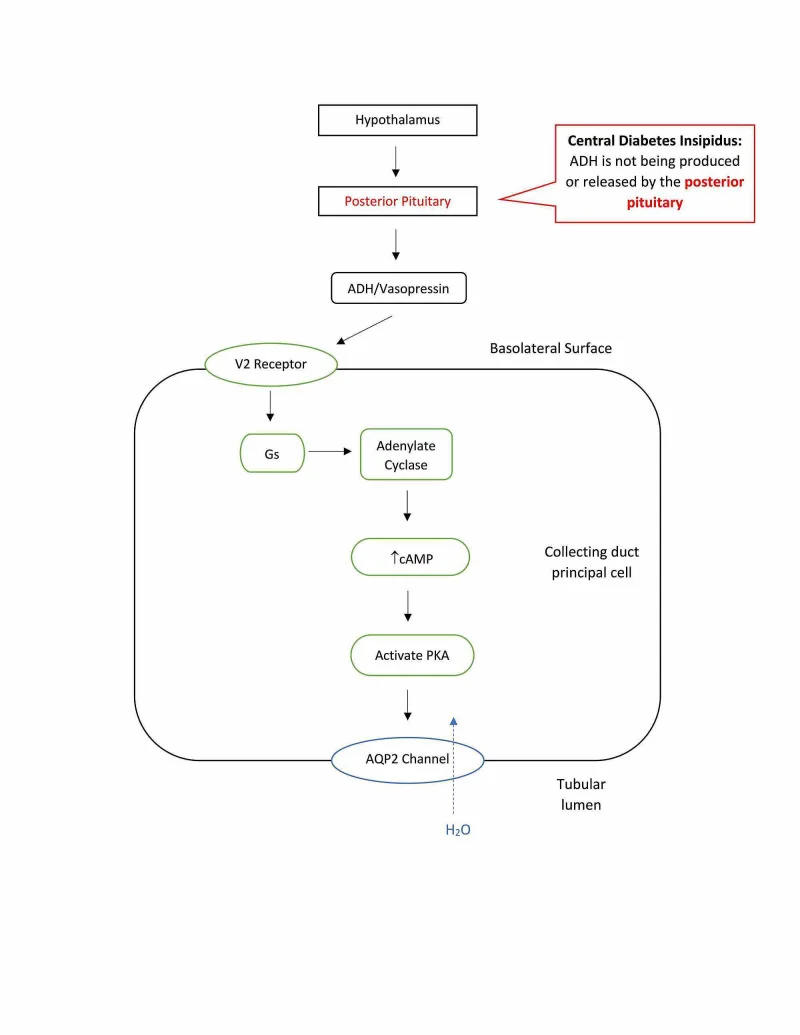
Long description
Flow diagram of central diabetes insipidus: failure of ADH (vasopressin) release from the posterior pituitary means no V2 receptor signalling, no cAMP and PKA activation and no AQP2 water channel insertion in the collecting duct principal cell.
- Polyuria. Central DI: urine output 3 to 30 L a day of very dilute urine. Nephrogenic: 3 to 20 L a day. [1][2]
- Dilute urine. Urine osmolality usually under 200-300 mOsm/kg (often under 200 mOsm/kg in nephrogenic DI), specific gravity usually under 1.005. [1][2]
- Nocturia. AVP would normally peak at night; without it the patient wakes repeatedly to void. Nocturia is almost always present in AVP-D and AVP-R. [1]
- Thirst is the safety valve. Most patients have an intact thirst mechanism and drink to match losses, so serum sodium stays near normal (roughly 136-147 mmol/L). [3]
- Hypernatraemia when thirst fails. Infants, older adults, patients with altered consciousness, those with adipsic DI, and anyone without free access to water cannot keep pace. They lose free water faster than intake, serum sodium rises, often well above 145 mmol/L and sometimes dramatically, and neurological symptoms (excitability, confusion, seizures, coma) may follow. [2][3]
- Chronic washout. Long-standing polyuria depletes the medullary osmotic gradient and down-regulates AQP2, so even normal kidneys become partially resistant to AVP. This is why a water-deprivation test can be hard to interpret in chronic primary polydipsia. [3]
- Most AVP-D is partial. Partial deficiency retains some concentrating ability, so the urine is not maximally dilute and the response to desmopressin is smaller. [4]
- "I am passing huge amounts of very pale, almost colourless urine" - the volume and the dilution.
- Drinking enormous amounts: carrying water at all times, waking at night to drink and to void.
- Most patients feel well if they can drink freely. The illness is discovered when they cannot (travel, surgery, intercurrent illness, or not being offered water).
- Thirst is usually for plain water, not sweet drinks.
- Associated posterior-pituitary problems may appear in central disease: headache, visual field defect, other hormone deficiencies.
The crucial distinction from primary polydipsia: primary polydipsia patients usually do not have nocturia and tend to wake to drink rather than to void; about 20% of patients with schizophrenia have polydipsia. [4]
- Volume status: often clinically euvolaemic if drinking freely; hypovolaemia and hypernatraemia if not.
- Signs of an underlying cause in central disease (visual fields, cranial-nerve findings, signs of a sellar mass).
- Infants and children: severe dehydration, vomiting, constipation, failure to thrive, growth retardation. Inherited forms often present before age 10, but timing varies from infancy to later childhood. [3]
- Older adults and patients with cognitive impairment: may not complain of thirst; dehydration may be the first sign.
- Postoperative patients: polyuria within hours of pituitary surgery, especially with intact consciousness and unrestricted fluid access.
Renal complications of untreated disease [2]
High urine output can produce ureteric and pelvic dilatation in severe congenital disease, and infants may fail to thrive. These are largely preventable if water is freely available.
The main question is whether a polyuric patient has diabetes insipidus (central or nephrogenic), primary polydipsia, or solute/osmotic diuresis. Each is separated by the urine osmolality, the serum sodium, and the response to water deprivation and desmopressin. [4]
| Cause | Typical clues | Urine osmolality pattern | Desmopressin response after water deprivation |
|---|---|---|---|
| Central DI (AVP-D) | Abrupt onset; nocturia almost always | Hypotonic, usually under 300 mOsm/kg (complete) or 300-800 (partial) | Rise of 50 to more than 100% (complete); 15-50% (partial) |
| Nephrogenic DI (AVP-R) | Lithium, hypercalcaemia, hypokalaemia; good thirst initially; hypernatraemia if no water access | Hypotonic, usually under 200-300 mOsm/kg | Little or no rise (under 50 mOsm/kg); up to 45% in partial |
| Primary (psychogenic) polydipsia | Psychiatric disease in some; wakes to drink, not to void; no nocturia usually; low serum sodium possible | Rises with deprivation (300-800, often up to 600-700) without significant plasma change | No meaningful rise (under 9%) |
| Osmotic/solute diuresis | Diabetes mellitus, recovery from renal failure, high sodium load, mannitol, radiocontrast, salt-wasting nephropathy | Over 300 mOsm/kg | Not tested this way; remove the solute |
| Appropriate diuretic effect | Drug history (loop/thiazide), polydipsia absent | Variable | Stop the drug |
Key clinical anchors (worth memorising):
- Serum sodium: in the setting of hypotonic polyuria, a value above 147 mmol/L strongly suggests DI, while a value under 135 mmol/L suggests primary polydipsia. Most patients present between 136 and 147 mmol/L and need dynamic testing. [3]
- Urine osmolality: under 300 mOsm/kg defines hypotonic polyuria; over 300 mOsm/kg suggests a solute diuresis. [3][4]
- Osmotic diuresis (most often uncontrolled diabetes mellitus) is the commonest cause of polyuria overall. Always check plasma glucose and exclude glucosuria before labelling a patient with DI.
- Cross-check with hypernatraemia in Renal: for correction of the water and sodium deficit itself, see the renal-genitourinary topic "Hypernatraemia". That page deals with the sodium problem; this page deals with the cause.
- Confirm polyuria with a 24-hour urine collection: output above 3 L a day or above 50 mL/kg in adults (above 2 L/m2 in children). If daily urine is under 2.5 L, further investigation for DI is usually not required.
- Exclude common confounders: diabetes mellitus, renal impairment, hypercalcaemia, hypokalaemia, drugs (diuretics, lithium, SGLT2 inhibitors), infection, prostate or bladder problems.
- Baseline. 24-hour urine volume and osmolality, serum electrolytes and osmolality, glucose. A urine osmolality over 700 mOsm/kg indicates significant AVP activity and effectively excludes DI. [4]
- Hypotonic polyuria (urine osmolality under 300 mOsm/kg) in the absence of solute diuresis is the trigger for dynamic testing.
This is the standard test. It is run as an inpatient in an endocrine unit because it can cause dangerous dehydration.
- Preparation: baseline weight, blood pressure, heart rate, plasma osmolality and electrolytes, urine osmolality. Correct electrolytes such as potassium and calcium first. Hold drugs that alter urine output (diuretics, SGLT2 inhibitors, glucocorticoids, NSAIDs) for 24 hours.
- Dehydration: nothing by mouth, usually up to 8 hours (can be longer or shorter). Overnight outpatient protocols exist (duration estimated from weight and urine output), but they demand supervision to exclude surreptitious drinking. Monitor weight, blood pressure and heart rate every hour, and urine output and osmolality every 2 hours, with plasma osmolality and sodium every 2 hours.
- Endpoints (stop and test): completed 8 hours of dehydration (up to 17 hours in some protocols); two consecutive urine osmolalities within 10% of each other plus 2% weight loss; weight loss over 3% (or 5% in some older protocols); serum sodium above the upper limit of normal (preferably above 150 mmol/L); orthostatic hypotension; intractable thirst.
- Desmopressin phase: if urine remains dilute, give desmopressin (2-4 mcg subcutaneously or intravenously; intranasal is not preferred because absorption is unpredictable) and collect urine hourly for 1-2 hours (the patient may eat and drink modestly).
Table of results (Merck Manual water-deprivation table, plus Frontiers thresholds). [1][2][3]
| Parameter | Normal | Complete central DI | Partial central DI | Complete nephrogenic DI | Partial nephrogenic DI | Primary polydipsia |
|---|---|---|---|---|---|---|
| Uosm after dehydration | Very high, often >700-800 | Very low (less than plasma osmolality) | Low, >=300 | Very low (less than plasma osmolality) | Very low (less than plasma osmolality) | High, 500-600 |
| Uosm rise after desmopressin | Minimal (under 5%) | 50 to >100% | 15-50% | under 50 (usually less than 50 mOsm/kg) | Up to about 45% | No real change |
| Plasma vasopressin/copeptin at end of deprivation | Appropriate | Low | Low-normal | High | High | Low |
Interpretation tips from specialist centres: [3][4]
- Some apply a simple rule: after deprivation, normal kidneys reach over 800 mOsm/kg; DI stays under 300 mOsm/kg.
- An intermediate result (300-800 mOsm/kg) suggests partial DI or primary polydipsia; after desmopressin a rise of under 50% points to partial DI.
- Prolonged polyuria (of any cause) can wash out the medulla and blunt responses. Results must be read with the clinical picture, not from a single binary cut-off.
- A doubling of urine osmolality after desmopressin is often accepted as evidence of intact renal response when the baseline is very low.
- Overall, the WDT is reported to have sensitivity 80-100% and specificity 67-100%.
Copeptin is the C-terminal peptide of provasopressin, secreted in equimolar amounts with AVP but much more stable in blood; it is a practical surrogate for AVP and is not routine in every laboratory.
- Baseline copeptin above 21.4 pmol/L diagnoses nephrogenic DI with 100% sensitivity and specificity.
- If below that, give hypertonic saline to raise plasma sodium above 150 mmol/L (some centres aim above 147 mmol/L) and repeat: copeptin under 4.9 pmol/L = central DI; at least 4.9 pmol/L = primary polydipsia. A multicentre prospective study gave this osmotic approach 96.5% accuracy (93.2% sensitivity, 100% specificity).
- After pituitary surgery, a plasma copeptin under 2.5 pmol/L within the first day predicts CDI with 97% specificity (positive predictive value 81%).
Imaging and further work-up for central disease [4]
- MRI of the sella and suprasellar regions for central DI: loss of the posterior-pituitary "bright spot", pituitary-stalk thickening, or a mass.
- Idiopathic or autoimmune CDI with a thickened stalk warrants follow-up imaging at 6-12 months in adults (every 6 months in younger patients) to exclude germinoma.
- Always test anterior-pituitary function in CDI, because craniopharyngioma-style lesions bring other deficits with them.
Approach to polyuria and suspected diabetes insipidus
- Polyuria (or a child with failure to thrive and dilute urine)Confirm output >3 L/day in adults, >50 mL/kg/day; exclude DM and drugs
- Baseline tests24-h urine volume and osmolality, serum sodium/osmolality, glucose, calcium, potassium
- Question: Urine osmolality?
Over 700 mOsm/kg
- Significant AVP activity: NOT DIInvestigate other causes
Under 300 mOsm/kg
- Hypotonic polyuria confirmedExclude solute diuresis; review serum sodium and meds
300-700 mOsm/kg
- Unclear: partial DI or primary polydipsiaProceed to dynamic testing
- Question: Serum sodium?
Above 147 mmol/L with hypotonic polyuria
- Diabetes insipidus likelyConfirm with WDT or copeptin
Under 135 mmol/L
- Primary polydipsia likely
136-147 mmol/L
- Dynamic testing neededMost patients sit here
- Water-deprivation test with desmopressinEndpoints as listed; 2-4 mcg SC/IV desmopressin given if urine stays dilute
- Question: Rise in urine osmolality after desmopressin?
50 to >100%
- Complete central DI (AVP-D)
15-50%
- Partial central DI
<50 mOsm/kg rise (complete) or up to 45% (partial)
- Nephrogenic DI (AVP-R)
No real change
- Primary polydipsia
- Copeptin (if available)Baseline >21.4 pmol/L = NDI; after hypertonic saline <4.9 pmol/L = central DI, >=4.9 = primary polydipsia
- Imaging + anterior-pituitary tests (central DI)MRI sella; repeat imaging if idiopathic
There is no staging system. Severity is described as:
- Complete vs partial deficiency or resistance, based on how much AVP is secreted or how much the kidney responds.
- Whether thirst is intact. Adipsic DI (no thirst) is life-threatening and carries a high mortality; patients with an intact thirst mechanism are in much safer territory.
- Acute versus chronic: acute polyuria can be severe, but if the cause is reversible (for example a transient postoperative insult) it may resolve within days.
- Particular risk groups: children, infants and older adults, because they cannot reliably recognise or communicate thirst.
Hospitalise any patient with hypernatraemic dehydration, altered consciousness, or no reliable thirst or access to water. Seek endocrinology input.
- If not in hypovolaemic shock: use a hypotonic intravenous fluid, usually 5% dextrose in water, because fluids with higher osmolality than the urine (such as half-isotonic saline) can worsen hypernatraemia in DI. Give only as much as needed, with close monitoring.
- Acute hypernatraemia (present 48 hours or less): lower serum sodium at 1-2 mmol/L per hour and restore normal in under 24 hours (5% dextrose in water at 3-6 mL/kg/hour if needed).
- Chronic hypernatraemia (more than 48 hours): limit correction to no more than 0.5 mmol/L per hour. A practical estimate of safe time is to double the difference between starting sodium and the target sodium. In adipsic DI, use low-molecular-weight heparin because thrombosis rates are high.
- Free water deficit: for men, 0.6 x (serum sodium - 140)/140 litres per kg body weight; for women, 0.5 x (serum sodium - 140)/140 litres per kg. Divide by the safe time (hours) for the hourly rate.
- Check a basic metabolic panel about every 4 hours until stable; high rates of dextrose can cause glucosuria and osmotic diuresis, needing insulin or a different hypotonic fluid. Transition to oral water as soon as possible.
- Hypovolaemic shock: treat volume first with isotonic saline, then correct the free-water deficit.
- If anterior hypopituitarism is present after surgery (for example cortisol deficiency), treat it at the same time.
The drug of choice is desmopressin (dDAVP), a synthetic vasopressin analogue.
- The amino group of cysteine has been removed, prolonging the half-life from about 5 minutes to 6-8 hours, so it can be given twice or three times daily.
- L-arginine is replaced by D-arginine, abolishing vasopressor activity.
Dose forms reported in the literature (mostly adult data):
| Formulation | Dose | Notes |
|---|---|---|
| Oral tablets | Starting 0.05 mg orally once or twice a day; usual 0.1-1.2 mg divided into 2-3 doses | Most convenient; onset about 1-2 h, duration 6-12 h; absorption reduced if taken within 90 minutes of a meal |
| Nasal spray | 10 mcg per 0.1 mL spray; usual 10-40 mcg daily (single or divided); children 10-30 mcg | Needs refrigeration; less effective if nasal mucosa is inflamed (coryza); metered spray |
| Sublingual melt | Women 27.7 mcg daily; men 55.3 mcg daily, 1 hour before bed | Limit water 1 hour before and 8 hours after the dose |
| Intravenous or subcutaneous injection | 1-2 mcg twice a day (some postoperative regimens use 0.25-0.50 mcg twice daily initially) | For hospitalised patients, post-trauma or postoperative; aqueous vasopressin (1-2 mcg) is an alternative with a 12-hour effect but rarely used long term |
- Usually a low dose at night is started first (to control nocturia) and titrated weekly until symptoms are stable over a month.
- The drug starts working within an hour and lasts 6-8 hours; inter-individual variation is large, especially by age and sex.
- Parenteral doses are much smaller than oral doses because bioavailability differs markedly (oral about 16-40% of intranasal).
- Hyponatraemia is the main hazard. After each dose, aquaresis should be allowed so excess water is excreted without electrolyte loss, and the dose should be reduced if hyponatraemia persists. Practical options: omit or delay a dose one to two times a week until 2-3 voids (aquaresis) occur; or delay each dose slightly.
If desmopressin is unavailable, second-line options include thiazide diuretics, carbamazepine, chlorpropamide, clofibrate and indapamide, but each has significant side effects. [3]
In infants and young children, thiazides have a special role. Thiazides paradoxically reduce urine volume by 25-50% (via mild extracellular volume contraction raising proximal tubular reabsorption) and are effective and safe early in life; treatment is usually switched to desmopressin as the diet moves to solid food. [1][3]
- Reverse the cause where possible: stop or adjust the offending drug (lithium may take weeks to improve, sometimes never fully recovers), correct hypercalcaemia and hypokalaemia, relieve obstruction.
- Ensure free water intake sufficient to match losses; serious sequelae are rare if patients can drink at will.
- Reduce the solute load with a low-salt, low-protein diet: protein no more than about 1.0 g/kg/day and sodium no more than 2.3 g/day (under 100 mEq/day), aiming at an osmotic load of about 15 mOsm/kg/day. This can be hard in growing children, where it may compromise nutrition. [3]
- Medications to cut urine output:
- Thiazide diuretics: hydrochlorothiazide 25 mg orally once or twice daily in adults, or 1-4 mg/kg/day divided into one or two doses in children; chlorothiazide liquid is useful in infants. In a classic low-sodium study, hydrochlorothiazide cut urine output to roughly half of baseline after 3 days. [2][3]
- Potassium-sparing diuretics such as amiloride (often combined with a thiazide; amiloride is also used specifically with lithium). [2][3]
- NSAIDs such as indomethacin, modestly reducing urine volume by 10-25%, perhaps by reducing renal blood flow and GFR. Use carefully (renal, gastric, cardiovascular risks). [1][2]
- Together, sodium restriction plus a thiazide plus an NSAID (such as indomethacin) can further reduce urine volume in AVP-R.
- Partial nephrogenic DI may respond to supraphysiologic doses of desmopressin (for example 80 mcg intravenously 3-4 times daily, 20 mcg intranasally 4 times daily, or 720 mcg daily orally). Only about a quarter of patients with congenital NDI respond, so this is reserved for patients with inadequate response or contraindications to diuretics. [3]
Additional agents mentioned in central disease [1]
Thiazides, NSAIDs, prostaglandin inhibitors and restriction of dietary sodium all reduce urine volume in partial central DI as well (urine volumes can fall 25-50% on a thiazide), and these drugs do not carry the fluid-retention risk of exogenous vasopressin. Chlorpropamide, carbamazepine and clofibrate can stimulate vasopressin release and are occasionally useful, but they are inferior to desmopressin and seldom used.
- Caused by placental vasopressinase (cysteine aminopeptidase) degrading AVP, usually in the second or third trimester; also seen when partial CDI becomes apparent because of the extra clearance.
- Serum sodium in pregnancy normally runs about 5 mmol/L lower than outside pregnancy, which can mask the diagnosis.
- Desmopressin is the treatment because it is not broken down by placental vasopressinase. It is FDA category B; small studies (3.4% fetal abnormality vs a population risk of about 4.5%, and 2 malformations among 53 desmopressin-exposed pregnancies) do not show a signal of major risk, but the numbers are small.
- Doses are usually the same or slightly higher than outside pregnancy; placental enzyme activity disappears 4-6 weeks after delivery, so gestational DI treatment can usually be withdrawn within 2 months.
- Desmopressin has minimal transfer to breast milk and does not affect infant water balance. Pre-existing CDI needs continued treatment through pregnancy.
- Early diagnosis is essential: infants with inherited NDI can develop permanent brain damage and intellectual disability, and growth failure can require gastrostomy feeding.
- Monitoring of growth, development and sodium, with careful titration to avoid water intoxication.
- Desmopressin dosing in infants from the literature (nasal spray diluted to 1-5 mcg, 0.02-0.08 mcg subcutaneously, or continuous low-dose infusion postoperatively) must be supervised by a paediatric endocrinologist.
- Absent thirst means fixed fluid targets. A common approach: set a target weight, fix 24-hour urine output at 1.5-2 L, take daily weights and calculate daily water intake as an obligate volume (about 1.5 L) plus (target weight - daily weight); check plasma sodium weekly.
- Thrombosis risk during hypernatraemic dehydration is high; screen for sleep apnoea.
This is an important, fully reversible water-sodium sequence seen after pituitary or suprasellar surgery and after traumatic brain injury, in roughly 1.1-3.4% of postoperative patients:
- Phase 1 - central DI (days 1-3). Abrupt polyuria (often within the first 12-24 hours) from sudden loss of AVP, lasting 5-7 days.
- Phase 2 - SIADH (days 7-8, lasting 2-14 days). Unregulated AVP release from degenerating posterior-pituitary tissue causes antidiuresis and, typically, hyponatraemia.
- Phase 3 - permanent central DI. When most of the AVP-secreting neurons have degenerated, DI returns.
The two polyuric phases look like central DI (dilute polyuria responding to desmopressin); the middle phase looks like SIADH and needs fluid restriction rather than more desmopressin. Correctly identifying the phase matters because the treatment changes. Other postoperative causes of polyuria (mobilisation of intraoperative fluids, hyperglycaemia, diuretics, growth-hormone diuresis after acromegaly surgery) must be excluded.
Recovery data: about half and about 80% of patients with transient postoperative DI recover within 7 and 90 days, respectively; permanent DI occurs in up to 0-12.5% after endoscopic and 0-10% after microsurgical approaches, and in up to about 90% of craniopharyngioma operations.
- Free water access is the cornerstone of survival. Ensure unlimited oral water and teach families; use fixed daily fluid targets where thirst is absent.
- Bedside diagnosis: very large volumes of very pale urine (more than 3 L/day), specific gravity under 1.005 and nocturia. Urine osmolality and serum sodium, when available, refine the diagnosis; without them, pattern recognition plus a desmopressin trial can help, but must be supervised because of hyponatraemia risk.
- When desmopressin is unavailable: central DI can be managed with second-line agents (thiazides, carbamazepine, chlorpropamide, clofibrate, indapamide) and strict water access; nephrogenic DI with thiazides, NSAIDs, amiloride, low salt and low protein, and water access. Referral for difficult cases is essential.
- Avoid dangerous correction: if IV fluids are limited, aim for slow correction (no more than 0.5 mmol/L per hour for chronic hypernatraemia) using the simplest hypotonic fluid available; oral water whenever possible.
- Education: sick-day rules, recognising dehydration, MedicAlert-style identification, and early help-seeking during vomiting illnesses, in order to prevent death in adipsic or paediatric DI.
- Prevention of brain injury in infants: feed on demand, monitor growth, identify inherited DI in families with infant polyuria.
- Diabetes insipidus is a water disorder: failed action of AVP, not sugar. Urine is large in volume and dilute, and thirst is intense.
- Central (AVP-D) and nephrogenic (AVP-R) DI have different causes and different treatment; telling them apart drives management.
- Polyuria definitions: above 3 L/day or above 50 mL/kg/day in adults; children above 2 L/m2/day.
- Hypotonic polyuria is a urine osmolality under 300 mOsm/kg; normal kidneys concentrate above 800 mOsm/kg when water-deprived.
- Serum sodium above 147 mmol/L (with hypotonic polyuria) suggests DI; under 135 mmol/L suggests primary polydipsia.
- The water-deprivation test with desmopressin separates complete vs partial central DI, nephrogenic DI and primary polydipsia; copeptin adds accuracy (baseline above 21.4 pmol/L = NDI; after hypertonic saline under 4.9 pmol/L = central DI).
- Desmopressin is first-line for central DI in many forms (oral, nasal, sublingual, parenteral, intravenous). Doses differ greatly by route; titrate slowly; watch for hyponatraemia.
- Nephrogenic DI is treated with free water, a low-salt low-protein diet, thiazides, amiloride and sometimes NSAIDs; address lithium, hypercalcaemia and hypokalaemia.
- Thiazides paradoxically reduce urine volume (about 25-50%) by creating mild extracellular volume depletion and increasing proximal reabsorption.
- The triphasic pattern after pituitary surgery (DI then SIADH then DI) needs correct phase identification.
- Gestational DI responds to desmopressin because placental vasopressinase does not degrade it.
- Adipsic DI is the dangerous extreme: fixed fluid targets and close monitoring.
- For hypernatraemia correction itself, cross-check with the renal-genitourinary topic "Hypernatraemia".
- Low-resource care depends above all on guaranteeing free water and preventing dangerous sodium shifts.
Answer each one in your head first, then open the answer.
A patient from a psychiatric ward passes 6 L of dilute urine a day, wakes to drink rather than to void, and has serum sodium 130 mmol/L. Is this diabetes insipidus?
Show answerHide answer
No. The picture fits primary (psychogenic) polydipsia: nocturia is usually absent, patients often wake to drink, and serum sodium tends to be low. Diabetes insipidus usually gives nocturia and a normal-to-raised serum sodium. A water-deprivation test would show intact concentration in primary polydipsia and little desmopressin response. [4]
A man on long-term lithium presents with polyuria. Urine osmolality is 180 mOsm/kg. After water deprivation it stays dilute, and 10 mcg intranasal desmopressin produces no meaningful rise. What is the diagnosis?
Show answerHide answer
Complete central DI on a water-deprivation test - what happens to urine osmolality after desmopressin?
What is the triphasic pattern after pituitary surgery, in order?
A pregnant woman develops massive polyuria in the third trimester. Why desmopressin, and not plain vasopressin?
A patient has serum sodium 152 mmol/L, urine 4 L/day of water-clear urine with specific gravity under 1.005, and a copeptin of 8 pmol/L without osmotic stimulation. What does the copeptin suggest?
Infant, failure to thrive, massive dilute urine, parents have similar history. Best first action?
- Endocrinology: posterior pituitary and hypothalamic disorders; syndrome of inappropriate antidiuretic hormone secretion (SIADH).
- Renal-genitourinary: hypernatraemia (cross-check for water/sodium correction); hypercalcaemia; hypokalaemia; polycystic kidney disease; tubulointerstitial disease.
- Neurosurgery: pituitary surgery and postoperative water-balance disorders; traumatic brain injury.
- Obstetrics: pregnancy and endocrine disorders; lactation.
- Carmichael JD. Arginine Vasopressin Deficiency (Central Diabetes Insipidus). Merck Manual Professional Version (full review Jun 2025, last updated May 2026). Accessed .
- Hechanova LA. Arginine Vasopressin Resistance (Nephrogenic Diabetes Insipidus). Merck Manual Professional Version (full review Mar 2026, last updated Oct 2026). Accessed .
- Flynn K, Hatfield J, Brown K, Vietor N, Hoang T. Central and nephrogenic diabetes insipidus: updates on diagnosis and management. Frontiers in Endocrinology (Lausanne) 2025;15:1479764. Accessed .
- Tomkins M, Lawless S, Martin-Grace J, Sherlock M, Thompson CJ. Diagnosis and Management of Central Diabetes Insipidus in Adults. J Clin Endocrinol Metab 2022;107:2701-2715. Accessed .
- Almalki MH, Ahmad MM, Brema I, et al. Management of Diabetes Insipidus following Surgery for Pituitary and Suprasellar Tumours. Sultan Qaboos Univ Med J 2021;21:354-364. Accessed .
Figure credits
Educational content for medical students and doctors. It is not a substitute for clinical judgement or local guidelines.