Hypopituitarism
Partial or complete failure of the pituitary gland to secrete one or more hormones: causes, diagnosis and replacement.
- Hypopituitarism is the partial or complete failure of the pituitary gland to secrete one or more hormones. It may be a single deficiency or multiple, permanent or, in some situations, transient.
- The most dangerous missing hormone is ACTH: without cortisol the patient can collapse and die in an adrenal crisis, and this must always be found and treated before anything else.
- Causes include pituitary adenomas and their treatment (surgery or radiotherapy), bleeding into a tumour (apoplexy), Sheehan syndrome, infiltrative or inflammatory disease, head injury, radiation and genetic defects.
- Diagnosis is biochemical: a morning cortisol, stimulation tests of the adrenal axis, free thyroxine with an inappropriately normal or low TSH, gonadal hormones, IGF-1 and, when needed, dynamic tests of growth hormone. MRI looks for the cause.
- Replacement follows the missing hormone in order: hydrocortisone first, then levothyroxine, then sex steroids, then growth hormone, with desmopressin for diabetes insipidus. Every patient needs an emergency steroid card, sick-day rules and a hydrocortisone injection kit.
By the end of this page you should be able to:
- Explain what hypopituitarism is and how primary, secondary and tertiary hormone failure differ.
- List the common causes and describe which hormones are usually lost first.
- Link each missing hormone to its clinical effects.
- Plan the investigation of a patient with suspected hypopituitarism, including the cut-offs that separate adrenal insufficiency from normal.
- Prescribe replacement therapy in the correct order, at the usual adult doses, with monitoring.
- Recognise pituitary apoplexy and manage an adrenal crisis and the "sick-day" adjustments.
- Outline the approach when tests or drugs are not available.
DefinitionHypopituitarism
Hypopituitarism is the state in which the pituitary gland fails to secrete one or more of its hormones. "Panhypopituitarism" means that all or nearly all axes are failing. A single-axis failure is called an isolated deficiency.
Two levels of failure look the same in the blood but sit at different anatomical sites. When the problem is in the pituitary itself (inability to release a stored hormone) the result is secondary deficiency and the target gland receives no signal. When the problem sits higher, in the hypothalamus (failure to release the hormone that tells the pituitary to act) the result is tertiary deficiency. In both, the pituitary hormone is low and the target-gland hormone is low. Imaging and clinical context separate them, not a single blood test.
A modern review gives a clear working definition: hypopituitarism includes all conditions causing partial or complete failure of the anterior and, less often, the posterior lobe of the pituitary to secrete hormones.[2] It can be transient when it follows compression (for example by a macroadenoma), inflammation (hypophysitis), surgery or functional suppression, and in those cases the axis can recover.[2]
Good-quality modern prevalence data are scarce. The most often quoted come from a Spanish study published in 2001, which found a prevalence of about 455 cases per million inhabitants and an incidence of about 42 new cases per million inhabitants per year.[3] If those figures are applied to a country such as Brazil, there would be roughly 100,000 affected people and about 8,500 new cases each year.[3] True rates are almost certainly higher, because newer causes such as traumatic brain injury, intracranial haemorrhage and drug-induced hypophysitis are recognised more often and the tests for them are used more widely.[3]
Hypopituitarism itself is associated with increased mortality, particularly from cardiovascular and cerebrovascular disease; two recent meta-analyses of observational studies confirmed this excess, with higher rates in women and in people who are younger at diagnosis.[3]
A pituitary tumour, usually a macroadenoma, and its treatment (surgery or radiotherapy) account for most cases in adult series.[2][3] Causes fall into groups that are shared between adults and children, with different frequencies.
| Group | Causes | Notes |
|---|---|---|
| Neoplastic | Pituitary adenoma (functioning and non-functioning), craniopharyngioma, meningioma, Rathke's cleft cyst, germinoma, metastases, pituitary carcinoma | Mass effect compresses normal cells; surgery and radiotherapy add damage |
| Infiltrative or inflammatory | Autoimmune (lymphocytic) hypophysitis, granulomatous disease (sarcoidosis, granulomatosis with polyangiitis), Langerhans cell histiocytosis, haemochromatosis, IgG4-related hypophysitis | Hypophysitis often begins with ACTH and TSH deficiency[2] |
| Vascular | Pituitary apoplexy, Sheehan syndrome, subarachnoid haemorrhage, intrasellar aneurysm | Sudden onset; apoplexy is an emergency |
| Traumatic | Traumatic brain injury, sports-related head trauma, pituitary stalk transection, skull-base fracture | Prevalence after TBI varies widely (16% to 69% in studies)[3] |
| Iatrogenic | Surgery, radiotherapy (pituitary, nasopharyngeal or cranial), immune checkpoint inhibitors | Somatotrophs most sensitive to radiation, then gonadotrophs, then thyrotrophs, then corticotrophs[2] |
| Genetic or congenital | PROP1, POU1F1 (PIT-1), HESX1, SOX2, LHX3/4 mutations; Kallmann, Prader-Willi and Bardet-Biedl syndromes | Often presents in childhood but may declare late |
Sheehan syndrome deserves separate mention because it remains a common cause in less-resourced settings. It is necrosis of the pituitary during or shortly after labour, usually after major haemorrhage or shock. The enlarged, vasodilated pituitary of pregnancy infarcts when the blood pressure falls. It classically presents months or years later with failure to lactate, followed by amenorrhoea and features of adrenal and thyroid failure.[3]
Pituitary adenomas cause deficiency by mass effect: a macroadenoma compresses the normal gland and distorts the stalk, raising prolactin from loss of dopamine delivery and eventually silencing the other axes.[2] Non-functioning adenomas are the most common tumour cause.
Immune checkpoint inhibitors (anti-CTLA-4 and anti-PD-1 drugs) are now a frequent drug cause of hypophysitis. It occurs in 11% to 12% of patients treated with an anti-CTLA-4 antibody such as ipilimumab, and is less common with anti-PD-1 and anti-PD-L1 agents. It tends to affect ACTH first and is usually permanent even after the drug is stopped.[3]
Radiation produces deficiency years after treatment, so monitoring should begin within one year of radiotherapy.[2]
Two useful general rules. First, hormone loss usually follows a sequence: growth hormone first, then gonadotrophins, then TSH, then ACTH.[2] Second, hypophysitis in particular tends to affect ACTH and TSH first, before the GH axis.[2]
Figure 1Neuroendocrinology Figure
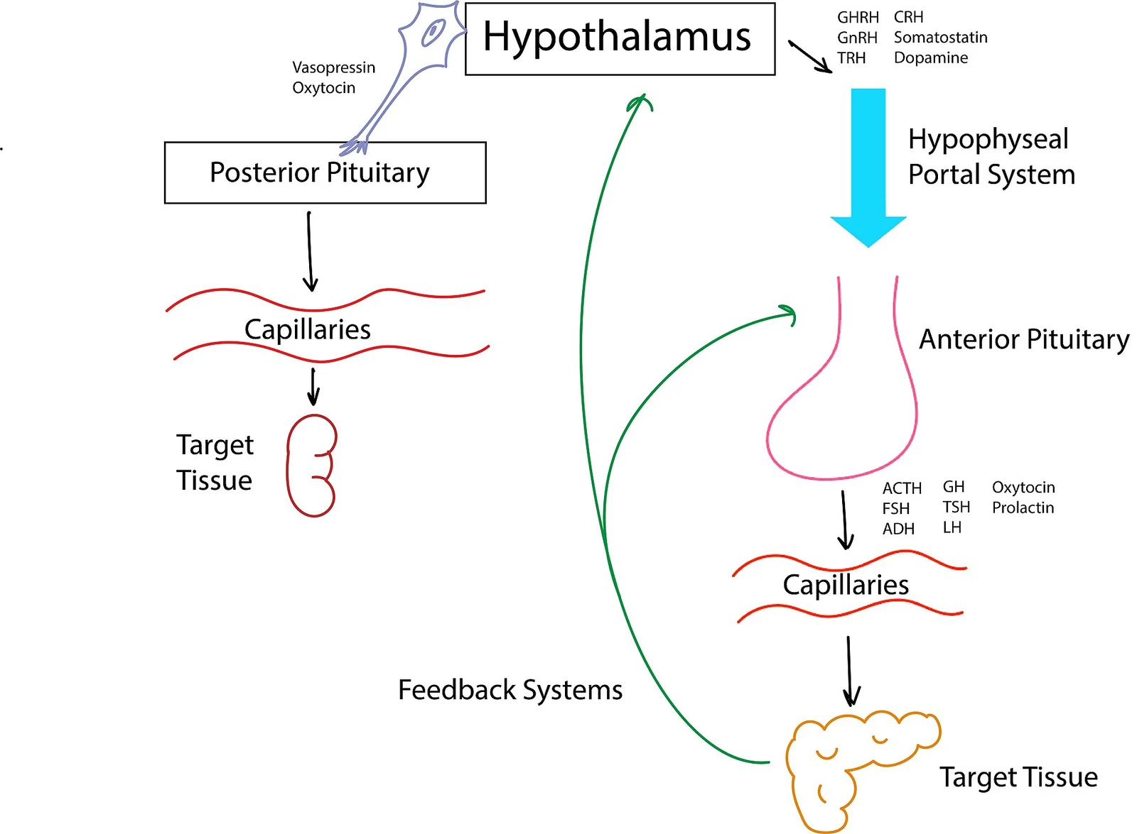
Long description
Hypothalamus, posterior and anterior pituitary hormones and their control

Everything the patient feels follows from the absence of the end-organ hormone, not from the missing pituitary signal itself:
- Loss of ACTH reduces cortisol. The renin-angiotensin system is intact, so aldosterone is preserved; there is no hyperkalaemia, unlike in Addison disease. The patient is pale, hypotensive and often hyponatraemic.
- Loss of TSH reduces free thyroxine; because the thyroid itself is healthy, this looks like hypothyroidism without the raised TSH of primary disease.
- Loss of LH and FSH silences the gonads: amenorrhoea and infertility in women, low testosterone, loss of libido and infertility in men.
- Loss of GH changes body composition (more fat, less lean mass), bone mass and lipids, and impairs quality of life.
- Loss of prolactin is mainly a problem after delivery, when milk is not produced.
- Loss of ADH prevents the kidneys concentrating urine, producing massive dilute polyuria.
| Missing pituitary hormone | Resulting condition | Clinical effects of the deficiency |
|---|---|---|
| ACTH | Secondary (central) adrenal insufficiency | Fatigue, anorexia, nausea, weight loss, hypotension, postural dizziness, hyponatraemia; high risk of adrenal crisis with stress; hypoglycaemia in children |
| TSH | Secondary (central) hypothyroidism | Cold intolerance, slowed metabolism, weight gain, bradycardia, dry skin, constipation; TSH is not raised |
| LH and FSH (gonadotrophins) | Secondary hypogonadism | Women: amenorrhoea, infertility, dyspareunia, poor bone mass. Men: low libido, erectile dysfunction, infertility, reduced muscle and bone mass |
| Growth hormone (GH) | Adult GH deficiency | Increased visceral fat, reduced lean mass, osteopenia with fractures, dyslipidaemia, reduced exercise capacity, impaired quality of life |
| Prolactin | Hypoprolactinaemia | Inability to lactate postpartum (main consequence); no other major syndrome in adults |
| ADH (vasopressin) | Central diabetes insipidus | Large volumes of dilute urine (can exceed 10 L per day), thirst, risk of severe dehydration if thirst is impaired |
There is no single "hypopituitarism" presentation. The clinical picture is the sum of whichever axes are lost, plus features of the cause, such as bitemporal hemianopia from a macroadenoma.
ACTH deficiency often dominates, and its symptoms (fatigue, nausea, anorexia, weight loss, dizziness) are vague, so many patients are diagnosed only after hospital admission for an adrenal crisis. The risk is real: long delays in diagnosing secondary adrenal insufficiency are common, and many cases are established only after an adrenal crisis.[3]
TSH deficiency produces the familiar hypothyroid picture, but without the raised TSH. Because TSH cannot be used to titrate treatment, free T4 is measured instead.
Gonadotrophin deficiency presents with menstrual upset in women and low libido or impotence in men. A young woman with unexplained amenorrhoea and low oestradiol in the setting of a pituitary or hypothalamic problem should have FSH and LH measured.
GH deficiency in adults is subtle: loss of vigour, reduced lean mass, abdominal fat gain, dyslipidaemia, low mood and poor concentration. These improve, at least in part, with GH replacement.
ADH deficiency produces central diabetes insipidus: polyuria with dilute urine (often above 3.5 L per day in a 70 kg adult) and polydipsia. It is dangerous only when thirst is impaired or water is unavailable, because dehydration can be fatal.
Red flagRed flags for an adrenal crisis
A patient with known or suspected hypopituitarism who presents unwell with hypotension (often a postural drop of at least 20 mmHg), nausea or vomiting, abdominal pain, fever, confusion or hypoglycaemia may be in adrenal crisis. Treat first and investigate afterwards.[5]
The main differentials are primary failure of the target glands (Addison disease, primary hypothyroidism, primary hypogonadism). These produce a high pituitary drive, so ACTH, TSH, LH or FSH would be raised rather than low. A mixed picture therefore points to hypopituitarism. The other differential is functional failure (energy deficit, malnutrition, excessive exercise, chronic opioid use, serious illness), which usually improves when the insult resolves.
Morning cortisol is the single most informative first test. Drawn at 08:00 to 09:00, a value below 100 nmol/L (about 3 micrograms/dL) is strongly predictive of secondary adrenal insufficiency, while a value above about 15 micrograms/dL excludes it. Values between 3 and 15 micrograms/dL need a stimulation test. These cut-offs change with the assay in use.[3]
When a stimulation test is available:
- Short Synacthen (cosyntropin) test: synthetic ACTH 250 micrograms IV or IM; cortisol measured at baseline, 30 and 60 minutes. A peak cortisol below 500 nmol/L (about 18 micrograms/dL) confirms secondary adrenal insufficiency. The test can be falsely negative within four weeks of a pituitary insult or in partial deficiency. It may not be available in every setting.[3]
- Insulin tolerance test (ITT): 0.1 units/kg IV of regular insulin; glucose and cortisol measured at baseline and at 30, 60, 90 and 120 minutes. A peak cortisol below 500 nmol/L (about 18 micrograms/dL; about 350 nmol/L with newer mass-spectrometry-standardised assays) confirms the diagnosis. ITT also assesses the GH axis. It is the gold standard but requires trained staff and monitoring, and is contraindicated in epilepsy, cardiac arrhythmia and cerebrovascular disease.[3]
- Glucagon stimulation is an alternative when ITT is unsafe, but it is less accurate.[3]
Clinical rule: if the patient is acutely unwell or an adrenal crisis is possible, never delay hydrocortisone for a test; draw a paired serum cortisol and plasma ACTH first and treat. Giving glucocorticoid in that situation does not compromise the diagnosis, which can be confirmed once the patient has recovered.[3][5]
Measure free T4 and TSH together. In secondary hypothyroidism, free T4 is low and TSH is normal or inappropriately low, because the pituitary cannot raise it. Free T4 falls before TSH changes in partial disease, so a low-normal free T4 with a low-normal TSH in a patient with other pituitary failure warrants close follow-up. TSH is also suppressed by glucocorticoid replacement and by acute illness, so it must never be used to titrate dose in central hypothyroidism.[3]
In women with oligomenorrhoea or amenorrhoea, oestradiol with FSH and LH inappropriately low or normal establishes secondary hypogonadism. In post-menopausal women, the key clue is a normal or low FSH and LH when they should be raised. In men, total testosterone measured before 10:00 after an overnight fast, in the absence of acute illness and drugs that affect testosterone (glucocorticoids, opioids, barbiturates), is paired with FSH and LH; two low results are needed if the first is borderline. There is no indication for stimulation testing.[3]
Prolactin should be measured in everyone: it is often raised from stalk compression (secondary hyperprolactinaemia), which is itself a cause of hypogonadism, and low prolactin postpartum explains failure to lactate.
Basal GH is useless because it is secreted in pulses. In a patient with a clear pituitary lesion and at least two other proven deficiencies, a low IGF-1 for age and sex is enough to diagnose adult GH deficiency without a stimulation test. Otherwise the gold standard is the ITT (GH peak below 3 micrograms/L; 5 micrograms/L during the transition from childhood to adulthood). The glucagon stimulation test is an alternative, and macimorelin is another if available. A normal IGF-1 does not exclude deficiency, and IGF-1 is lowered by oral oestrogens, uncontrolled hypothyroidism, malnutrition and liver disease.[3]
MRI of the pituitary and hypothalamus with gadolinium is the imaging study of choice: it looks for adenoma, empty sella, infiltrate or metastasis, and confirms a structural cause. It is also the investigation of choice for pituitary apoplexy, where it establishes the diagnosis in over 90% of patients.[4] Other useful tests include renal and liver function, full blood count (anaemia, eosinophilia in adrenal failure), electrolytes, and a visual-field assessment if a mass is present.
Investigating suspected hypopituitarism
- SuspicionVague ill-health, hypotension, amenorrhoea or subfertility, or a known pituitary or parasellar lesion, radiation or head injury
- Morning cortisol (08:00-09:00)under 3 micrograms/dL (100 nmol/L) strongly predicts adrenal insufficiency; over 15 micrograms/dL makes it unlikely
- Question: Cortisol between 3 and 15 micrograms/dL?
Yes
- Short Synacthen test (or ITT if safe)Peak cortisol under 18 micrograms/dL (500 nmol/L) confirms secondary adrenal insufficiency
No
- Act on the cortisol resultTreat if under 3 micrograms/dL; move on to the other axes if over 15 micrograms/dL
- ThyroidFree T4 with TSH. Free T4 low with normal or low TSH means secondary hypothyroidism
- Gonadal axisOestradiol or total testosterone with FSH and LH
- GH axisIGF-1 for age and sex; ITT or glucagon test if the diagnosis is uncertain
- Prolactin and ADHProlactin (high suggests stalk compression); water deprivation or copeptin if polyuria
- MRI of pituitary and hypothalamusFind the cause; urgent MRI if apoplexy suspected
Remember again: if adrenal crisis is suspected, blood for cortisol and ACTH should be drawn and hydrocortisone given immediately. Hydrocortisone does not interfere with a later Synacthen test beyond 18 to 24 hours, but clinical safety comes first.[3][5]
The general principle is to replace the target hormone of the missing axis, in this order: hydrocortisone first, then levothyroxine, then sex steroids, then growth hormone, with desmopressin for diabetes insipidus.[1] The order matters clinically: giving levothyroxine before cortisol can precipitate an adrenal crisis, because thyroid hormone accelerates cortisol clearance and unmasked hypoadrenalism collapses.[2][3] Growth hormone can also unmask subclinical adrenal insufficiency, and both GH and sex steroids alter thyroid replacement requirements.[3]
Hydrocortisone is the glucocorticoid of choice. The usual adult daily dose is 15 to 20 mg in two or three divided doses, with half or two-thirds of the total taken immediately on waking and the rest at lunch and late afternoon, the last dose at least 4 to 6 hours before bedtime.[2][3] Dose is titrated to clinical state, not to blood tests: the guideline favours clinical assessment of under- or over-replacement over cortisol day-curves.[2] Where hydrocortisone is unavailable, the options are prednisone or prednisolone 2.5 to 5 mg once in the morning (1 mg prednisone roughly equals 4 mg hydrocortisone).[3] Dexamethasone should be avoided for routine replacement, and any daily dose above about 20 mg hydrocortisone equivalent should be avoided long term because of a worse metabolic profile and increased mortality in hypopituitary patients.[2][3]
Mineralocorticoid is not needed in secondary adrenal insufficiency, because aldosterone is regulated by renin and angiotensin rather than by ACTH.[3]
Exam trapHydrocortisone before thyroxine
In combined ACTH and TSH deficiency, always start hydrocortisone before (or, with the same urgency, alongside) levothyroxine. Starting levothyroxine in a patient with untreated adrenal insufficiency can precipitate an adrenal crisis by accelerating cortisol clearance. The same applies when GH replacement is begun: cortisol has to be adequate first.[2][3]
Levothyroxine replacement in secondary hypothyroidism follows the same principles as in primary disease, but TSH cannot be used to assess adequacy. The target is a free T4 in the mid to upper half of the normal reference range, with blood drawn before the daily dose. The average requirement is about 1.6 micrograms/kg/day, but it is lower in older adults, in malnutrition and in those who are less active. It rises when oral oestrogen (which increases thyroxine-binding globulin) is started, from roughly 1.3 to 1.8 micrograms/kg/day. Triiodothyronine and thyroid extracts are not recommended for long-term replacement.[2][3]
Men: testosterone replacement is given to men with a clear biochemical deficiency and no contraindications, to prevent the symptoms and complications of deficiency. The target is the mid-normal range, roughly 450 to 600 ng/dL.[3] Choice of formulation depends on preference, pharmacokinetics, cost and availability:
- Testosterone undecanoate 1000 mg IM, repeated at 6 weeks, then every 10 to 14 weeks [3]
- Testosterone enanthate or cypionate 100 to 200 mg IM every 2 to 4 weeks, or 100 mg weekly [3]
- Transdermal testosterone gel 50 to 100 mg (1%) once daily [3]
Testosterone must not be given to men seeking fertility; in that situation it is stopped and hCG (with FSH if needed) or pulsatile GnRH is used to stimulate spermatogenesis. Contraindications include breast or prostate cancer (or PSA above 4 ng/mL, or above 3 ng/mL in high-risk men), untreated severe obstructive sleep apnoea, uncontrolled heart failure, recent myocardial infarction or stroke (within 6 months), elevated haematocrit and thrombophilia. Monitoring uses testosterone (mid-interval or trough depending on preparation), haematocrit and PSA during the first year.[2][3]
Women: in pre-menopausal women with hypogonadism, combined oestrogen-progestogen (or unopposed oestrogen after hysterectomy) is given to control vasomotor symptoms and to protect bone, lipids and cardiovascular health; the goal in older women is simply control of hot flushes. Typical doses are transdermal estradiol 50 to 100 micrograms per day by patch twice weekly, or micronized oestradiol 1 to 2 mg per day by mouth, with cyclic progestogen 2.5 to 10 mg for 7 to 10 days each month if the uterus is present. Estrogen raises thyroxine-binding globulin and may require a higher levothyroxine dose. The route and regimen depend on risk profile, preference and cost. Routine DHEA or testosterone in women is not recommended, although a short trial of low-dose testosterone may help selected women with hypoactive sexual desire disorder.[2][3]
Replacement is given to all adults with proven deficiency for whom treatment is intended. Doses are not weight-based in adults, because that causes fluid retention and arthralgia. Starting doses are about 0.1 mg/day for adults aged 60 years or more, 0.2 mg/day for young men, 0.3 mg/day for young women and 0.4 to 0.7 mg/day during the transition from paediatric care, given as a daily subcutaneous injection at bedtime. The dose is titrated monthly to an IGF-1 between the median and the upper limit of normal for age and sex. Maintenance is usually 0.4 to 0.6 mg/day, with wide individual variation by sex, age, body mass index and (in women) route of oestrogen replacement. Contraindications are active malignancy, uncontrolled diabetes, diabetic retinopathy and intracranial hypertension.[3]
Because GH reduces 11β-hydroxysteroid dehydrogenase type 1 activity, GHD treatment can unmask subclinical secondary adrenal insufficiency, and free T4 may fall; cortisol and free T4 should be checked about 6 weeks after starting, with adjustment of replacement. GH replacement is continued during cancer surveillance as for the general population once tumour growth is controlled.[3]
Prolactin deficiency has no routine replacement in adults; its main role is lactation. Recombinant human prolactin has been used experimentally in lactation insufficiency but is not standard therapy.
Desmopressin (DDAVP), a synthetic analogue of vasopressin, is the standard treatment. A therapeutic trial of 10 to 20 micrograms intranasally may control polyuria for up to 16 hours.[2] Complete deficiency can produce polyuria exceeding 10 L per day. Where thirst is intact, a deliberate slight undertreatment is safe: patients can maintain water balance by drinking, whereas overshooting the dose risks hyponatraemia. Polyuria above 50 mL/kg body weight per 24 hours (about 3.5 L in a 70 kg adult) defines the problem. Partial disease may need no treatment. Monitoring covers serum sodium, urea, creatinine, uric acid, potassium and symptom control. Patients should carry a medical alert bracelet, because ADH deficiency with impaired thirst or reduced consciousness can be fatal within hours. Blood and electrolytes should be taken after pituitary surgery to see whether function recovers before committing to long-term DDAVP.[2]
Classical pituitary apoplexy is a life-threatening medical emergency caused by haemorrhage and/or infarction of a pituitary tumour. It presents with sudden severe headache, vomiting, visual disturbance (often a bitemporal hemianopia), ophthalmoplegia, fever, neck stiffness and reduced consciousness, and the picture mimics subarachnoid haemorrhage or meningitis. A high index of suspicion is essential because prompt management is life- and vision-saving.[4]
Acute secondary adrenal insufficiency occurs in about two-thirds of patients and is the main source of mortality. Blood should be drawn for cortisol, TSH, free T4, prolactin, IGF-1, GH, LH, FSH, testosterone or oestradiol, electrolytes, renal and liver function, full blood count and clotting, and empirical hydrocortisone started in anyone with haemodynamic instability, altered consciousness, reduced visual acuity or severe visual field defects.[4]
Emergency glucocorticoid regimen: hydrocortisone 100 mg IM bolus, then 50 to 100 mg IM every 6 hours; or 100 to 200 mg IV as a bolus followed by 2 to 4 mg per hour by continuous IV infusion. Continuous infusion is preferred to intermittent IV dosing because much of an intermittent dose is filtered into the urine. Fluid and electrolyte balance needs careful attention.[4] [5]
MRI is the investigation of choice and confirms the diagnosis in over 90% of patients; a pituitary CT is used if MRI is contraindicated or unavailable. CT head is still needed first to exclude subarachnoid haemorrhage or meningitis when the picture is unclear.[4]
Surgical intervention through the transsphenoidal route is considered in patients stabilised on steroids who have severely reduced visual acuity, severe and persistent visual field defects, or a deteriorating level of consciousness. Studies show greater improvement in visual outcomes with early surgery (within about 8 days). Patients without these criteria are managed conservatively with continued monitoring. Urgent referral to the combined neurosurgical and endocrine team is mandatory; thereafter follow-up at 4 to 6 weeks and then 6- to 12-monthly.[4]
Induction of labour and other major stresses can precipitate apoplexy, and an endocrine emergency card is worth carrying for any known pituitary adenoma.
An adrenal crisis is a life-threatening emergency from cortisol deficiency, triggered by infection, trauma, surgery, severe vomiting or diarrhoea, sudden withdrawal of glucocorticoid, or failure of sick-day rules. The Society for Endocrinology guidance on adrenal crisis applies to secondary as well as primary disease.[5]
Presentation includes fatigue, low blood pressure with a postural drop, dizziness, collapse, abdominal pain, nausea and vomiting (more prominent in primary disease), fever, confusion and hypoglycaemia. Hyponatraemia, normochromic anaemia, eosinophilia and pre-renal failure are expected laboratory findings. Drug history for glucocorticoids is essential.[5]
Management (do not delay treatment to complete investigations):
- Hydrocortisone 100 mg as an immediate bolus IV or IM.
- Then 200 mg per 24 hours as a continuous IV infusion, or 50 mg IV/IM every 6 hours.
- Rapid IV rehydration: 1000 mL isotonic saline in the first hour, then 4 to 6 L over 24 hours, monitoring for fluid overload in renal impairment and in older adults. [5]
- Treat hypoglycaemia with slow IV dextrose.
- Contact an endocrinologist for review, investigation of the cause and later tapering.[5]
Diagnosis can be tied down afterwards; there are no adverse consequences of starting life-saving hydrocortisone, and diagnosis can be safely established once the patient has clinically recovered.[5] A short Synacthen test can be performed if the patient is haemodynamically stable, but confirmation can wait until after recovery.[5]
Preventing a crisis matters as much as treating one. The following are the standard sick-day rules, and every patient, their partner and their carers should be taught them in writing.[2][5]
- Minor illness (fever requiring bed rest and/or antibiotics) or minor surgery: double or triple the usual daily oral glucocorticoid dose for 2 to 3 days, then return to baseline. [2]
- Major illness or major surgery: hydrocortisone 50 to 100 mg every 6 hours parenterally, then taper to double the normal oral dose for a couple of days once fully conscious and eating and drinking normally. [2]
- Uncomplicated outpatient dental work under local anaesthesia and most radiological studies: no extra glucocorticoid needed.
- Moderately stressful procedures (barium enema, endoscopy, arteriography): 100 mg hydrocortisone IV just before the procedure.
- Severe stress or trauma: use a prefilled hydrocortisone syringe from an emergency kit.
Further practice points:
- The cause of crisis is often failure to apply sick-day rules 1 and 2, or sudden withdrawal of chronic glucocorticoid.[5]
- Provide a steroid emergency card and encourage medical alert bracelets or necklaces.
- Provide an emergency kit of 100 mg hydrocortisone sodium succinate for injection, and teach the patient and carer to use it. Check expiry dates regularly.
- Review the patient regularly by an endocrinologist (monthly at first, then every 6 to 12 months).
Pregnancy: the hydrocortisone dose usually does not need to change in the first two trimesters, but it may need to rise by 20% to 40% in the third trimester because free cortisol rises normally. Levothyroxine should be monitored every 4 to 6 weeks, with dose increases; a common tactic is to double the weekend dose once pregnancy is confirmed, then fine-tune. DDAVP is continued if needed, with slight dose adjustments. Growth hormone replacement is stopped during pregnancy because its safety is not established.[2]
Children: transition from paediatric to adult care is a risky period for both recognition and dosing. IGF-1 thresholds for GH are higher in the transition, and partial deficiencies should be reassessed with adult cut-offs before lifelong treatment.
Older adults: cortisol and T4 requirements may be lower; GH starting doses are halved; adrenal crisis is particularly dangerous in the frail and in renal impairment.
The prognosis depends on the cause. Patients with craniopharyngiomas have more morbidity than those with other causes, with or without GH replacement.[2] Women with hypopituitarism carry higher relative risks of type 2 diabetes, myocardial infarction, cerebral infarction and fractures than men.[2] Most excess mortality is avoidable and is driven by untreated or under-treated adrenal insufficiency: adrenal crises, and untreated or over-treated exposure to glucocorticoids.
Complications include:
- Adrenal crisis and sudden death
- Infertility and sexual dysfunction
- Osteoporosis and fractures, especially with untreated hypogonadism and GH deficiency
- Dyslipidaemia, visceral adiposity and metabolic syndrome (GH deficiency and excess glucocorticoid)
- Diabetes insipidus, with risk of severe dehydration if access to water or thirst is impaired
- Post-radiation deficiency appearing years later
Because deficiency can evolve, a single normal test at diagnosis is not enough. Re-check axes after surgery, during the first year after radiotherapy, at transition from paediatric care, and whenever the clinical picture changes. Patients who have had a pituitary tumour treated need ongoing imaging to detect recurrence, because recurrence can occur years later. Screening for hypopituitarism after traumatic brain injury and subarachnoid haemorrhage is increasingly recommended, because both are under-recognised causes.
Hypopituitarism can be managed safely with modest resources, provided the diagnosis is considered. Adapted from practical guidance:[3]
- Testing: a morning cortisol with the 3 and 15 microgram/dL cut-offs is the safest available test of the adrenal axis; treat below 3 and do not investigate above 15. Where Synacthen is unavailable (as in some countries), fall back on clinical judgment, the ITT if staff and monitoring are available, or the glucagon stimulation test, which uses a cheaper and more available drug.
- Drugs: hydrocortisone and cortisone acetate may be unavailable; prednisone and prednisolone, which are inexpensive and widely available, are acceptable, with equivalence 1 mg prednisone to 4 mg hydrocortisone. Avoid dexamethasone for routine replacement. Growth hormone is expensive and rarely available; GH testing is therefore often pointless unless replacement can be funded. Testosterone enanthate, transdermal patches and oral natural oestrogens are usually the affordable options for sex steroid replacement. [3]
- Imaging: a sellar CT, where MRI is unavailable, still finds macroadenomas and large infiltrative lesions; refer for MRI when apoplexy or a small tumour is suspected.
- Education: invest in patient and family education. Mortality in these settings comes mainly from adrenal crisis that was not recognised in time. A steroid card, sick-day rules in the local language and a plan for where to obtain injected hydrocortisone on a weekend night save lives. Some countries' patient organisations provide free educational kits and identification bracelets.
- Causes specific to the setting: Sheehan syndrome and traumatic brain injury are common; ask about obstetric haemorrhage in any woman presenting years later with amenorrhoea and asthenia, and about head injury in all young men.
- Do not over-test or over-treat: a borderline cortisol and an unclear clinical picture is safer when watched and re-tested than when treated indefinitely without review.
- Hypopituitarism is failure of one or more pituitary axes. It may be isolated or combined, and transient or permanent.
- ACTH deficiency is the most dangerous and is treated first. Hydrocortisone does not need mineralocorticoid because aldosterone is renin-driven.
- The usual order of hormone loss is GH, then gonadotrophins, then TSH, then ACTH, but hypophysitis and drugs often break this rule.
- Pituitary tumours and their treatment (surgery, radiotherapy) are the most common adult causes.
- Morning cortisol below 3 micrograms/dL (100 nmol/L) strongly suggests secondary adrenal insufficiency; above 15 micrograms/dL excludes it. Peak cortisol below 500 nmol/L (18 micrograms/dL) on Synacthen or ITT confirms it.
- Secondary hypothyroidism shows low free T4 with normal or low TSH, which must never be used to titrate levothyroxine. Target free T4 in the mid to upper half of the reference range, average dose 1.6 micrograms/kg/day.
- Sex steroid replacement: testosterone to mid-normal levels in men (avoid if fertility is wanted), and oestrogen-progestogen in young women for symptom control and bone and vascular protection.
- GH is replaced at low, individually titrated bedtime doses, with IGF-1 monitoring and a 6-week check of cortisol and free T4.
- Desmopressin controls central diabetes insipidus; slight deliberate undertreatment avoids hyponatraemia when thirst is intact.
- Hydrocortisone must be started before (or with) levothyroxine to avoid precipitating an adrenal crisis.
- Every patient needs an emergency hydrocortisone kit, sick-day rules and a steroid card; the emergency dose is 100 mg IV or IM and then 200 mg over 24 hours.
- Pituitary apoplexy is an emergency: empirical hydrocortisone, MRI as the imaging of choice, and surgery for severe or deteriorating visual deficits.
- In low-resource settings, prednisone or prednisolone, glucagon stimulation testing, clinical judgment and patient education carry most of the load.
Answer each one in your head first, then open the answer.
A 34-year-old woman delivered at home six years ago after a major postpartum haemorrhage. Since then she has not menstruated and cannot breastfeed, and she now complains of fatigue, cold intolerance and dizziness on standing. What is the most likely diagnosis and which axis is the most immediately dangerous?
Show answerHide answer
Sheehan syndrome, causing hypopituitarism. Secondary adrenal insufficiency (ACTH deficiency) is the most immediately dangerous because it can precipitate an adrenal crisis, and should be investigated and treated before the thyroid axis.
A patient has a morning cortisol of 7 micrograms/dL and a free T4 of 8 pmol/L with a TSH of 1.2 mU/L. How should these results be interpreted?
Show answerHide answer
The cortisol is in the indeterminate range (3 to 15 micrograms/dL) and needs a short Synacthen test; a peak cortisol below 18 micrograms/dL (500 nmol/L) would confirm secondary adrenal insufficiency. The free T4 is low with a normal TSH, indicating secondary hypothyroidism. Neither TSH nor the cortisol value alone rules the other axis in or out.
Why must hydrocortisone be started before levothyroxine in a patient with combined ACTH and TSH deficiency?
Show answerHide answer
Levothyroxine accelerates cortisol clearance (and GH replacement can do the same). In a patient with unrecognised adrenal insufficiency, starting thyroxine first can precipitate an adrenal crisis. The rule extends to starting GH: cortisol must be adequate first.
A man with secondary hypogonadism wants to father children. Can he stay on testosterone replacement?
Show answerHide answer
No. Testosterone suppresses the gonadal axis; he should be switched to hCG, with FSH added if sperm counts are inadequate, or to pulsatile GnRH in the rare centre that offers it. Testosterone is restarted afterwards if conception is not achieved and not desired.
Which investigations are first-line and which are confirmatory for adult GH deficiency?
Show answerHide answer
First-line is IGF-1 for age and sex, and in a patient with a clear pituitary lesion plus at least two other deficiencies a low IGF-1 alone is diagnostic. Confirmatory tests are the insulin tolerance test (GH peak below 3 micrograms/L, or 5 in the transition period) or the glucagon stimulation test if ITT is contraindicated.
A patient on hydrocortisone 15 mg daily develops a febrile illness with vomiting. What adjustment should be made and when is hospital care needed?
Show answerHide answer
For a febrile illness needing bed rest, double or triple the oral hydrocortisone dose for 2 to 3 days. Once vomiting prevents oral dosing, the patient needs a self-injected or urgently administered 100 mg hydrocortisone IM or IV and hospital care, because this is a possible adrenal crisis with rehydration and further hydrocortisone.
What is the emergency hydrocortisone regimen for pituitary apoplexy and what is the preferred imaging?
Show answerHide answer
Hydrocortisone 100 mg IM bolus then 50 to 100 mg IM every 6 hours (or 100 to 200 mg IV bolus then 2 to 4 mg per hour by continuous infusion); MRI is the investigation of choice, confirming the diagnosis in over 90% of patients.
Clinical conditions that often run alongside: secondary adrenal insufficiency, central diabetes insipidus, secondary hypothyroidism, pituitary adenoma, pituitary apoplexy, prolactinoma, acromegaly, traumatic brain injury, Sheehan syndrome, craniopharyngioma and immune-checkpoint-inhibitor hypophysitis. Stronger cross-links will be added once those topics are verified individually.
- Fleseriu M, Hashim IA, Karavitaki N, Melmed S, Murad MH, Salvatori R, Samuels MH. Hormonal Replacement in Hypopituitarism in Adults: An Endocrine Society Clinical Practice Guideline. J Clin Endocrinol Metab. 2016;101(11):3888-3921. doi:10.1210/jc.2016-2118. Accessed .
- Alexandraki KI, Grossman AB. Management of Hypopituitarism. J Clin Med. 2019;8(12):2153. doi:10.3390/jcm8122153. Accessed .
- Garmes HM, Boguszewski CL, Miranda PAC, et al. Management of hypopituitarism: a perspective from the Brazilian Society of Endocrinology and Metabolism. Arch Endocrinol Metab. 2021;65(2):212-230. doi:10.20945/2359-3997000000335. Accessed .
- Baldeweg SE, Vanderpump M, Drake W, et al. SOCIETY FOR ENDOCRINOLOGY ENDOCRINE EMERGENCY GUIDANCE: Emergency management of pituitary apoplexy in adult patients. Endocr Connect. 2016;5(5):G12-G15. doi:10.1530/EC-16-0057. Accessed .
- Arlt W, et al. SOCIETY FOR ENDOCRINOLOGY ENDOCRINE EMERGENCY GUIDANCE: Emergency management of acute adrenal insufficiency (adrenal crisis) in adult patients. Endocr Connect. 2016;5(5):G1-G3. doi:10.1530/EC-16-0054. Accessed .
Figure credits
Educational content for medical students and doctors. It is not a substitute for clinical judgement or local guidelines.