Cushing syndrome
Excess cortisol from any cause: how it produces the Cushingoid phenotype, how to screen and find the source, and how to treat it.
- Cushing syndrome is the clinical picture of prolonged exposure to excess glucocorticoid, most often from prescribed corticosteroids, and less often from a pituitary, ectopic or adrenal tumour.
- The patient looks Cushingoid: central obesity with a moon face and buffalo hump, thin skin, purple striae wider than 1 cm, easy bruising and proximal muscle weakness.
- The diagnosis is biochemical, not radiological: after excluding exogenous steroids, screen with urine free cortisol (two or three measurements), late-night salivary cortisol, or a 1 mg overnight dexamethasone suppression test, and confirm in a second test.
- Once two tests agree, measure plasma ACTH to split the cause into ACTH-dependent (pituitary Cushing disease or ectopic ACTH) and ACTH-independent (adrenal).
- Surgery on the source is first-line treatment; it is supported or replaced by medication such as osilodrostat, ketoconazole, metyrapone, mifepristone and pasireotide, and occasionally by bilateral adrenalectomy.
By the end of this page you should be able to:
- Define Cushing syndrome and distinguish its endogenous causes from iatrogenic glucocorticoid excess.
- Explain every major feature from prolonged glucocorticoid action on tissues.
- Decide whom to test, choose and interpret the three screening tests, and avoid their common pitfalls.
- Use ACTH, imaging and inferior petrosal sinus sampling (IPSS) to locate the source.
- Outline treatment of each cause, the drug choices and their monitoring, perioperative glucocorticoid cover and the follow-up plan.
- Outline management when modern tests or drugs are unavailable.
DefinitionCushing syndrome
Cushing syndrome is the clinical syndrome produced by prolonged exposure of tissue to excess glucocorticoid. It may be iatrogenic, from prescribed glucocorticoids, or endogenous, from excess cortisol production by the adrenal glands or excess adrenocorticotropic hormone (ACTH) by the pituitary or an ectopic tumour.
Cushing disease is the subset caused by an ACTH-secreting pituitary adenoma and is the most common endogenous form. [3] "Cushing syndrome" is used for the whole clinical picture regardless of cause; in practice both labels are referred to as hypercortisolism.
Endogenous Cushing syndrome is uncommon. European population-based studies reported an incidence of two to three cases per million inhabitants per year, and the most common single cause overall remains exogenous corticosteroid therapy. [1]
| Category | Mechanism | Typical cause |
|---|---|---|
| Iatrogenic | Prescribed glucocorticoid bypasses the hypothalamic-pituitary-adrenal (HPA) axis | Oral, injected, inhaled, topical or depot glucocorticoids; and glucocorticoid-like drugs such as megestrol acetate |
| ACTH-dependent | An ACTH-secreting tumour drives both adrenals | Pituitary Cushing disease; ectopic ACTH syndrome from a carcinoid, small cell lung cancer, pancreatic or other neuroendocrine tumour |
| ACTH-independent | One or both adrenals produce cortisol without ACTH | Adrenal adenoma; adrenal carcinoma; bilateral macronodular or primary pigmented nodular hyperplasia (including Carney complex) |
This split is the spine of the whole topic. ACTH-dependent disease accounts for most endogenous cases; the adrenal causes form the remainder. An ACTH measurement taken only after the diagnosis is confirmed tells you which half you are in.
Endogenous Cushing syndrome is rare. [1] It is found more often in women and can occur at any age; pituitary Cushing disease peaks in women in their third or fourth decade, and adrenal adenomas are relatively more common in children and older adults. Because the features mimic common disease, it is estimated that many mild cases are never diagnosed. Screening studies have found unsuspected Cushing syndrome in about 0.5 to 1 percent of unselected hypertensive patients and up to about 5 to 6 percent of obese women with diabetes, hypertension or polycystic ovary syndrome. [1]
This rarity matters in two ways. First, the pre-test probability in an unselected patient is very low. Second, the choice of whom to test determines whether the result can be trusted.
The aetiology follows the classification.
Exogenous (iatrogenic) glucocorticoids. Prescribed corticosteroids taken for asthma, autoimmune disease, dermatological disease and many other indications are the single most common cause of Cushing syndrome worldwide. [1] Potency of the preparation, dose, route (oral, rectal, inhaled, topical, injected or depot), duration and concomitant cytochrome P450 (CYP3A4) inhibitors that prolong steroid half-life all determine whether Cushingoid features develop. [1] Topical bleaching creams, herbal "tonics", joint or epidural injections and, in high doses, megestrol acetate can be overlooked if the history is shallow. [1]
ACTH-dependent causes. A pituitary corticotroph adenoma (Cushing disease) drives both adrenals through ACTH. An ectopic ACTH source, usually a well-differentiated neuroendocrine tumour or rarely small cell lung cancer, produces a similar picture, often with darker skin from high ACTH and marked hypokalaemia from the 11-deoxycorticosterone effect of torrential cortisol.
ACTH-independent causes. A single adrenal adenoma, adrenal carcinoma or bilateral nodular hyperplasia (macronodular or primary pigmented nodular) produces cortisol without ACTH. Adrenal incidentalomas discovered on imaging for another reason reveal subtle hypercortisolism in a substantial fraction, quoted as about 2 to 18 percent depending on referral bias and test used. [1]
Risk factors worth noting. Familial disease raises a specific possibility: patients with Carney complex, multiple endocrine neoplasia type 1 (MEN1) or familial isolated pituitary adenoma (FIPA) can develop Cushing syndrome in childhood or early adulthood and need surveillance in an endocrinology centre. [1] Repeated fractures or infections, treatment-resistant hypertension, diabetes with central obesity in a young person and unexplained osteoporosis are the clinical triggers that should make you ask "could this be hypercortisolism?". [1]
Figure 1The hypothalamic-pituitary-adrenal (HPA) axis
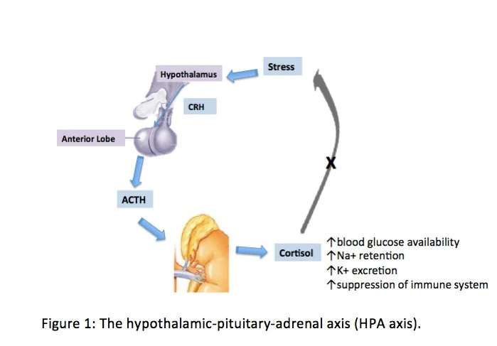
Long description
Hypothalamic-pituitary-adrenal axis: CRH, ACTH, cortisol and feedback

Cortisol is the end product of the HPA axis and the body's main glucocorticoid. Its actions explain the whole syndrome in six lines.
- Protein catabolism. Glucocorticoids break down muscle, skin, bone matrix and capillary collagen. This gives proximal myopathy, thin skin, easy bruising and purple striae when the dermis tears across expanding fat.
- Fat redistribution. Fat is deposited in the face, supraclavicular fossae, dorsocervical pad ("buffalo hump") and visceral depots, while fat elsewhere may be lost. Hence the moon face with peripheral thinning and central obesity.
- Anti-insulin action. Glucocorticoids drive hepatic gluconeogenesis and antagonise insulin in muscle and fat, producing glucose intolerance and frank diabetes.
- Sodium retention and potassium wasting. Cortisol occupies the mineralocorticoid receptor (11-beta-hydroxysteroid dehydrogenase type 2 normally prevents this); hence hypertension and hypokalaemia, worst in ectopic ACTH syndrome.
- Bone. Cortisol suppresses osteoblasts and intestinal calcium absorption, with increased resorption. Osteoporosis and vertebral fractures can be the presenting finding.
- CNS and mood. Early hypercortisolism produces insomnia, irritability, impaired short-term memory and depression; severe cases can produce psychosis. The hippocampal effects are well described.
The same mechanisms make Cushing syndrome a useful model of steroid toxicity in general: almost every feature has a direct glucocorticoid cause.
The onset is insidious, over months to years. Patients describe weight gain concentrated centrally with a rounding face and change of collar size, fatigue, loss of strength in rising from a chair or climbing stairs, back pain, new or worse hypertension or diabetes, depression, poor sleep and menstrual irregularity. In children the hallmark is weight gain with decreasing growth velocity. [1]
Some clues are more discriminatory than others. Reddish-purple striae wider than 1 cm, facial plethora, proximal myopathy (or proximal muscle weakness), easy bruising with trivial injury, and unexplained osteoporosis in a young person are the features that best separate Cushing syndrome from ordinary obesity or depression. [1]
Figure 2Cushing's syndrome (vector image)
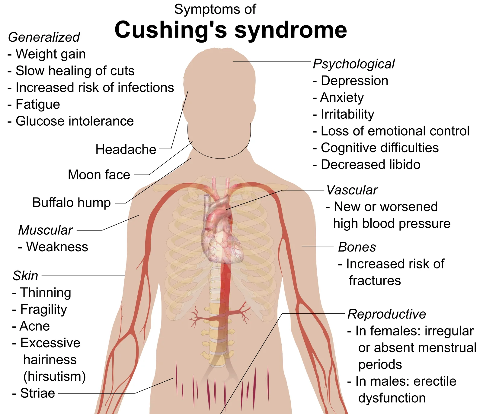
Long description
Illustration of the features of Cushing syndrome

- General: moon face (facial fullness), dorsocervical and supraclavicular fat pads, central obesity, peripheral thinning.
- Skin: thin skin, easy bruising, poor wound healing, acne, hirsutism in women, purple striae wider than 1 cm on the abdomen, flanks, axillae or thighs.
- Muscle and bone: proximal weakness (difficulty rising from sitting without using arms), vertebral osteoporosis, kyphosis.
- Other: hypertension, peripheral oedema, unexplained hypokalaemia, signs of diabetes.
- Children: decreased height percentile with increasing weight, virilisation or pseudoprecocious puberty.
- Complications may dominate: infection, pulmonary embolism, fracture, psychosis.
A few features from the history and examination in older photographs can make the diagnosis obvious: compare a five-year-old photograph with today's appearance.
Cushing syndrome is late to be diagnosed because its features are common: obesity, depression, type 2 diabetes, hypertension and menstrual irregularity. The wider differentials below need a short period of observation rather than one test.
Pseudo-Cushing states (non-neoplastic hypercortisolism) include pregnancy, depression, alcohol dependence, glucocorticoid resistance, morbid obesity, poorly controlled diabetes, physical stress, malnutrition or anorexia, intense chronic exercise and hypothalamic amenorrhoea. [1] These can produce mildly elevated cortisol and abnormal screening tests. They are handled by treating the underlying condition, repeating tests over 3 to 6 months, and avoiding treatment of hypercortisolism without a secure diagnosis. [3]
Conditions that mimic specific features include polycystic ovary syndrome (hirsutism and menstrual disturbance), alcoholic liver disease (striae and malnutrition), familial lipodystrophy and simple obesity with striae. A sex hormone, alcohol or liver panel and a 24-hour UFC that is normal settle most of these.
The mass already on an image. An adrenal incidentaloma or a pituitary lesion found incidentally may or may not be functioning; do not assume every 4 mm pituitary microadenoma seen on MRI explains a patient's obesity unless two screening tests agree.
Exogenous glucocorticoid exposure must be excluded first, with a really thorough drug history including oral, rectal, inhaled, topical, depot and intra-articular preparations and over-the-counter products. [1]
Endogenous Cushing syndrome should be suspected and tested for in the following groups, once exogenous use is excluded: [1]
- patients with unusual features for age (for example osteoporosis or hypertension in a young adult);
- patients with multiple and progressive features, particularly those with high discriminatory value;
- children with a falling height percentile and rising weight;
- patients with an adrenal incidentaloma compatible with an adenoma.
Widespread testing of other groups is discouraged because false-positive results are common and harmful. [1] At the other end, about 2 to 3.3 percent of patients with poorly controlled diabetes have had surgically confirmed Cushing syndrome in some case series, and screening studies report it in 0.5 to 1 percent of hypertensive patients. [1] Where suspicion is high but a test is normal, re-test in 6 months if features progress. [1]
Patients with familial syndromes that predispose to Cushing syndrome (Carney complex, MEN1) should be screened under endocrinology supervision. [1]
The Endocrine Society 2008 diagnostic guideline recommends starting with one test of high diagnostic accuracy from this list, chosen for the patient: [1]
- 24-hour urinary free cortisol (UFC). Collect two or three complete samples. [1]
- Late-night salivary cortisol. Collect between 2300 and 2400 on two separate evenings. [1] [3]
- 1 mg overnight dexamethasone suppression test (DST). Give 1 mg between 2300 and 2400, and measure serum cortisol at 0800 to 0900 the next morning, using the 1.8 microg/dL (50 nmol/L) cutoff. [1]
Normal reference orientation. Late-night salivary cortisol in normal subjects is below 145 ng/dL (4 nmol/L) when measured by validated assays. [1] After 1 mg dexamethasone a normal subject suppresses serum cortisol to below 1.8 microg/dL (50 nmol/L); many laboratories still use the older "less than 5 microg/dL" (140 nmol/L) cut-off, which is more specific but less sensitive. [1] Using the stricter 1.8 cut-off gives sensitivity over 95 percent but specificity of about 80 percent; raising the cut-off to 5 microg/dL (140 nmol/L) pushes specificity above 95 percent. [1]
Pitfalls that cause false positives or false negatives (check all of these before telling a patient they have Cushing syndrome): [1]
- Exogenous glucocorticoids in any form, including skin creams, "tonics", joint injections and megestrol acetate.
- Oral oestrogens raise cortisol-binding globulin; up to 50 percent of women on the oral contraceptive pill fail an overnight DST. Withdraw oestrogen for 6 weeks where practical or interpret a UFC instead. [1]
- CYP3A4 inducers (phenytoin, carbamazepine, rifampicin, phenobarbital, primidone, ethosuximide, pioglitazone) accelerate dexamethasone metabolism and cause false non-suppression. [1]
- CYP3A4 inhibitors (itraconazole, ritonavir, fluoxetine, diltiazem, cimetidine, aprepitant) and liver or renal failure slow dexamethasone metabolism and cause false suppression. [1]
- UFC falls with renal impairment, becoming falsely low below a creatinine clearance of 60 mL/min, and rises with high fluid intake (5 L a day or more) and with severe obesity, depression, alcohol and malnutrition. [1]
- Salivary cortisol is affected by smoking, bleeding gums, blood contamination of the swab, shift work, crossed time zones, samples taken during stress, and bedtime that is consistently long after midnight. [1]
- Late-night and sleeping cortisol tests assume a normal circadian rhythm and are not interpretable in night-shift workers or acute illness. [1] [3]
- In pregnancy, UFC rises up to 3-fold by term and dexamethasone testing is unreliable; use UFC with a value over 3 times the upper limit of normal. [1]
- In anticonvulsant users, the DST gives false positives; use salivary, blood or urine cortisol instead. [1]
- Cyclic Cushing syndrome needs UFC or salivary cortisol collected at the time of symptoms; DSTs may be normal when the disease is quiescent. [1]
- Assay differences are large; interpret every result against the laboratory's own normal range and retest with a second test before telling the patient anything. [1]
If the first test is abnormal in a patient with a moderate clinical suspicion, refer to an endocrinologist and repeat with a different screening test. Concordant abnormal results in two different tests mean "go and confirm the cause"; concordant normal results essentially exclude Cushing syndrome; discordant results need time, repeat testing, an endocrine second opinion, and sometimes a longer low-dose DST (2 mg a day for 48 hours). [1]
Imaging and IPSS are tools to find the source, and should not be used to screen. [3]
Once two screening tests agree, measure plasma ACTH to split the cause. [1]
- Suppressed ACTH with high cortisol points to an adrenal source (adenoma, carcinoma or hyperplasia): the HPA axis is switched off by autonomous cortisol.
- Normal or high ACTH points to ACTH-dependent disease (pituitary Cushing disease or ectopic ACTH).
Then image appropriately. Pituitary MRI is the imaging of choice for an ACTH-secreting adenoma. With standard 1.5 tesla MRI only about 50 percent of microadenomas are clearly depicted, and about a third of patients with Cushing disease have a normal scan. [3] Higher-field 3 tesla imaging is preferred where available, and thin-slice dynamic sequences may show a lesion; even then, a pituitary incidentaloma can be mistaken for the cause. [3] Initial imaging in suspected ectopic ACTH syndrome is a neck-to-pelvis thin-slice CT to look for a neuroendocrine primary or metastatic disease, particularly in a man with very high UFC and profound hypokalaemia. [3]
When the source is still unclear, the 2021 consensus gives the practical rules that many centres use: [3]
- no single test or combination of tests absolutely differentiates pituitary from ectopic ACTH secretion; [3]
- a pituitary lesion 10 mm or larger with concordant dynamic testing is enough to proceed as Cushing disease without IPSS; [3]
- lesions smaller than 6 mm should prompt IPSS; [3]
- for lesions 6 to 9 mm expert opinion differs and most centres still perform IPSS. [3]
IPSS (bilateral inferior petrosal sinus sampling) is the gold standard to exclude ectopic ACTH and should be performed only in hypercortisolaemic patients in a specialist centre. A central (pituitary)-to-peripheral ACTH gradient below 2 before, or below 3 after, stimulation suggests an ectopic source; false negatives and positives occur, and prolactin measurement can rescue a misleading result. [3] Because IPSS is invasive and carries procedural risk, it is reserved for patients who are biochemically proven to be in the active phase, and for "difficult" cases where MRI is negative or shows a small equivocal lesion. [3]
Diagnosing Cushing syndrome and finding its source
- Exclude exogenous glucocorticoid useTake a full drug history including topical, inhaled, depot and over-the-counter products.
- Screen with one high-accuracy test24-hour urinary free cortisol, late-night salivary cortisol, or the 1 mg overnight dexamethasone suppression test; if abnormal, repeat with a different test.
- Two screening tests agree: measure plasma ACTHConcordant normal results essentially exclude the syndrome; discordant results need repeat testing and an endocrine opinion.
| Cause | Screening cortisol tests | Plasma ACTH | Source-finding step |
|---|---|---|---|
| Pituitary (Cushing disease) | Abnormal | Normal or high | Pituitary MRI; IPSS when the lesion is small or MRI is negative |
| Ectopic ACTH syndrome | Abnormal, often very high urinary free cortisol | Normal or high | Neck-to-pelvis CT; IPSS to exclude a pituitary source |
| Adrenal adenoma, carcinoma or hyperplasia | Abnormal | Suppressed | Adrenal imaging |
The history-and-physical tests (high-dose dexamethasone suppression test and CRH stimulation) add information but no single test reaches 100 percent specificity; results may be discordant in up to a third of patients. [3] CRH is not available in many countries. These tests can guide the decision, but imaging plus IPSS settles it.
Exam trapIPSS is not a screening test
IPSS should never be requested in an undiagnosed patient. It is only used after two screening tests have established Cushing syndrome, because the central-to-peripheral gradient overlaps that of healthy people and of pseudo-Cushing states. [3]
There is no single international "score"; diagnosis is a combination of two concordant screening tests plus a cause-specific test. [3] Objective biochemical targets used in the literature are: post-transsphenoidal remission defined as morning serum cortisol below 55 nmol/L (about 2 microg/dL) or UFC below the upper limit of normal; recurrence usually declared when late-night salivary cortisol or UFC rises again. [3] [2]
Severity is clinical rather than laboratory: life-threatening hypercortisolism, severe infection, pulmonary embolism, acute psychosis, profound hypokalaemia or rapidly progressive bone disease push a patient into emergency treatment within 24 to 72 hours. [2]
The principles are simple and come from both the 2015 Endocrine Society treatment guideline and the 2021 consensus update: normalise cortisol (or its action), remove or control the cause, and treat every comorbidity. [2] [3]
Life-threatening complications are treated in parallel with, and not sequentially after, cortisol control. [2] If infection, pulmonary thromboembolism, cardiovascular decompensation or acute psychosis are present, treat the hypercortisolism within 24 to 72 hours, together with the appropriate antibiotics or anticoagulation. [2]
Surgical resection of the causal lesion is the first-line treatment for all endogenous causes, unless surgery is impossible or unlikely to reduce cortisol. [2]
- Pituitary Cushing disease: transsphenoidal selective adenomectomy by an experienced pituitary surgeon. In experienced hands remission is achieved in about 80 percent of microadenomas and 60 percent of macroadenomas. [3] Refer to a Pituitary Tumour Centre of Excellence; case volume matters, and published surveys associate more than 200 transsphenoidal procedures with the lowest complication rates. [3] [2]
- Adrenal adenoma or carcinoma: unilateral adrenalectomy by an experienced adrenal surgeon (curative in almost all benign adenomas). [2]
- Ectopic ACTH syndrome: localise and resect the ectopic tumour with nodal dissection where appropriate; resection cures about 76 percent of patients in observational series. [2]
- Bilateral adrenal disease (macronodular, primary pigmented nodular, McCune-Albright): bilateral adrenalectomy is the definitive treatment. [2]
Perioperative care after pituitary surgery. Measure serum sodium repeatedly during days 5 to 14 (hyponatraemia peaks then), free T4 and prolactin within 1 to 2 weeks for overt hypopituitarism, and obtain a baseline pituitary MRI 1 to 3 months after successful transsphenoidal surgery. [2]
Thromboprophylaxis. Cushing syndrome confers a VTE risk more than 10-fold higher than non-functioning pituitary adenomas and 18-fold higher than in healthy people, and the risk persists into the postoperative months. [3] Evaluate risk factors for venous thrombosis and give perioperative prophylaxis, preferably with low-molecular-weight heparin over oral anticoagulants (long half-life, no quick reversal). [2] [3] Early ambulation, compression stockings and periodical reassessment are the rest of the plan. [3]
A shared decision between a multidisciplinary team and the patient, weighing preferences, treatment goals, tumour size, urgency, drug interactions and cost. [2] [3]
Repeat pituitary surgery is reasonable, particularly with evidence of incomplete resection or an MRI-visible lesion. [2] [3]
Radiotherapy or radiosurgery is used for persistent or recurrent disease after transsphenoidal surgery, or for aggressive tumours; conventional radiotherapy 45 to 50 Gy in fractions or stereotactic radiosurgery around 20 Gy; hypopituitarism occurs in 25 to 50 percent. [3] Confirm medical control of cortisol first, because radiation takes months to work. [2]
Bilateral adrenalectomy (BLA) gives immediate cortisol control and is used when medical therapy cannot normalise cortisol promptly, in very severe disease, or sometimes in women planning pregnancy. Laparoscopic BLA in large series has had a 10 to 18 percent complication rate and under 1 percent mortality. [3] It commits the patient to lifelong glucocorticoid and mineralocorticoid replacement, and post-BLA the risk of Nelson syndrome (progressive corticotroph tumour growth, inferred from rising ACTH and imaging) is a long-term concern in 25 to 40 percent of patients after 5 to 10 years. [3] BLA therefore requires plasma ACTH monitoring and serial pituitary imaging, starting about 6 months after surgery, with pasireotide reported to suppress ACTH in established Nelson syndrome. [2] [3]
Medical therapy is second-line: it controls cortisol while waiting for radiation, while preparing for surgery, in persistent or recurrent disease, or in patients who are not operated on. [2] [3] Adrenal steroidogenesis inhibitors are generally used first because their effect is predictable; tumour-directed drugs (pasireotide, cabergoline) are reserved for cases where residual tumour is visible and shrinkage is a goal; mifepristone is used only by experienced clinicians. [3]
| Drug | Target | Usual dose | Main points |
|---|---|---|---|
| Ketoconazole | Adrenal steroidogenesis | 400 to 1200 mg a day by mouth, twice daily | 64 percent UFC normalisation in retrospective series; escape in 15 to 25 percent; needs gastric acid (avoid proton pump inhibitors); hepatotoxicity in 10 to 20 percent, usually within 6 months; monitor liver enzymes; drug interactions; hypogonadism in men [3] |
| Osilodrostat | Adrenal steroidogenesis | 2 to 7 mg a day twice daily by mouth, up to 30 mg twice daily | 86 percent UFC normalisation in a phase 3 randomised withdrawal study; risk of hypocortisolism, hypokalaemia, hypertension, androgenic effects such as hirsutism, QT prolongation [3] |
| Metyrapone | Adrenal steroidogenesis | 500 mg to 6 g a day, every 6 to 8 hours | 47 percent UFC normalisation at 12 weeks in a prospective study; rapid onset (hours to days); hirsutism and hypokalaemia in women; 11-deoxycortisol cross-reacts in some cortisol immunoassays [3] |
| Pasireotide | Pituitary somatostatin receptor | 0.3 to 0.9 mg per mL twice daily subcutaneously, or 10 to 30 mg monthly intramuscularly (long-acting) | 15 to 26 percent UFC normalisation short-acting, 40 percent long-acting; tumour shrinkage in some; high risk of hyperglycaemia requiring patient selection and glucose monitoring; QT prolongation [3] |
| Mifepristone | Peripheral glucocorticoid receptor | 300 to 1200 mg a day by mouth | Improves glycaemia and blood pressure; no biochemical marker of efficacy (cortisol stays high); risk of hypokalaemia, hypertension, adrenal insufficiency; clinicians must be experienced; drug interactions [3] |
| "" |
The heterogeneity of the table (which lumps a glucocorticoid-receptor blocker with steroidogenesis inhibitors) reflects the fact that no single agent suits all patients. Selection depends on severity of hypercortisolism, need for speed, presence of residual tumour, diabetes, hypertension, liver function, cost and availability. [3]
Clinical pearlSpeed matters
In severe disease, cortisol must drop quickly. Osilodrostat and metyrapone act within hours; ketoconazole within days; etomidate intravenously is reserved for inpatient, very severe cases. If hypercortisolism is extreme and unresponsive, bilateral adrenalectomy should be undertaken before irreversible complications occur. [3]
Preoperative medical therapy has sparse evidence but is used by many experts when surgery is delayed or when a patient has life-threatening complications requiring rapid cortisol control. [3]
Monitoring response. Use a combination of UFC, late-night salivary cortisol and clinical features (weight, blood pressure, glucose, quality of life). Change treatment after 2 to 3 months at maximum tolerated dose if cortisol has not normalised; consider combination therapy if there is partial response, and switch if resistance is clear. [3]
Adrenal crisis during therapy. Steroidogenesis inhibitors can cause adrenal insufficiency and withdrawal symptoms; patients need education, an emergency steroid card and clear instructions about when to stop and resume treatment. [2] [3]
After successful transsphenoidal or adrenal surgery the HPA axis is suppressed and the patient is, in effect, adrenal insufficient. Replace with hydrocortisone 10 to 12 mg per square metre per day in divided doses, twice or three times daily, with the first dose as early as possible after waking. [2] The axis recovers in adults about 6 to 12 months after resection of an ACTH-producing tumour and about 18 months after unilateral adrenalectomy. [2] In children recovery averaged 12.6 plus or minus 3.3 months in one large series. [2] Recovery can be assessed with morning cortisol (before the day's dose) every 3 months, followed by ACTH stimulation when the level reaches about 7.4 microg/dL (200 nmol/L); the axis has recovered when baseline or stimulated cortisol is about 18 microg/dL (500 nmol/L) or greater. [2] Warn the patient about the steroid withdrawal syndrome: anorexia, nausea, weight loss, fatigue, myalgia and arthralgia that can take 1 year or longer to resolve despite physiological replacement, and reassure them frequently. [2] [3]
Bilateral adrenalectomy, of course, requires lifelong glucocorticoid and mineralocorticoid replacement with a stress-dosing plan and a medical alert. [2]
In Cushing disease, recurrence after surgery is 23 percent for microadenomas and 33 percent for macroadenomas in one series, with 15 to 66 percent cumulative recurrence 5 to 10 years after successful surgery. [2] Hence lifelong monitoring: after HPA axis recovery, check late-night salivary cortisol annually, since it becomes abnormal before UFC or the DST. [2] [3] Recurrence can occur more than 10 years later, and patients drop out of follow-up, which is where most harm occurs. [1] [2]
Treat comorbidities for life: hypertension, diabetes, dyslipidaemia, osteoporosis (including bisphosphonates where persistent hypercortisolism leaves bone fragile), infection prevention with targeted vaccination, thrombosis risk, psychological distress. [2] [3]
- Cardiovascular disease, venous thromboembolism and infection are the main causes of excess mortality in active disease; cardiovascular risk may not fully normalise after cure. [2] [3]
- Vertebral fractures in 30 to 50 percent of patients, largely tracking severity of hypercortisolism. [3]
- Type 2 diabetes in up to 30 percent, dyslipidaemia in 16 to 64 percent. [3]
- Proximal myopathy, often persistent despite cure. [3]
- Infection risk is higher in severe hypercortisolism than in mild to moderate disease. [2]
- Growth hormone deficiency in many cured children and some adults, needing assessment 3 to 6 months after surgery in children and 12 months in adults. [2] [3]
- Impaired quality of life and psychiatric morbidity can last for years. [2] [3]
Standardised mortality ratio (SMR) is increased 1.7- to 4.8-fold in active Cushing syndrome and the hazard ratio reaches 2.8 to 16 with persistent or recurrent hypercortisolism. [2] Successful treatment improves but may not fully normalise SMR and cardiovascular or bone risk, so treat the comorbidities for life. [2] [3] Before effective treatment, median survival was 4.6 years. [2]
Primary prevention is preventing inappropriate glucocorticoid use: prescribe the lowest effective dose for the shortest duration, use inhaled and topical agents preferentially, screen for hidden sources such as mixed formulations or imported creams, and review drug lists before any DST. [1] Secondary prevention is familial: Carney complex, MEN1 and FIPA patients need regular screening for Cushing syndrome by endocrinology, with cardiac and skin surveillance. [1]
Pregnancy. Use UFC, since dexamethasone suppression is unreliable and UFC can rise up to threefold by term; only a value over 3 times the upper limit of normal is diagnostic. Ketoconazole is avoided in pregnancy and metyrapone, cabergoline or transsphenoidal surgery are selected by experts; none of the drugs is licensed, and higher cortisol cut-offs apply. [1] [3]
Children. Endogenous Cushing syndrome before 18 years is very rare. Growth failure with weight gain is the classic presentation. UFC, late-night salivary cortisol and 1 mg DST are used, but the Dex-CRH test is not useful in children and IPSS has a smaller role than in adults. [3] Transsphenoidal surgery is first-line; thromboprophylaxis is reserved for selected paediatric cases; recovery of adrenal function takes about 12 months; growth hormone is assessed at 3 to 6 months. [3] [2]
Renal failure. Use the 1 mg overnight DST rather than UFC; UFC becomes falsely low below a creatinine clearance of 60 mL/min. [1]
Epilepsy and enzyme inducers. Avoid DST and use circadian cortisol tests. [1]
Elderly patients. Presentations may be dominated by osteoporosis, hypertension and diabetes; exercise caution when interpreting salivary cortisol in diabetic hypertensive men. [1]
- In places without salivary cortisol assays, UFC or a carefully executed 1 mg overnight DST, with simultaneous dexamethasone level if possible, will establish or refute the diagnosis. Most ordinary chemistry laboratories can measure serum cortisol. [1] [3]
- When MRI field strength or quality is limited, a negative scan does not exclude a pituitary adenoma; rely on two concordant screening tests and, where needed, a regional referral for IPSS. [3]
- IPSS is unavailable in many countries; high-dose dexamethasone plus CRH (where CRH can be obtained), MRI and whole-body CT can substitute, and neck-to-pelvis CT is the best first step in a man with high UFC and hypokalaemia. [3]
- Ketoconazole is the cheapest and most widely available steroidogenesis inhibitor and is usually the first choice where cost or availability limits other drugs, but requires twice-daily dosing, gastric acid, and liver-function monitoring. Metyrapone acts faster but is often more expensive; osilodrostat is not yet widely available outside centres with good access. [2] [3]
- Bilateral adrenalectomy is low-tech and definitive where drug supply or monitoring is unreliable; it mandates a reliable lifelong steroid supply and a stress-dose education programme.
- Refer patients with Nelson syndrome features (rising ACTH, enlarging sellar mass after adrenalectomy) early; where pasireotide is unavailable, conventional radiotherapy or repeat surgery is used. [3]
- Where neither surgeon nor coagulation support exist, focus on treating complications, controlling comorbidities and arranging referral; excess mortality without treatment remains substantial. [2]
- Cushing syndrome is prolonged glucocorticoid excess; iatrogenic steroid therapy is the most common cause.
- The features are all explained by glucocorticoid actions: protein catabolism, fat redistribution, insulin resistance, mineralocorticoid effects and bone loss.
- Reddish-purple striae wider than 1 cm, facial plethora, proximal weakness, easy bruising and unexplained osteoporosis are the most discriminatory findings.
- Diagnosis requires exclusion of exogenous steroids first and two concordant screening tests (UFC, late-night salivary cortisol, 1 mg overnight DST), ideally under endocrine supervision.
- Pitfalls (oestrogen, drugs, renal failure, fluid intake, shift work, pregnancy, cyclic disease, assay variability) can flip results; check them before diagnosing.
- ACTH separates ACTH-dependent from ACTH-independent disease; MRI plus, where needed, IPSS finds the source.
- Surgery on the source is first-line; medical therapy, repeat surgery, radiation and bilateral adrenalectomy are second-line or adjunctive.
- All patients need lifelong glucocorticoid replacement plans after remission, complication care and recurrence surveillance.
Answer each one in your head first, then open the answer.
A 48-year-old woman with treatment-resistant hypertension, central obesity and purple striae asks whether her oral contraceptive could have caused all this. What do you say, and what is the first test you would order?
Show answerHide answer
Oral contraceptives do not cause Cushing syndrome, but they do raise cortisol-binding globulin and can produce false positive overnight 1 mg dexamethasone suppression tests in up to 50 percent of women. Stop oestrogen for 6 weeks if possible and use a 24-hour urinary free cortisol (or re-test off the pill) rather than declaring a diagnosis from a single DST. If the UFC is normal and two different tests agree, Cushing syndrome is very unlikely; explain the obesity and hypertension on their own merits while keeping the patient under review.
A 30-year-old man presents with a moon face, proximal weakness and serum potassium of 2.6 mmol/L. UFC is 10 times the upper limit of normal and plasma ACTH is markedly raised. What are the two most likely sources and how do you tell them apart?
Show answerHide answer
Pituitary Cushing disease or an ectopic ACTH-secreting tumour (often small cell lung cancer or a bronchial or pancreatic neuroendocrine tumour), because ACTH is clearly raised. MRI of the pituitary is the first imaging, with standard 1.5 tesla showing only about half of microadenomas; if it shows a tumour 10 mm or larger with concordant dynamic tests, proceed as Cushing disease. For a lesion under 6 mm, or no lesion, arrange IPSS in a specialist centre, remembering that no single laboratory test is perfectly specific and the central-to-peripheral gradient threshold is below 2 before or below 3 after stimulation.
Why can ACTH be normal in Cushing disease and why does that not exclude it, and what is the sampling trap with late-night salivary cortisol?
Show answerHide answer
ACTH is usually normal to modestly raised rather than floridly high in pituitary Cushing disease because the corticotroph adenoma retains partial feedback; ectopic ACTH syndrome tends to drive much higher levels. The late-night salivary cortisol assumes a normal circadian rhythm, so it is uninterpretable in shift workers, people crossing time zones, acutely ill patients and chronic smokers or people with bleeding gums who contaminate the swab.
A patient has just undergone transsphenoidal surgery for Cushing disease and is discharged. What does the daily glucocorticoid prescription look like and for how long, and what must you warn them about?
Show answerHide answer
Hydrocortisone 10 to 12 mg per square metre per day in divided doses, first dose on waking, then taper according to 3-monthly morning cortisol and 18 microg/dL (500 nmol/L) stimulated recovery thresholds. The HPA axis recovers in 6 to 12 months after transsphenoidal surgery in adults. Warn about the steroid withdrawal syndrome: anorexia, nausea, weight loss, fatigue, muscle and joint aches, skin flaking and low mood, which can last a year or more and can occur before the axis has fully recovered. Provide an emergency steroid card, stress-dose instructions and contact details.
In a centre with no IPSS and no salivary cortisol assay, how would you diagnose and treat a suspected ectopic ACTH syndrome in a man with UFC 15 times the upper limit, severe hypokalaemia and weight loss?
Show answerHide answer
Confirm hypercortisolism with two UFC collections and a 1 mg overnight DST, measure ACTH, then do neck-to-pelvis thin-slice CT and, if needed, whole-body cross-sectional imaging. Control cortisol rapidly with metyrapone or osilodrostat (ketoconazole as a cheaper option) while searching for and resecting the tumour if found; use bilateral adrenalectomy only if the source cannot be found or controlled. Arrange referral, because untreated severe ectopic ACTH syndrome has high mortality from infection, thrombosis and hypokalaemia. [3]
(Plain text only, until those topics are published and verified: primary hyperaldosteronism; screening and evaluation of hypertension; osteoporosis; type 2 diabetes mellitus; depression.)
""
- Nieman LK, Biller BMK, Findling JW, Newell-Price J, Savage MO, Stewart PM, Montori VM. The diagnosis of Cushing's syndrome: an Endocrine Society Clinical Practice Guideline. The Journal of Clinical Endocrinology and Metabolism 2008;93(5):1526-1540. Accessed .
- Nieman LK, Biller BMK, Findling JW, Murad MH, Newell-Price J, Savage MO, Tabarin A. Treatment of Cushing's syndrome: an Endocrine Society Clinical Practice Guideline. The Journal of Clinical Endocrinology and Metabolism 2015;100(8):2807-2831. Accessed .
- Fleseriu M, Auchus R, Bancos I, et al. Consensus on diagnosis and management of Cushing's disease: a guideline update. The Lancet Diabetes and Endocrinology 2021;9(12):847-875. Accessed .
Figure credits
Educational content for medical students and doctors. It is not a substitute for clinical judgement or local guidelines.