Acromegaly
Chronic GH excess: causes, presentation, diagnosis with IGF-1 and GH suppression, surgery, medicines, radiation and screening.
- Acromegaly is a slow, multisystem disease caused by long-standing excess growth hormone (GH) and its mediator insulin-like growth factor 1 (IGF-1), almost always from a benign pituitary somatotroph adenoma.
- The overgrowth is acral and facial (enlarged hands, feet, nose, jaw), but the illness is really a systemic one: hypertension, type 2 diabetes, sleep apnoea, cardiomyopathy, arthropathy and colonic polyps.
- Diagnosis is biochemical first: a raised age-adjusted serum IGF-1, confirmed when GH fails to fall below 0.4 micrograms per litre during an oral glucose tolerance test.
- Pituitary MRI then shows the adenoma and shows which structures it threatens.
- The goals are to normalise IGF-1, control adenoma growth and treat every complication. Transsphenoidal surgery by an experienced surgeon is first-line; medical therapy with somatostatin analogues, pegvisomant or cabergoline and, less often, radiotherapy follows if needed. [3]
- With modern treatment, biochemical control is achievable in most patients and mortality in controlled disease is close to that of the general population.
By the end of this page you should be able to:
- Explain why GH excess produces acral overgrowth, coarse features and systemic disease, and name the common causes.
- Recognise the presentation early, including the comorbidity clusters that appear years before classic features.
- Plan the biochemical and imaging investigation and state the cut-offs that define a normal response to glucose.
- Describe the stepwise management and the place of surgery, somatostatin analogues, pegvisomant, cabergoline and radiotherapy.
- List the complications that need structured screening and how follow-up is organised.
- Adapt investigation and treatment where advanced tests or drugs are not available.
DefinitionAcromegaly
Acromegaly is the clinical syndrome produced by chronic excess of growth hormone (GH) and insulin-like growth factor 1 (IGF-1) in adults whose epiphyseal growth plates have closed. It usually results from a pituitary somatotroph adenoma. When the same hormone excess begins before puberty, while growth plates are still open, the result is gigantism rather than acromegaly. [1]
GH is secreted by somatotroph cells of the anterior pituitary and acts on the liver and other tissues to release IGF-1, which mediates most of the growth and metabolic effects. Both hormones are normally tightly regulated by hypothalamic growth hormone-releasing hormone (GHRH, stimulatory) and somatostatin (inhibitory). In acromegaly that control is lost.
Classification is by where the excess signal arises:
- Pituitary somatotroph adenoma, the cause in the great majority of patients. About 70% are macroadenomas (larger than 10 mm), often invasive into the cavernous sinus. [1]
- Mixed somatotroph-lactotroph adenoma, in which GH and prolactin are both secreted; prolactin is often raised and the adenoma may be larger.
- Ectopic GHRH secretion, in which a neuroendocrine tumour (most often pancreatic or bronchial) drives the pituitary somatotrophs. This accounts for under 1% of acromegaly. [1]
- Ectopic GH secretion, from pancreatic, bronchial or ovarian tumours, or non-Hodgkin lymphoma, or ectopic somatotroph tissue in the sphenoid sinus or clivus. Very rare. [1]
- Syndromic and hereditary forms: multiple endocrine neoplasia type 1 (MEN1), MEN4, McCune-Albright syndrome and Carney complex, in which acromegaly often presents at a younger age. [1]
Hereditary and familial GH-secreting adenomas are uncommon but matter clinically, because they present earlier and may recur.
Acromegaly is rare. A systematic review and meta-analysis of studies from 1955 to 2016 reported a pooled prevalence of 5.9 (95% confidence interval 4.4 to 7.9) cases per 100 000 people and an incidence of 0.38 (95% confidence interval 0.32 to 0.44) cases per 100 000 person-years. [2] Older and newer regional reports vary considerably: early studies quoted 30 to 60 cases per million, while more recent estimates are 70 to 90 per million. [2] The apparent rise is probably due to better IGF-1 and GH assays, wider use of pituitary MRI and improved survival, rather than a true increase in the number of somatotroph tumours.
Diagnostic delay is still long. Patient-directed questionnaires record a mean delay of about 14 years from first symptom to diagnosis, and cohort studies 6 to 10 years. [1] [6] A Swedish cohort found that 24% of patients waited over 10 years and that this group had more comorbidity and excess mortality. [2] Women are diagnosed about 4.5 years later than men and accumulate more complications. [1]
The cause of most sporadic adenomas is not established. The leading view is monoclonal expansion of a somatotroph with activating mutations in signalling genes; this is background from reviews and the clinical implication is that the adenoma grows slowly and secretes autonomously.
Risk factors and settings that point to a specific cause:
- No known modifiable risk factor exists in sporadic disease. Female sex shows a modest excess in several but not all cohorts.
- Family history or early onset should raise the possibility of MEN1, MEN4, McCune-Albright syndrome or Carney complex. Children and young adults carry a higher genetic predisposition, for example aryl hydrocarbon receptor-interacting protein (AIP) mutations in familial isolated pituitary adenoma. [1]
- Pituitary incidentaloma. Many adenomas are now found by MRI done for headache or other reasons; not all secrete excess hormone, but every such mass needs an endocrine assessment.
- Occupational or medical exposure is not an established cause.
There is no role in prevention beyond treating the adenoma and its complications.
Figure 1Endocrine growth regulation
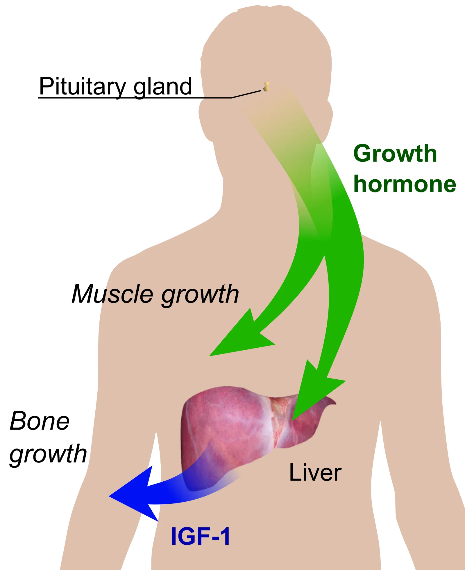
Long description
Growth hormone from the pituitary gland acts on muscle and on the liver, where it stimulates IGF-1, which in turn drives bone growth.

GH and IGF-1 are anabolic and anti-insulin growth signals. Their chronic excess explains every feature of the disease:
- GH is secreted in pulses and acts directly on bone, muscle, fat, heart, kidney and many other tissues. It is lipolytic and anti-insulin.
- IGF-1, produced mainly by the liver, is the mediator for linear and soft-tissue growth. IGF-1 levels reflect average GH secretion and are relatively stable through the day, which is why a random IGF-1 is the screening test.
- Linear growth stops after epiphyseal fusion, but joints and soft tissues continue to respond, which is why an adult enlarges in hands, feet, jaw and viscera rather than in height.
- Glucose intolerance and diabetes occur because GH opposes the action of insulin: it reduces peripheral glucose uptake and increases hepatic glucose production. [1]
- Sodium and water retention and the anti-natriuretic effect of GH raise vascular volume and contribute to hypertension. [2]
- Soft-tissue overgrowth in the upper airway (tongue, pharynx, vocal cords) narrows the airway and causes obstructive sleep apnoea. [1]
- Bone and cartilage enlarge (mandible, frontal bones, hands) but their microarchitecture may become abnormal, so joint pain and deformity persist even after biochemical control. [4]
- Cardiac growth: GH/IGF-1 stimulate myocardial hypertrophy and can produce a cardiomyopathy. Clinically significant cardiomyopathy has become less common with earlier diagnosis and control. [1]
Structural changes in joints, bone and heart do not fully regress once established, even when IGF-1 is normalised. Biochemical control prevents progression and reduces mortality, but prevention depends on early treatment.
Acromegaly is recognised late because its onset is insidious and the changes are gradual. Symptoms are multisystem and often present for years before anyone notices the face:
- Acral enlargement: increasing ring, shoe or hat size; tight gloves or rings; coarsening of facial features noticed on old photographs.
- Jaw and dental change: prognathism, underbite, widening of the gap between teeth, ill-fitting dentures.
- Soft-tissue symptoms: sweating, oily skin, skin tags, generalised increase in body bulk with proximal weakness.
- Sleep: snoring, sleep apnoea, daytime sleepiness, morning headache.
- Rheumatological: joint pain and stiffness, carpal or cubital tunnel syndrome, back pain; women more often present with carpal tunnel and headache. [1]
- Endocrine and metabolic: weight gain, fatigue, thirst, polyuria (diabetes), menstrual disturbance or erectile dysfunction.
- Visual: bitemporal hemianopia, diplopia or headaches from mass effect of a large adenoma.
- Gastrointestinal: constipation, change in bowel habit; women more often complain of constipation. [1]
- Psychological and cognitive: low mood, depression, reduced quality of life even after control.
- Sex differences: men more often present with prognathism, sleep apnoea and cardiovascular features; women are diagnosed later and carry more complications at presentation. [1]
Comorbidity clusters are often the first clue: hypertension, type 2 diabetes and dyslipidaemia may precede the diagnosis by years, as may snoring or carpal tunnel syndrome. When several of these occur together in a person without obesity, especially under 60, acromegaly should be considered. [1]
- Hands: large, spade-like; wide, thick digits; skin thickened.
- Feet: enlarged, often with widened forefoot.
- Face: coarse features, prominent supraorbital ridges, broad nose, thickened lips, prognathism, macroglossia; the oval face becomes square.
- Jaw: underbite, widened spacing (diastema).
- Chest/back: kyphosis may develop; chest is broad.
- Neck: goitre is common.
- Skin: oily, hyperhidrosis, acanthosis nigricans, skin tags.
- Voice: deepened.
- Joints: reduced range, deformity.
- Visual fields: formal perimetry if mass effect suspected.
- Cardiovascular: hypertension, signs of left ventricular dysfunction or valvular disease; listen for cardiomyopathy.
- Signs of hypopituitarism: if the adenoma is large, other axes may be lost (loss of libido, fatigue, postural hypotension).
Old photographs and shoe or ring sizes are invaluable.
In a recent synthesis of cohorts, acral enlargement was present in around 90% of patients at diagnosis and facial changes in 65 to 70%. Functional symptoms are commoner earlier in the illness than visible change. [1] Diagnostic delay ranges from 6 to 10 years on average in modern cohorts. [1]
The differential has two parts: conditions that mimic the metabolic and soft-tissue picture, and conditions that can be confused with a pituitary mass on imaging.
| Condition | Features shared with acromegaly | Feature that separates it |
|---|---|---|
| Pseudoacromegaly (insulin resistance, severe obesity, pachydermoperiostosis) | Coarse features, enlarged hands, joint change | Normal IGF-1 and GH suppression on OGTT. [1] |
| Familial tall stature / gigantism | Increased height and large hands in youth | Closes at puberty; IGF-1 and GH normal. [1] |
| Untreated primary hypothyroidism | Coarse skin, macroglossia, lethargy | Raised TSH, low free T4, low IGF-1. [1] |
| Other causes of hyperprolactinaemia | Menstrual disturbance, galactorrhoea, pituitary mass | Prolactin raised, GH and IGF-1 normal; MRI shows adenoma but biochemistry normal. [1] |
| Hereditary acromegaloid features (Nora disease) | Facial coarsening, large extremities | No biochemical GH excess; different inheritance. [1] |
| Non-functioning pituitary macroadenoma | Mass effect, hypopituitarism, visual field defect | GH and IGF-1 normal; may cause mild IGF-1 changes only. [1] |
On MRI, the main differential of a sellar mass is a true somatotroph adenoma versus a non-functioning adenoma, Rathke's cleft cyst, craniopharyngioma or meningioma. Biochemistry resolves most cases; histology resolves the rest.
The modern approach is to measure serum IGF-1 first and, when it is raised or when the clinical picture is strongly suspicious, to perform an oral glucose tolerance test (OGTT) to test whether GH suppresses.
- IGF-1 should be measured using an assay calibrated to the WHO international standard, with age-adjusted reference intervals. It is the recommended first test because it integrates GH secretion over days and can be taken at any time of day without fasting. [1]
- IGF-1 may be misleading in puberty, uncontrolled diabetes, liver disease, renal insufficiency, malnutrition, pregnancy, oral oestrogen therapy and obesity. Repeat or use dynamic testing if the result and the clinical picture disagree. [1]
- OGTT (gold standard confirmatory test): give 75 g oral glucose; measure GH at 0, 30, 60, 90 and 120 minutes. In health, GH falls below 0.4 micrograms per litre with an ultrasensitive assay. Failure of suppression supports acromegaly. A random GH above 0.4 micrograms per litre is usually present at diagnosis. [1]
- About one third of patients show a paradoxical rise in GH during the OGTT; this does not exclude the diagnosis. [1]
- Very rarely, IGF-1 is high while GH suppresses below 0.4 micrograms per litre, in microadenomas or very early disease. [1]
- The older Endocrine Society 2014 guideline threshold of GH above 1 microgram per litre during OGTT has been superseded by the lower cut-off made possible by modern assays, and the 2024 consensus now places greater weight on IGF-1 above 1.3 times the upper limit of normal in a patient with characteristic signs. [1] [5]
- The 2014 Endocrine Society guideline recommended raised IGF-1 for age with confirmation by GH above 1 microgram per litre during OGTT, and set out the therapeutic algorithm now built on by later consensus statements. [1] [5]
- Additional tests: prolactin (mixed adenoma); IGF-binding protein 3 is not useful. There is no benefit to GH profiling except in diabetes, where OGTT is not recommended. Soluble Klotho and related assays are research tools. [1]
Special populations matter:
- In uncontrolled diabetes, IGF-1 can be falsely normal or low. Control the diabetes first if acromegaly is still suspected. [1]
- In older adults, IGF-1 and GH are physiologically lower; obesity suppresses GH despite active disease.
- In pregnancy, avoid OGTT and use IGF-1 interpreted with pregnancy reference ranges.
- In children and adolescents, strict age- and puberty-adjusted IGF-1 ranges are needed; gigantism has higher GH levels and more aggressive adenomas. [1]
- Pituitary MRI with gadolinium is standard. Microadenomas (10 mm or less) usually appear as hypoenhancing lesions; macroadenomas (larger than 10 mm) extend suprasellar and into the cavernous sinus and threaten the optic chiasm. [1]
- Use 3-tesla imaging with dynamic contrast-enhanced sequences where available; they improve detection of small or "MRI-negative" lesions. [1]
- About 70% of patients have a macroadenoma at diagnosis. [1]
- Functional imaging (PET with 11C-methionine or 18F-fluoroethyltyrosine PET/MRI) is reserved for MRI-negative but biochemically active disease. [1]
- CT of the skull base is used when MRI is impossible, for surgical planning of bony anatomy.
- When MRI shows no discrete adenoma but acromegaly is biochemically proven, distinguish ectopic GHRH-driven hyperplasia from ectopic GH secretion and repeat high-resolution imaging. [1]
High-resolution imaging also has treatment implications: T2-weighted hypointensity predicts a better response to somatostatin analogues in some series, although this is not consistent. [1]
- Visual fields (formal perimetry) if there is any mass effect.
- Cardiovascular: blood pressure, ECG, and echocardiography when hypertension, left ventricular hypertrophy, valvular disease or heart failure is suspected. GH/IGF-1 excess is strongly associated with cardiovascular morbidity, and cardiomyopathy, though less common now, must be excluded. [1]
- Glucose assessment: fasting glucose and HbA1c; OGTT if diabetes is suspected.
- Lipids and sleep study (polysomnography) if sleep apnoea is suspected; over half of patients are affected.
- Bone: assess fracture risk and vertebral morphology; active disease increases vertebral fracture risk.
- Colonoscopy: see screening.
Investigating suspected acromegaly
- Clinical suspicionClassic features, comorbidity cluster, or incidental pituitary mass
- First test: serum IGF-1 (age-adjusted)Can be taken at any time of day; fasting not required
- If IGF-1 is raised or clinical picture is strongly suggestiveProceed to OGTT (75 g glucose)
- Question: Does GH suppress below 0.4 micrograms per litre?
No
- Biochemical acromegalyConfirm with repeat IGF-1 if discordant
- Pituitary MRI with gadolinium
- Assess as belowVisual fields, cardiovascular, metabolic, colon
Yes
- Acromegaly unlikelyIf IGF-1 is still high, repeat IGF-1 and reconsider confounders (diabetes, liver, renal, obesity, oestrogen)
- If MRI shows no adenomaConsider ectopic GHRH or GH secretion; functional imaging
Acromegaly is a clinicobiochemical diagnosis. The modern criteria combine:
- characteristic clinical features or signs attributable to GH/IGF-1 excess, and
- IGF-1 above 1.3 times the age-adjusted upper limit of normal, with or without failed GH suppression. [1]
Where results are equivocal (IGF-1 mildly raised, classic features absent, or discordant IGF-1 and GH), repeat IGF-1, ensure good control of confounders, and use the OGTT. A few patients have normal IGF-1 and suppressible GH despite features: this may be early disease, resolved acromegaly after pituitary apoplexy, or an acromegaloid condition (insulin resistance, hypothyroidism, lipodystrophy, pachydermoperiostosis). [1]
Remission after treatment is defined as age-adjusted normal IGF-1 measured 3 months after surgery, confirmed by a nadir GH below 0.4 ng per mL during OGTT. Discordant IGF-1 and GH results occur in over one quarter of patients after surgery; an isolated mildly elevated IGF-1 should be repeated, since it may normalise over 3 to 6 months. [3] [4]
- Normalise IGF-1 (and, where used, GH) to reduce mortality and morbidity.
- Remove or control the adenoma and prevent mass-effect complications.
- Recognise and treat every complication (diabetes, hypertension, sleep apnoea, cardiomyopathy, colon polyps, arthropathy, fractures).
- Preserve normal pituitary function where possible.
Mortality is roughly doubled in uncontrolled acromegaly (standardised mortality ratio 2.1, 95% confidence interval 1.5 to 2.9) but is not significantly different from the general population in controlled disease (0.9, 95% confidence interval 0.6 to 1.3). [2] Biochemical control is therefore the single strongest predictor of outcome.
Transsphenoidal resection by an experienced neurosurgeon is the first-line treatment for most patients. [3] It decompresses the sella, reduces tumour volume and lowers GH rapidly, and a dedicated centre has few permanent endocrine complications.
- Surgery achieves remission in a proportion that depends on tumour size and invasion. Preoperative imaging and (rarely) histology guide selection; women, particularly postmenopausal women, have larger, more invasive adenomas and lower surgical remission rates than men. Age itself does not predict outcome. [4]
- Preoperative medical therapy with a somatostatin analogue may be used for severe comorbidities that make intubation unsafe (severe sleep apnoea, heart failure), and to shrink a large adenoma before operation in some centres. The benefit on long-term surgical outcome has not been uniformly shown. [3]
- Early postoperative IGF-1 measured at 6 weeks is a useful early indicator of disease activity, though definitive assessment is at 3 months. [4]
- When surgery is not appropriate: patients who refuse operation, those with unacceptable surgical risk or uncontrolled comorbidities, centres without a pituitary neurosurgeon, or adenomas that are largely unresectable (mostly cavernous sinus). Use primary medical therapy. [3]
Medical therapy is used when surgery does not achieve remission, when it is contraindicated, or as primary therapy in selected patients. The choice depends on IGF-1 level, tumour growth, comorbidities (especially diabetes) and patient preference.
| Drug (class) | Administration | Expected biochemical control | Main cautions |
|---|---|---|---|
| Octreotide LAR (long-acting somatostatin analogue) | Intramuscular every 4 weeks | 30-60% [3] | Gastrointestinal and biliary side-effects. [3] |
| Lanreotide autogel (long-acting somatostatin analogue) | Deep subcutaneous every 4 to 8 weeks; 120 mg every 4 weeks as initial primary therapy in some series | 30-60% [3] | As octreotide; can be self-injected. [3] [4] |
| Octreotide capsules (oral somatostatin analogue) | 20 mg twice daily, taken at least 1 hour before or 2 hours after meals; adjust in 20 mg steps every 2 weeks | 58% in previously controlled patients [3] | Gastrointestinal; bioavailability falls 90% with a meal. [3] |
| Pasireotide LAR (multireceptor somatostatin analogue) | 40 mg intramuscular monthly, increase to 60 mg after 3 months if uncontrolled | 20% in patients uncontrolled on first-generation analogues [3] | Hyperglycaemia in 65-75%; QT prolongation; HbA1c rises 0.6 to 0.7%. [3] |
| Pegvisomant (GH receptor antagonist) | Subcutaneous daily (total weekly dose can be given less often); dose titrated to IGF-1 | 60-70% in real-world series [3] | No anti-tumour effect; lipodystrophy under 3%; liver enzymes up to 6%; useful in diabetes. [3] [4] |
| Cabergoline (dopamine agonist, off label for acromegaly) | Oral; titrate up (mean dose in responders about 2.5 plus or minus 1.4 mg per week) | Modest as monotherapy; best with mild IGF-1 elevation or mixed GH/prolactin adenoma | Not approved for acromegaly in all countries (not FDA-approved); as for prolactinoma. [3] |
| Paltusotine (oral non-peptide somatostatin agonist) | 40 mg once daily, adjust 20 to 60 mg after IGF-1 at 2 to 4 weeks | 83% in patients previously controlled on injectable analogues [3] | Gastrointestinal; taken 6 hours after a meal. [3] |
Somatostatin analogues (octreotide and lanreotide) are the standard first medical step. They bind somatostatin receptor subtype 2 on the adenoma and lower GH and IGF-1 and may shrink tumours in 50 to 80% of patients. They control 30 to 60% of unselected patients; first-generation analogues are effective in about 21 to 43% depending on whether they are used as primary or adjuvant therapy. [3] Predictors of resistance include high baseline IGF-1, higher body weight, younger age, sparsely granulated large adenomas with high T2 signal and low somatostatin-receptor expression. [3]
Pasireotide is a multireceptor analogue with higher affinity for subtype 5. It helps some patients resistant to octreotide or lanreotide but carries a high risk of hyperglycaemia (new or worsening in 65 to 75%) and should be preferred in patients with normal glucose metabolism; most hyperglycaemia can be managed with oral agents. [3]
Pegvisomant has no anti-tumour effect and is the drug of choice when diabetes is poorly controlled, because it improves glucose metabolism independent of IGF-1 control. It also improves quality of life. Combine it with a somatostatin analogue when there is residual adenoma or growth concern. [3] [4] Pegvisomant monotherapy is associated with adenoma growth in 2 to 7% of patients, so MRI must be repeated. [3]
Cabergoline is an option in patients with mild IGF-1 elevation after surgery (under about 1.5 times the upper limit of normal) and in mixed GH/prolactin adenomas, at around 2.5 mg per week; it can add control on top of an analogue or pegvisomant. [3]
Combination strategies are increasingly used and are evidence-based: low-dose octreotide or lanreotide plus weekly pegvisomant 40 to 160 mg is effective and cost-efficient. [3]
Radiotherapy is usually third-line: after unsuccessful surgery and resistance or intolerance to medical therapy, or for aggressive or growing adenomas. [3] Stereotactic radiosurgery and fractionated stereotactic techniques target the residual tumour precisely.
- Antisecretory efficacy is 50 to 70%, but maximal effect takes 3 to 5 years, so medical therapy must continue in the interim. [3]
- Hypopituitarism develops in up to one third of patients with normal pituitary function, confirming the need for lifelong hormone monitoring. [4]
- Conformal, fractionated and proton-beam techniques exist for larger or ill-defined remnants.
- Radiotherapy use has fallen sharply as medical therapy has improved.
- IGF-1 every 3 to 6 months in the first year after surgery, with MRI at 3 months or later, then IGF-1 every 6 to 12 months for 5 years, and imaging guided by biochemistry and histology. [3]
- Monitor and treat hypertension, diabetes, lipids, sleep apnoea, fractures, arthropathy and visual fields long-term. Lifelong monitoring is required because recurrence and late complications occur. [3]
- The ACROSTUDY registry (2 077 patients on pegvisomant) supported the goal of normal IGF-1 to avoid the excess cardiovascular, cerebral, malignant and respiratory mortality of active disease. [3]
- 10-year ACROSTUDY follow-up on pegvisomant reported 73% biochemical control with low rates of transient transaminase rises and 6.8% with MRI-visible tumour growth. [4]
Refer to (or seek early advice from) a pituitary centre when the IGF-1 is raised and GH does not suppress; when MRI shows a sellar mass; when vision is threatened; when the patient is not controlled on first-line medical therapy; or when surgery is being considered. Outcomes are best in high-volume centres.
Untreated or late-diagnosed acromegaly produces multisystem damage:
- Cardiovascular: hypertension in 30 to 60% of registry cohorts, often with left ventricular hypertrophy and (now less common) cardiomyopathy or heart failure; arrhythmia; valvular disease. [2] Cardiovascular disease accounted for much of the excess mortality in older cohorts. [2]
- Metabolic: diabetes in about one third of patients, with prevalence rising with duration of GH excess; insulin resistance, dyslipidaemia. [2]
- Respiratory: obstructive sleep apnoea (very common), restrictive defects from chest cage and spinal deformity.
- Musculoskeletal: arthropathy, carpal tunnel syndrome, vertebral fractures (which can progress after control); prognathism and malocclusion.
- Neoplastic: colonic polyps are clearly more common than in controls (67% versus 24% in one comparison), but studies have not shown a clear difference in histological subtypes or in clinically significant colon cancer rates. After screening colonoscopy at diagnosis, further testing is performed as in the general population. [4] Thyroid nodules are common, but thyroid cancer rates are not higher than in the general population. [4]
- Neurological: bitemporal hemianopia or cranial nerve palsies from mass effect.
- Endocrine: hypopituitarism from large adenomas or after surgery/radiotherapy; hyperprolactinaemia in mixed tumours.
- Reproductive: menstrual disturbance, galactorrhoea, erectile dysfunction.
- Quality of life: fatigue, joint pain, reduced physical functioning that often persist even after biochemical control.
Complications are independent predictors of mortality and must be treated even when the hormone excess is controlled. [2]
Historically acromegaly shortened life by 2 to 3 times. Modern pooled data show mortality in uncontrolled disease is still twice the general rate, whereas in controlled disease it is not significantly different from the general population. [2] The leading cause of death has shifted from cardiovascular disease in older cohorts to malignancies (mainly age-related) in newer ones. [2] Diagnostic delay, hypopituitarism (especially ACTH deficiency with over-replacement of hydrocortisone), hypertension and diabetes all worsen prognosis and are the actionable targets. Sex differences are emerging: recent data suggest higher mortality risk in women, who are diagnosed later. [2]
There is no primary prevention. Secondary prevention is early recognition: a low threshold for IGF-1 testing in any adult with a combination of unexplained hypertension, diabetes, sleep apnoea, carpal tunnel syndrome, change in shoe or ring size, or early arthropathy, especially under 60 and without obesity.
Population screening is not cost-effective because of the low prevalence. Single-comorbidity screening (for example in all patients with diabetes) has a very low yield (about 1% or less). [1]
Screening that is recommended for diagnosed patients:
- Colonoscopy at diagnosis, then bowel screening as for the general population. [4]
- Echocardiography (and other cardiac assessment) when there is hypertension, arrhythmia, unexplained ventricular hypertrophy or symptoms.
- Sleep study if snoring or sleepiness.
- Bone assessment for fracture risk, including vertebral imaging.
- Visual fields if there is a large or growing adenoma.
- Thyroid ultrasound only if a nodule is palpable; do not over-investigate thyroid nodules, because thyroid cancer is not more common than in the general population. [4]
Children and adolescents. Acromegaly in childhood is gigantism if the growth plates are open. These patients are usually more aggressive, with higher GH, larger adenomas and earlier progression, and carry a higher genetic load (AIP, MEN1, McCune-Albright, Carney complex). IGF-1 must be interpreted with strict age- and puberty-adjusted ranges. [1]
Pregnancy. Management should be individualised; most pregnant women need no or minimal treatment because placental GH replaces pituitary GH. Surgery is reserved for progressive visual field loss. Somatostatin analogues are generally stopped when pregnancy is confirmed; pegvisomant is not used routinely. Glucose tolerance needs monitoring, particularly where analogues have been used. [5]
Older adults. Presentation is often "apathetic": weight loss, confusion, new atrial fibrillation and few adrenergic features rather than coarse acral change. Amiodarone and iodinated contrast are common iatrogenic causes of thyroid (not GH) excess and should not be confused. Prompt diagnosis matters because cardiovascular complications are common. [2] (Older data on acromegaly specifically are sparse, but the same attention to cardiovascular risk applies.)
People with diabetes. Pegvisomant is preferred for biochemical control because it improves glucose metabolism; somatostatin analogues, especially pasireotide, can worsen glycaemia. [3] [4]
- Serum IGF-1 assays and pituitary MRI may not be available or affordable. In such settings, a clinical diagnosis with raised GH and failed suppression on a locally available glucose load (if assay available), plus skull imaging (CT if MRI is unavailable), is the practical fallback. [3]
- Surgery requires a pituitary neurosurgeon; if this is unavailable locally, refer early before compressive complications develop.
- Long-acting somatostatin analogues are often unavailable or unaffordable. Oral cabergoline is cheaper and more available; use it for mild IGF-1 elevation or mixed adenomas, and for patients who cannot access analogues. [3]
- Pegvisomant is rarely available outside high-income settings; biochemical control relies more on surgery and (where affordable) octreotide. [3]
- Where GH and IGF-1 cannot be repeated often, monitor clinically (symptom burden, ring and shoe size, blood pressure, glucose) and treat comorbidities aggressively, which is where most mortality benefit lies. [2]
- Build relationships with a regional pituitary centre for second opinions and for surgery. Sending preoperative and postoperative hormone assessments by whatever route is available keeps care safe.
East Africa
In many East African settings, MRI and IGF-1 testing are limited to referral centres, pituitary neurosurgery is scarce, and long-acting somatostatin analogues are costly. Cabergoline, widely used for prolactinomas, is a practical and affordable option for mild disease. Early referral before mass-effect visual loss occurs is the single action that saves sight and life. [3]
- Acromegaly is chronic GH and IGF-1 excess, almost always from a pituitary somatotroph adenoma (about 70% macroadenomas).
- Causes include sporadic adenoma, mixed GH/prolactin adenoma, ectopic GHRH or GH (under 1%) and hereditary or syndromic forms.
- Acral enlargement, coarse facies and prognathism are the signature features, but sleep apnoea, carpal tunnel, hypertension, diabetes, arthropathy and colonic polyps often precede them by years.
- IGF-1 is the first-line test; a normal OGTT response lowers GH below 0.4 micrograms per litre with modern assays. The 2024 consensus places more weight on IGF-1 above 1.3 times the upper limit of normal.
- MRI shows the adenoma, its invasion and its threat to vision.
- Transsphenoidal surgery by an experienced surgeon is first-line; primary medical therapy if surgery is not suitable or fails.
- Somatostatin analogues (octreotide, lanreotide) control 30 to 60%; pasireotide helps resistant disease but causes hyperglycaemia in 65 to 75%.
- Pegvisomant is preferred in diabetes, has no anti-tumour effect and needs MRI monitoring; weekly pegvisomant 40 to 160 mg plus a low-dose analogue is an effective combination.
- Cabergoline is an affordable option for mild IGF-1 elevation and mixed adenomas, at about 2.5 mg per week in responders.
- Radiotherapy is third-line: 50 to 70% efficacy after 3 to 5 years, with hypopituitarism in up to one third. [4]
- Complications must be screened and treated: colonoscopy at diagnosis, then general population screening; cardiac assessment for hypertrophy or heart failure.
- Biochemical control normalises mortality; uncontrolled disease doubles it. Long-term follow-up is lifelong.
Answer each one in your head first, then open the answer.
A 48-year-old man presents with enlarged hands, prognathism and sweating. IGF-1 is 2.4 times the upper limit of normal. How do you confirm acromegaly and what is the first-line definitive treatment?
Show answerHide answer
Confirm with a 75 g oral glucose tolerance test: acromegaly is supported when GH fails to fall below 0.4 micrograms per litre with an ultrasensitive assay (a paradoxical rise occurs in about one third of patients and does not exclude the diagnosis). Next arrange pituitary MRI with gadolinium, visual fields and cardiovascular and metabolic assessment. First-line definitive treatment is transsphenoidal surgery by an experienced pituitary neurosurgeon, unless the adenoma is largely unresectable or he is unfit for surgery. [1] [3]
A 52-year-old woman has type 2 diabetes that is poorly controlled and acromegaly confirmed after surgery failed. Which drug would you choose first, and why?
Show answerHide answer
Pegvisomant. It blocks the GH receptor peripherally and improves glucose metabolism independent of IGF-1 control, whereas somatostatin analogues, especially pasireotide, can worsen glycaemia. It has no anti-tumour effect, so repeat pituitary MRI is required (adenoma growth is seen in 2 to 7%). [3] [4]
What IGF-1 and GH criteria define remission three months after surgery?
After making the diagnosis, what screening tests should be arranged for complications?
Show answerHide answer
Colonoscopy at diagnosis (then as for the general population); blood pressure and cardiovascular assessment with echocardiography if hypertrophy or heart failure is suspected; fasting glucose or HbA1c; a sleep study if snoring or sleepiness; and bone and vertebral assessment. Thyroid nodules are managed as in the general population, because thyroid cancer is not more common in acromegaly. [2] [4]
Which patients are most likely to benefit from cabergoline, and at what dose?
Show answerHide answer
Patients with mild IGF-1 elevation after surgery (under about 1.5 times the upper limit of normal) or mixed GH/prolactin adenomas. The mean dose in responders is about 2.5 plus or minus 1.4 mg per week; an initial study titrated to 0.5 mg daily. Cabergoline is not FDA-approved for acromegaly. [3]
Why does radiotherapy come after surgery and medical therapy, and what are its main limitations?
A 35-year-old woman is found to have acromegaly but no adenoma is visible on pituitary MRI. What should you consider?
Show answerHide answer
Ectopic GHRH secretion (under 1% of acromegaly) or ectopic GH, and very small adenomas under 2 to 3 mm. Check plasma GHRH, image chest and abdomen with targeted cross-sectional imaging and 68 gallium-DOTATATE PET where available. Distinguish true early disease from syndromic or mimic conditions. [1]
How is acromegaly managed differently in a low-resource setting?
Show answerHide answer
Rely on surgery if a surgeon is available; use cheaper oral cabergoline for mild disease; avoid or minimise expensive analogues and pegvisomant; monitor clinically and treat comorbidities (especially hypertension and diabetes) aggressively, with early referral before compressive visual loss. [2] [3]
- Pituitary adenoma and hypopituitarism
- Hyperprolactinaemia
- Diabetes mellitus (type 2)
- Hypertension
- Obstructive sleep apnoea
- Osteoporosis and vertebral fractures
- Carpal tunnel syndrome
- Colonic polyps and bowel screening
Measured or claimed numbers above come from the sources listed in the front matter and saved in the accompanying claims ledger.
- Yogi-Morren D, Chanson P. Acromegaly diagnosis.. J Clin Endocrinol Metab 2026;111(Suppl 1):S38-S50. doi:10.1210/clinem/dgag189. Accessed .
- Esposito D, Johannsson G. Changing understanding of acromegaly epidemiology and early mortality risk.. J Clin Endocrinol Metab 2026;111(Suppl 1):S1-S12. doi:10.1210/clinem/dgag150. Accessed .
- Castinetti F, Ioachimescu AG. Current treatment landscape of acromegaly.. J Clin Endocrinol Metab 2026;111(Suppl 1):S64-S76. doi:10.1210/clinem/dgag152. Accessed .
- Fleseriu M, Biller BMK, Freda PU, et al. A Pituitary Society update to acromegaly management guidelines.. Pituitary 2021;24(1):1-13. doi:10.1007/s11102-020-01091-7. Accessed .
- Katznelson L, Laws ER Jr, Melmed S, et al. Acromegaly: an endocrine society clinical practice guideline.. J Clin Endocrinol Metab 2014;99(11):3933-3951. doi:10.1210/jc.2014-2700. Accessed .
- Akirov A, Masri-Iraqi H, Dotan I, Shimon I. The Biochemical Diagnosis of Acromegaly.. J Clin Med 2021;10(5):1147. doi:10.3390/jcm10051147. Accessed .
Figure credits
- Figure 1: Mikael Häggström. When using this image in external works, it may be cited as: Häggström, Mikael (2014). "Medical gallery of Mikael Häggström 2014". WikiJournal of Medicine 1 (2). DOI:10.15347/wjm/2014.008. ISSN 2002-4436. Public Domain. or By Mikael Häggström, used with permission., via Wikimedia Commons, Public domain, Public domain.
Educational content for medical students and doctors. It is not a substitute for clinical judgement or local guidelines.