Osteoporosis
Osteoporosis: low bone mass and microarchitectural deterioration that lead to fragility fractures; risk factors, screening, diagnosis, and treatment.
- Osteoporosis is the most common metabolic bone disease: bone density and microarchitecture are reduced, so the skeleton becomes fragile and breaks after little or no trauma.
- It is silent until a fracture occurs. A fracture in an adult aged 50 years or older, particularly of the hip, spine or wrist, should be treated as a warning sign, not as bad luck.
- The WHO criterion for the diagnosis is a bone mineral density (BMD) T-score of −2.5 or below at the hip or lumbar spine on dual-energy X-ray absorptiometry (DXA); a fragility fracture in an adult is an equivalent diagnosis.
- Causes include age, menopause, genetics, lifestyle and many drugs and diseases; each patient needs a check for secondary causes.
- Treatment rests on adequate calcium and vitamin D, exercise and falls prevention, plus drugs that reduce bone breakdown or build bone. All drugs treat; none cure.
By the end of this page you should be able to:
- Explain bone remodelling and how calcium and vitamin D keep blood calcium steady.
- Define osteoporosis by DXA T-score and by fragility fracture, and distinguish it from osteopenia.
- Take a focused history that captures the major risk factors and secondary causes.
- Plan screening and BMD testing using USPSTF and Bone Health and Osteoporosis Foundation (BHOF) criteria.
- Explain FRAX and when a 10-year risk above a threshold justifies treatment.
- List the tests that rule out secondary causes of bone loss.
- Compare the drug classes, with doses, benefits and the cautions for each.
- Outline a bisphosphonate drug holiday, follow-up and management in low-resource settings.
Osteoporosis is a disease in which bone mass declines and the tiny bony struts inside bone (trabeculae) become thinner, perforated and disconnected. The result is bone that is less able to absorb the everyday loads of walking, lifting and standing, so fractures occur from forces that healthy bone would absorb. A hip fracture in older age is a landmark event: only between 40% and 60% of people regain their previous mobility and ability to carry out activities of daily living after a hip fracture, and excess mortality in the following year ranges from 8.4% to 36% [2]. Vertebral fractures are more often silent, yet a vertebral fracture carries a five-fold higher risk of another vertebral fracture and a two- to three-fold higher risk of fracture elsewhere [1].
The disease is under-recognised. In the United States, about 12.6% of community-dwelling residents aged 50 years or older have osteoporosis, with a far higher proportion in women aged 65 years or older (27.1%) than in men (5.7%) [2]. Even after a fracture, many patients leave hospital without any plan to assess or treat their skeleton.
DefinitionOsteoporosis
Osteoporosis is a disease characterised by low bone density, deterioration of bone tissue, disrupted bone microarchitecture, compromised bone strength and fracture. The diagnosis is made when the bone mineral density (BMD) at the hip or lumbar spine is 2.5 standard deviations or more below that of a young adult reference population (a T-score of −2.5 or below), or when an adult sustains a fragility fracture. [1]
The World Health Organization (WHO) T-score cut-offs applied to DXA are:
- Normal: T-score at −1.0 and above.
- Low bone mass (osteopenia): T-score between −1.0 and −2.5. [1]
- Osteoporosis: T-score at or below −2.5 [1].
A fragility fracture is a fracture that occurs from a fall from standing height or lower, a force that would not break healthy bone [2]. An adult who suffers a hip, vertebral or wrist fracture therefore has osteoporosis even if a later DXA reading improves, because the history of an adult fracture defines the disease. In younger adults and in anyone with atypical features, the Z-score (comparison with age-matched peers) is used instead of the T-score, and osteoporosis is supported by additional evidence.
Most episodes of osteoporosis build over decades. Girls acquire between 40% and 50% of their total bone mass during the early teenage years, so peak bone mass in early adulthood is the single strongest protection against later fracture [1]. Mineral density is usually stable until the early forties, then begins a gradual fall that accelerates in women to roughly 2% per year for the ten years after the menopause [1]. Older adults have higher fracture rates than younger people with the same BMD, because bone quality declines and falls become more frequent [2].
Genes account for an estimated 60% to 80% of total adult bone mass, but modifiable factors (calcium and vitamin D intake, physical activity, smoking, alcohol, body weight, medications and chronic disease) matter greatly for the individual [1]. Among Caucasian adults in the United States aged 50 years or older, an estimated 50% of women and 20% of men will suffer an osteoporotic fracture in the remainder of their lives [1]. Falls occur in about one third of adults aged 65 years or older, which is why fall prevention is part of fracture prevention [1].
Many factors can be changed; others cannot. Lifestyle factors include low calcium intake, vitamin D insufficiency, inadequate physical activity, smoking (active or passive), excess alcohol, frequent falling, immobilisation and excess vitamin A [1]. Genetically determined low peak bone mass, a parental history of hip fracture, cystic fibrosis, osteogenesis imperfecta, Ehlers–Danlos syndrome and several other inherited disorders raise risk. Hypogonadal states (anorexia nervosa, premature menopause before the age of 40 years, hypogonadism, hyperprolactinaemia, androgen insensitivity and the female athlete triad) are a major cause in younger patients [1].
Osteoporosis in an adult is often a sign of an underlying process. Endocrine causes include Cushing syndrome, diabetes mellitus (types 1 and 2), hyperparathyroidism, hyperthyroidism and obesity [1]. Disorders of the gut that reduce absorption include coeliac disease, inflammatory bowel disease, pancreatic disease, bariatric or gastric surgery and malabsorption syndromes [1]. Rheumatological and autoimmune diseases such as rheumatoid arthritis, systemic lupus erythematosus and ankylosing spondylitis reduce BMD, as do haematological disorders including multiple myeloma, monoclonal gammopathies, lymphomas and sickle cell disease [1]. Chronic kidney disease, HIV infection, sarcoidosis, Parkinson disease, stroke and multiple sclerosis are further examples, along with medications [1].
Drugs are an important and sometimes overlooked cause. Glucocorticoids at 5 mg per day or more of prednisone (or equivalent) for three or more months are the classic example. Other implicated drugs include androgen deprivation therapy and aromatase inhibitors, anticonvulsants (phenobarbital, phenytoin, valproate), methotrexate, cyclosporine A and tacrolimus, medroxyprogesterone acetate, tamoxifen used before the menopause, thiazolidinediones, excess thyroid hormone replacement, selective serotonin reuptake inhibitors, proton pump inhibitors, unfractionated heparin and cancer chemotherapeutic agents [1].
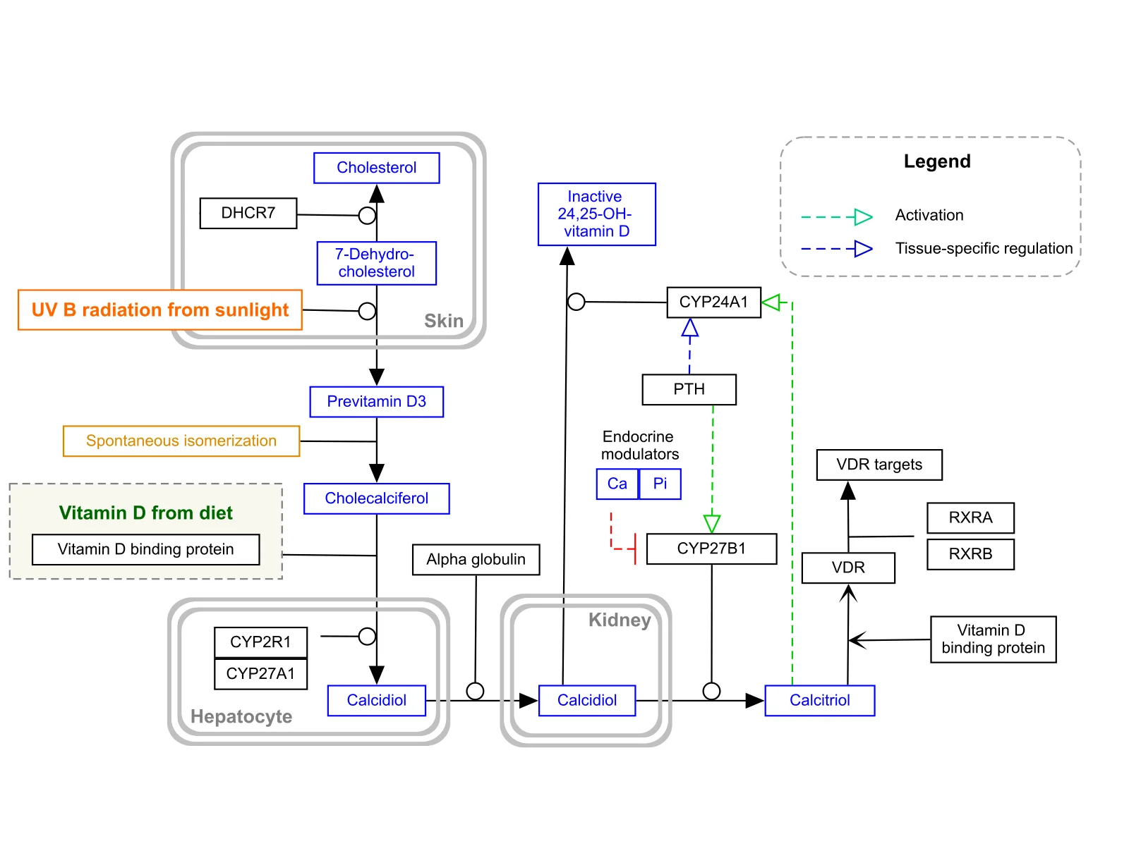
Figure 1Vitamin D synthesis metabolism pathway WikiPathways WP1531
Long description
Vitamin D synthesis and metabolism pathway: sunlight converts 7-dehydrocholesterol in the skin to vitamin D3, which is hydroxylated in the liver and then the kidney to active calcitriol, with PTH and calcium regulating the kidney step.

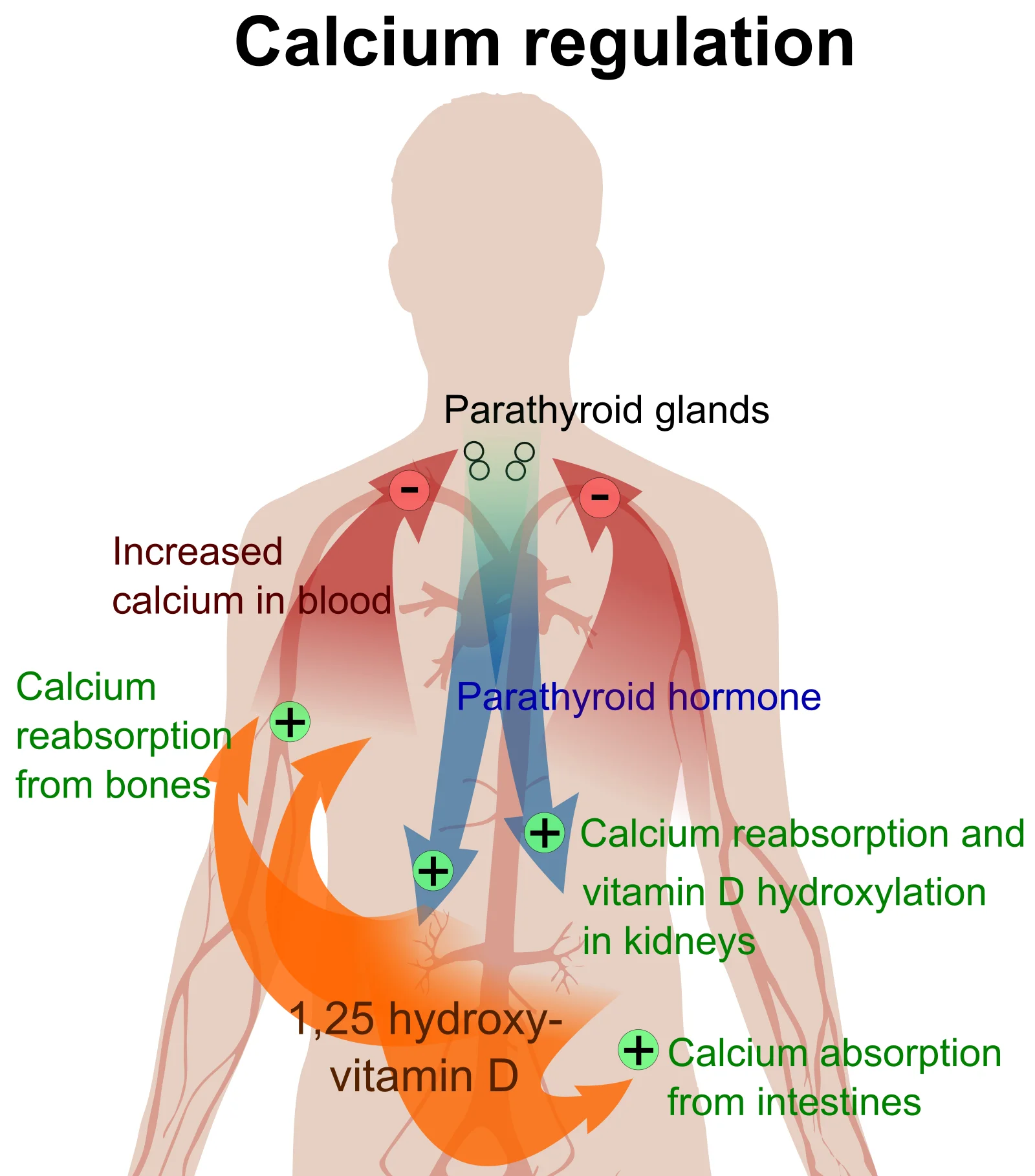
Figure 2Calcium regulation
Long description
Calcium regulation: low blood calcium raises parathyroid hormone, which increases calcium release from bone, calcium reabsorption and vitamin D activation in the kidney, and calcium absorption from the gut.

Bone is living tissue. Throughout life, old or damaged bone is removed and replaced in a coupled process called bone remodelling:
- Activation. Osteoclasts, the bone-resorbing cells, are recruited to a site of microdamage. They attach to bone and seal off a shallow pocket.
- Resorption. The osteoclasts release acid and enzymes that dissolve the mineral and digest the matrix, excavating a cavity (Howship lacuna).
- Reversal. A brief pause follows, during which growth factors are released from the degraded matrix.
- Formation. Osteoblasts, the bone-forming cells, fill the cavity with osteoid, a protein scaffold rich in type I collagen.
- Mineralisation. Calcium and phosphate crystallise within the osteoid, restoring mineralised bone.
In healthy young adults, resorption and formation are equal. The balance is regulated by hormones and by mechanical loading. Osteocytes, the cells embedded in mineralised bone, act as sensors of mechanical strain: where strain is high, osteocytes produce less sclerostin (a cytokine that inhibits formation), so bone is laid down. Romosozumab, an antibody against sclerostin, exploits this pathway. Osteocytes also produce RANKL (receptor activator of nuclear factor kappa-B ligand), the principal signal that drives osteoclast formation; denosumab blocks RANKL. Teriparatide and abaloparatide (parathyroid hormone analogues) stimulate both formation and resorption, with a net gain in bone when given intermittently once daily. Parathyroid hormone given continuously (as in primary hyperparathyroidism) does the opposite and harms the skeleton.
In osteoporosis, the coupling breaks down: resorption runs ahead of formation with each remodelling cycle. Because trabecular bone has a much larger surface-area-to-mass ratio than cortical bone, it is depleted first; this is why vertebral bodies (rich in trabecular bone) show the earliest changes, and why vertebral fractures precede hip fractures.
Calcium balance is defended by a hormonal network:
- Parathyroid hormone (PTH) is released when blood calcium falls. PTH mobilises calcium from bone by stimulating osteoclasts (via RANKL on osteoblasts), increases calcium reabsorption in the kidney, and stimulates the renal synthesis of active vitamin D, which raises calcium absorption from the gut.
- Vitamin D is obtained from skin synthesis (UVB) and diet. It is hydroxylated in the liver to 25-hydroxyvitamin D (25(OH)D), the storage form that is measured clinically, and then in the kidney to 1,25-dihydroxyvitamin D (calcitriol), the active hormone. Calcitriol increases intestinal absorption of calcium and phosphate and, with PTH, supports normal bone mineralisation.
- Calcitonin, secreted by thyroid C cells when calcium is high, lowers it modestly and rapidly by inhibiting osteoclasts; its long-term role is small.
When calcium or vitamin D is insufficient, blood calcium is defended by PTH-driven bone resorption, which over years lowers BMD and contributes to osteoporosis. Adequate intake and sufficiency of vitamin D are therefore part of treatment, not an afterthought.
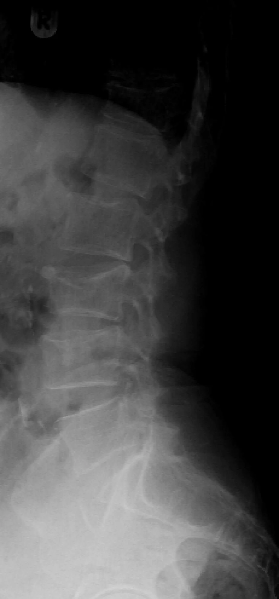
Osteoporosis is usually asymptomatic until a fracture occurs. Gradual height loss, an increasingly kyphotic posture and chronic back pain can reflect multiple vertebral compression fractures. Acute back pain after bending or lifting, worsening on standing and coughing, suggests a new vertebral fracture.
Examination should include measured height (ideally with a wall-mounted stadiometer, without shoes), review of previous heights (height loss of 1.5 in or more in an adult, or prospective height loss of 0.8 in or more, flags silent vertebral fractures [1]), kyphosis, tenderness over the spine, assessment of gait and balance, and a search for causes of secondary osteoporosis. The primary fracture prevention and secondary fracture prevention thresholds that follow show how the findings guide management [1].
A low BMD or a fracture must be distinguished from other causes of bone fragility. Osteomalacia and rickets (from vitamin D or phosphate deficiency) produce poorly mineralised osteoid rather than low bone volume; biochemical tests are usually abnormal. Paget disease of bone shows focal, overactive remodelling with enlarged, deformed bones and a markedly raised alkaline phosphatase. Hyperparathyroidism increases bone resorption and raises calcium and PTH. Malignancy (including multiple myeloma) can cause pathological fracture, weight loss and abnormal protein studies. Osteogenesis imperfecta presents in childhood with multiple fractures and blue sclerae. Osteoporosis remains by far the most common explanation in an older adult, but secondary causes should be excluded before starting drug treatment.
Figure 3Compressionfracture
Long description
Lateral spine radiograph showing a vertebral compression fracture with loss of vertebral body height.

The first question is usually risk, not testing: are there risk factors, a previous fracture, conditions or medications that favour bone loss, or features of secondary osteoporosis? Then:
- DXA of the hip and lumbar spine is the preferred method for establishing and confirming the diagnosis, predicting fracture risk and monitoring treatment. It reports BMD in g/cm2 and as T- and Z-scores [1].
- Vertebral imaging (lateral thoracic and lumbar radiographs or DXA-based vertebral fracture assessment) is used to detect silent vertebral fractures; the criteria are set out with screening below [1].
- Fracture risk assessment with FRAX® (see below) combines BMD and clinical factors to estimate 10-year fracture probability.
Before starting treatment, underlying contributors should be sought. The BHOF guide proposes a basic panel:
- Full blood count
- Albumin, and albumin-adjusted calcium
- Renal function, phosphorus and magnesium
- Liver function tests
- 25(OH) vitamin D
- Parathyroid hormone
- Total testosterone and gonadotrophins in men (especially those aged 50–69 years)
Selected patients should also have serum protein electrophoresis (and immunofixation and free light chains), a thyroid function test, tissue transglutaminase antibodies with IgA, iron studies, a prolactin level, 24-hour urinary calcium and creatinine, urinary protein electrophoresis and, when Cushing syndrome is suspected, salivary or urinary free cortisol [1]. These tests identify treatable causes such as coeliac disease, hyperthyroidism and myeloma that would not respond to, or would be worsened by, osteoporosis drugs [1].
For meaningful interpretation, DXA should be performed by trained staff on a calibrated instrument; repeat scans should be done on the same machine at the same facility whenever possible [1].
FRAX® is a country-calibrated tool that predicts the 10-year probability of hip fracture and of major osteoporotic fracture (hip, spine, wrist or proximal humerus) in people aged 40 to 90 years, using clinical factors with or without BMD at the femoral neck [2]. Risks predicted by BMD alone or by FRAX alone are similar, and both are less accurate than FRAX with BMD [2]. FRAX is the most studied risk tool and has been incorporated into 120 guidelines since 2016; clinicians should be aware that race-specific calibrations can underestimate risk in some groups and that FRAX does not include falls, frailty, diabetes or lumbar spine BMD [2].
To put the numbers in context, a 65-year-old white woman with body mass index 25 and no risk factors has a 10-year hip fracture probability of 1.3% and a major osteoporotic fracture probability of 9.3% on FRAX without BMD [2].
BHOF recommends starting pharmacological treatment in postmenopausal women and men aged 50 years or older who have:
- a T-score of −2.5 or below at the femoral neck, total hip, lumbar spine or 33% radius; or
- osteopenia (T-score between −1.0 and −2.5) at the femoral neck or total hip with a 10-year hip fracture probability of at least 3% or a 10-year major osteoporotic fracture probability of at least 20% on the US-adapted FRAX model [1].
Secondary prevention (treating to prevent a further fracture) follows a fracture of the hip or vertebra regardless of BMD, and a fracture of the proximal humerus, pelvis or distal forearm in a person with osteopenia [1].
Non-drug measures are the foundation for every patient:
- Calcium: 1000 mg/day for men aged 50–70 years, and 1200 mg/day for women aged 51 years or older and men aged 71 years or older, from food first and supplements if intake is inadequate [1].
- Vitamin D: maintain serum 25(OH)D between 30 and 50 ng/mL. A supplement of 800–1000 units a day is usually enough in adults aged 50 years or older to achieve this; some adults with malabsorption need more. Note that a level of 20 ng/mL may be enough for a generally healthy person, but 30 ng/mL is advised in metabolic bone disease [1].
- Exercise: weight-bearing and resistance exercise, with balance and muscle-strengthening training, improves BMD and reduces falls.
- Falls: review sedating and hypotensive drugs, polypharmacy, vision, gait, foot problems and orthostatic hypotension; assess the home environment and refer for hazard remediation.
- General: stop smoking, limit alcohol, maintain healthy weight and support patients after a fracture with analgesia, graded return to activity and rehabilitation.
- Coordinate care with a fracture liaison service so that patients who fracture are assessed and started on treatment before discharge.
The drugs approved for osteoporosis are bisphosphonates, denosumab, romosozumab, parathyroid hormone analogues (teriparatide and abaloparatide), raloxifene, calcitonin salmon and oestrogen-related therapy (oestrogen/progestin or conjugated estrogens with bazedoxifene) [2]. Choice depends on fracture risk, prior fracture, side-effect profile, preferences and kidney function; all require adequate calcium and vitamin D intake. All antifracture therapies treat but do not cure the disease; bone loss resumes when they are stopped, sooner for non-bisphosphonates and later for bisphosphonates. Keep the diagnosis of osteoporosis even if a later DXA T-score is above −2.5 [1].
Bisphosphonates (alendronate, risedronate, ibandronate, zoledronic acid) remain first-line for most patients. They bind to hydroxyapatite and inhibit osteoclast-mediated resorption. Typical doses are:
- Alendronate 70 mg by mouth once weekly, or 10 mg daily (prevention 35 mg weekly or 5 mg daily) [8].
- Zoledronic acid 5 mg intravenously once a year for treatment (or every two years for prevention), given over at least 15 minutes [9].
For other oral bisphosphonates, use the standard labelled dose and follow local guidance.
Cautions: esophageal irritation (take on waking with plain water and remain upright for at least 30 minutes), hypocalcaemia, renal impairment (alendronate is not recommended when creatinine clearance is below 35 mL/min [8]), osteonecrosis of the jaw and atypical femoral fracture after prolonged use. If a patient on a bisphosphonate reports persistent thigh or groin pain, or jaw pain or delayed healing after dental work, investigate promptly [1][8][9].
Denosumab is a monoclonal antibody to RANKL given as 60 mg by subcutaneous injection every six months [4]. It is a potent antiresorptive and is particularly useful where oral bisphosphonates are not tolerated or kidney function limits bisphosphonate use (no dose adjustment is needed, but patients with advanced chronic kidney disease are at higher risk of hypocalcaemia and need specialist supervision) [4].
The key caution is the rebound after stopping. Denosumab suppresses bone turnover; when injections stop, bone turnover rises above the pretreatment level by 9 months, BMD returns to baseline within 18 months, and multiple vertebral fractures can occur as early as 7 months (on average 19 months) after the last dose, especially in anyone who has already had a vertebral fracture [4]. For this reason, denosumab must be followed by another antiresorptive drug (usually a bisphosphonate) if it is discontinued, and a dose should never simply be missed [4]. Denosumab also carries warnings for hypocalcaemia, osteonecrosis of the jaw and atypical femoral fracture, and serious infection [4].
Teriparatide and abaloparatide are parathyroid hormone–related analogues that stimulate bone formation. Teriparatide is given as 20 micrograms subcutaneously once daily, and its use for more than two years in a patient’s lifetime should be considered only if high fracture risk persists [3]. Abaloparatide is given as 80 micrograms subcutaneously once daily, and its use beyond two years has not been evaluated [5]. Both require supplemental calcium and vitamin D where dietary intake is inadequate, and both should be avoided in patients with open epiphyses, Paget disease, skeletal malignancy or metastases, prior skeletal irradiation, or hereditary disorders predisposing to osteosarcoma, because of the risk of osteosarcoma [3][5]. [4]
Romosozumab is a sclerostin inhibitor given as 210 mg subcutaneously once a month for 12 doses, as two 105 mg injections, with calcium and vitamin D supplementation [6]. It increases bone formation and, to a lesser extent, decreases resorption. It carries a boxed warning that it may increase the risk of myocardial infarction, stroke and cardiovascular death; it should not be started in anyone who has had a myocardial infarction or stroke in the preceding year, cardiovascular risk should be reviewed before starting, and treatment should be stopped if a myocardial infarction or stroke occurs during therapy [6]. After 12 monthly doses, an antiresorptive drug should follow [6].
Raloxifene is a selective oestrogen receptor modulator given as 60 mg by mouth once daily [7]. It reduces vertebral fracture risk and breast cancer risk, but it raises the risk of venous thromboembolism and fatal stroke in women at high cardiovascular risk, so it is not suitable for anyone with a history of venous thromboembolism and should be stopped at least 72 hours before prolonged immobilisation [7].
Menopausal hormone therapy (oestrogen, with or without progestin) reduces fracture risk and may be appropriate for recently menopausal women with menopausal symptoms, but it is not used for the primary or secondary prevention of cardiovascular disease or for breast cancer risk, and the benefits and risks should be discussed individually [7].
In addition to raloxifene, conjugated estrogens with bazedoxifene are among the oestrogen-related options listed in guidelines [2]. Calcitonin salmon is an older antiresorptive. Neither is a routine first choice today; calcitonin in particular should be used sparingly because of concerns about long-term safety with high-dose use.
| Drug or class | Typical adult dose | Key cautions |
|---|---|---|
| Alendronate (bisphosphonate) | 70 mg orally once weekly (or 10 mg daily); 5/35 mg for prevention [8] | Esophageal irritation, hypocalcaemia, renal impairment, osteonecrosis of the jaw, atypical femoral fracture [8] |
| Zoledronic acid (bisphosphonate) | 5 mg intravenously once a year (or every 2 years for prevention), over at least 15 minutes [9] | Renal impairment (contraindicated if CrCl under 35 mL/min), hypocalcaemia, ONJ, atypical fracture [9] |
| Risedronate/ibandronate (bisphosphonates) | Standard licensed oral or intravenous dose per product label | Similar class cautions; see local formulary |
| Denosumab | 60 mg subcutaneously every 6 months [4] | Rebound after stopping; transition to another antiresorptive; hypocalcaemia; ONJ; atypical fracture; serious infection [4] |
| Teriparatide | 20 micrograms subcutaneously once daily [3] | Limit totalling 2 years of lifetime use; avoid in high osteosarcoma risk; orthostatic hypotension; hypercalcaemia [3] |
| Abaloparatide | 80 micrograms subcutaneously once daily [5] | Not evaluated beyond 2 years; avoid in high osteosarcoma risk; orthostatic hypotension; hypercalcaemia [5] |
| Romosozumab | 210 mg subcutaneously once monthly for 12 doses (two 105 mg injections) [6] | Boxed warning: MI, stroke, cardiovascular death; not if MI/stroke in preceding year; ONJ; atypical fracture [6] |
| Raloxifene | 60 mg orally once daily [7] | Venous thromboembolism; fatal stroke risk; not with past VTE; stop 72 hours before prolonged immobilisation [7] |
| Menopausal hormone therapy | Individualised menopausal dose (not for CVD prevention) [2] | Thromboembolism and contraindications; discuss benefits and risks individually |
Approach to suspected osteoporosis after a fracture or low BMD
- Suspected fragilityAdult aged 50+ with low-trauma fracture, height loss, or low BMD on screening
- Confirm and stratifyDXA of hip and lumbar spine; apply WHO T-score; calculate FRAX where appropriate
- Question: Is there a fragility fracture of hip/vertebra or T-score under 2.5?
Yes
- Osteoporosis diagnosedInvestigate and treat secondary causes before drug therapy
- Start or optimise bone-active therapy + calcium/vitamin D/exercise/falls control
No (osteopenia)
- Check FRAXTreat if 10-year hip risk is at least 3% or major fracture risk is at least 20%
- Otherwise manage risk factors; revisit
Bisphosphonates remain in bone, so their antifracture effect lasts for years after a course of treatment. After five years of oral or three years of intravenous bisphosphonate, a clinician can consider a drug holiday in patients who are no longer at high risk (T-score of −2.5 or better, and no new fractures) [1]. A holiday is not appropriate after denosumab, teriparatide, abaloparatide or romosozumab, because their effects wear off quickly and, for denosumab, stopping without follow-on therapy risks rebound fracture [1][4]. BMD should be rechecked every one to two years while on treatment and after stopping or changing therapy, and the need for continued or resumed treatment reassessed regularly [1].
A vertebral fracture is both a diagnosis and an event:
- Treat acute pain with short rest, heat or ice, simple analgesia and early mobilisation; avoid prolonged bed rest.
- Image the spine to distinguish a new fracture from old collapse and to assess height loss.
- Start or optimise osteoporosis treatment: a vertebral fracture is an indication for pharmacological therapy regardless of BMD [1].
- Investigate and treat secondary causes.
- Build strength and posture with physiotherapy; orthotics are rarely needed.
- New or worsening neurological symptoms (leg weakness, numbness, sphincter disturbance) or severe unremitting pain require urgent imaging and referral.
Repeat BMD testing one to two years after starting or changing therapy, and at appropriate intervals thereafter depending on the patient [1]. More frequent testing is appropriate in higher-risk people (very low initial BMD, multiple fractures, older age); less frequent testing may suffice once BMD is stable and the patient is fracture-free. Bone turnover markers can, in selected patients, confirm a biological response within months, well before BMD changes.
Complications of osteoporosis itself are the fractures: vertebral (often silent), hip (the most disabling) and wrist, along with fractures of the pelvis, proximal humerus and ribs. A vertebral fracture increases the risk of another vertebral fracture five-fold and of fracture elsewhere two- to three-fold [1]. Hip fractures carry an 8.4% to 36% excess mortality in the following year [1]. Vertebral collapse can lead to kyphosis (hyperkyphosis), reduced lung volumes, abdominal discomfort, constipation and weight loss. Complications of treatment arise mainly with antiresorptive drugs: osteonecrosis of the jaw, atypical femoral fracture, hypocalcaemia and, with denosumab, rebound vertebral fractures if the drug is stopped without follow-on therapy [1][4].
Osteoporosis is treatable but not curable. Treatment lowers both vertebral and non-vertebral fracture risk: in trials of screening, the pooled relative risk of hip fracture was 0.83 and of major osteoporotic fracture 0.94 over 3.7 to 5 years [2]. Bisphosphonates reduce vertebral fracture risk by about half in placebo-controlled trials (pooled relative risk 0.51) and hip fracture risk with a pooled relative risk of 0.67 [2]. Denosumab also reduced radiographic vertebral, hip and non-vertebral fractures in the pivotal trial (relative risk 0.32, 0.60 and 0.80 respectively) [2]. [1]
Prognosis depends on age, prior fracture, comorbidity and continuation of therapy. Because the disease and the elevated fracture risk persist even if BMD improves, management is long-term and should be reassessed rather than stopped casually. Any fracture after the age of 50 years is a sentinel event that should trigger prompt evaluation and treatment.
Prevention starts in childhood: adequate nutrition, physical activity and avoidance of smoking in adolescence all build peak bone mass, but in adulthood the aim is to identify adults at high risk [1]. The USPSTF recommends:
- Screening women aged 65 years or older (Grade B). [2]
- Screening postmenopausal women younger than 65 years who have one or more risk factors and are assessed to be at increased risk (Grade B). [2]
- The evidence is currently insufficient to assess screening in men (Grade I statement), so clinicians should use clinical judgement and consider men with risk factors [2].
BHOF recommends BMD testing in:
- women aged 65 years or older and men aged 70 years or older, regardless of other risk factors;
- younger postmenopausal women, women in the menopausal transition and men aged 50–69 years who have clinical risk factors for fracture;
- any adult with a fracture after the age of 50 years;
- adults with conditions or medications associated with low bone mass or bone loss [1].
USPSTF recommendations apply to adults aged 40 years or older without known osteoporosis or fragility fracture and do not cover secondary osteoporosis from conditions (such as hyperthyroidism) or from chronic glucocorticoid use [2].
DXA is the standard screening test. Screening can include DXA with or without fracture risk assessment. Repeat BMD testing at intervals of four to eight years does not add much to fracture prediction; interval testing is better individualised by baseline T-score and age [2]. Vertebral fracture imaging (plain radiograph or DXA VFA) is used to detect subclinical vertebral fractures in women aged 65 years or older with femoral neck T-score of −1.0 or below, women aged 70 years or older and men aged 80 years or older with T-score of −1.0 or below at the lumbar spine, total hip or femoral neck, men aged 70–79 years with T-score of −1.5 or below, and adults aged 50 years or older with adult fractures, height loss, long-term glucocorticoid use or hyperparathyroidism [1].
Older adults have the highest fracture risk and the greatest benefit from treatment. Atrial fibrillation, swallowing difficulty and renal impairment all affect drug choice; zoledronic acid is given slowly and renal function must be checked, and alendronate should not be used if creatinine clearance is below 35 mL/min [8][9].
Men have lower lifetime fracture risk but higher excess mortality after hip fracture. BHOF recommends evaluating men similarly and consulting the Endocrine Society guideline for detailed management [1]; hypogonadism should be treated where it is the cause.
Pregnancy and younger adults: osteoporosis in premenopausal women or in men under 50 years points to a secondary cause or to severe risk factors (for example, anorexia nervosa or long-term steroids). Raloxifene and bisphosphonates are contraindicated in pregnancy, and denosumab and the anabolic agents are avoided or used with specialist advice [7]. This page deals mainly with postmenopausal osteoporosis; specialist referral is wise in younger patients.
Patients on glucocorticoids start to lose bone within months. Anyone expected to take 7.5 mg or more of prednisone a day (or equivalent) for at least six to twelve months, with low BMD or additional risk factors, should receive bisphosphonates or another approved drug plus calcium and vitamin D [1][8][9].
Many of the tools and drugs above are not available everywhere. Priority then is:
- DXA may be unavailable. BMD measured by older methods and clinical risk assessment by FRAX are the usual footing.
- Serum 25(OH)D assays, PTH, electrophoresis and bone turnover markers may be unavailable or expensive. A careful history (causes of malabsorption, steroid use, premature menopause, suspected multiple myeloma) and a modest set of labs suffice to exclude the common secondary causes.
- Vertebral imaging may be considered on clinical grounds (age alone, height loss, known glucocorticoid use) even when DXA is not available [1].
- Calcium and vitamin D, exercise and falls prevention are universally accessible and give real benefit.
- Where oral bisphosphonates are available, alendronate is generic and cheap, and is the practical first-line choice. Intravenous zoledronic acid requires an infusion setting and monitoring of renal function.
- Denosumab is feasible only if a reliable six-monthly injection programme and a follow-on antiresorptive can be arranged.
- Anabolic agents are expensive and usually restricted; if they cannot be obtained, antiresorptive therapy is a reasonable alternative.
- Referral pathways for vertebral fractures with neurological compromise or for hip fracture should be agreed before they are needed.
- Osteoporosis is low bone mass and microarchitectural deterioration leading to fragility fractures; it is silent until a fracture occurs.
- The diagnosis is a DXA T-score of −2.5 or below at the hip or lumbar spine, or an adult fragility fracture.
- Postmenopausal bone loss in women is roughly 2% per year for the ten years after the menopause; peak bone mass is largely genetic and is built in youth.
- Calcium intake targets are 1000 mg/day (men 50–70 years) and 1200 mg/day (women 51 years or older and men 71 years or older); vitamin D sufficiency is 30–50 ng/mL (20 ng/mL may be enough in healthy people). [1]
- Falls occur in about one third of adults aged 65 years or older and are a major part of fracture risk.
- FRAX estimates 10-year fracture probability; treatment is justified when hip fracture probability is at least 3% or major fracture probability at least 20% in osteopenia. [1]
- Secondary causes must be excluded with a basic laboratory panel.
- Bisphosphonates are first-line; alendronate 70 mg weekly and zoledronic acid 5 mg yearly are common choices.
- Denosumab 60 mg every six months is effective but rebounds if stopped; follow it with another antiresorptive.
- Teriparatide 20 micrograms daily, abaloparatide 80 micrograms daily and romosozumab 210 mg monthly are anabolic options, with limits on duration.
- A bisphosphonate holiday is considered after 5 years (oral) or 3 years (intravenous) of therapy; it does not apply to denosumab or anabolic agents.
- A vertebral fracture requires prompt pain relief, imaging and drug treatment.
- Secondary causes are common and should be treated in their own right; they include glucocorticoids at 5 mg/day or more for three months or longer, hypogonadism, hyperparathyroidism, hyperthyroidism, coeliac disease, myeloma and several drug classes.
- The disease persists even if BMD recovers, so treatment is reassessed rather than stopped abruptly.
Answer each one in your head first, then open the answer.
A 70-year-old woman has a DXA T-score of −2.8 at the femoral neck and has never fractured. She is otherwise well. Which statement best describes her situation and management?
Show answerHide answer
She has osteoporosis by BMD criterion (T-score below −2.5). She does not yet meet the secondary prevention threshold for drug treatment, but because her T-score is −2.5 or below she is within the primary prevention indication for pharmacological treatment. First steps are calcium 1200 mg/day, vitamin D to 30–50 ng/mL, weight-bearing and balance exercise, falls review, and a bone-active drug (usually a bisphosphonate), plus a check for secondary causes if clinically relevant. [1][2] [9]
Which of the following is the strongest reason never to simply stop denosumab?
Show answerHide answer
On stopping denosumab, bone turnover rises above the pretreatment level by 9 months, BMD returns to baseline within 18 months, and multiple vertebral fractures can occur as early as 7 months after the last dose. The drug is therefore continued or, if discontinued, followed immediately by another antiresorptive (usually a bisphosphonate). [4]
A 62-year-old postmenopausal woman has a femoral neck T-score of −1.8. Her FRAX 10-year hip fracture probability is 3.5%. Does she need drug treatment?
Show answerHide answer
Yes. Although she has osteopenia, her 10-year hip fracture probability is at least 3% on FRAX, which crosses the threshold for starting pharmacological treatment (along with calcium, vitamin D, exercise and falls prevention). [1]
Name the principal secondary causes of osteoporosis covered in this page and two sensible investigations to exclude them.
Show answerHide answer
Principal secondary causes: glucocorticoid therapy, hypogonadism, hyperparathyroidism, hyperthyroidism, coeliac disease, multiple myeloma and many drugs (including anticonvulsants, aromatase inhibitors, proton pump inhibitors and excess thyroid replacement). Useful investigations include 25(OH) vitamin D with parathyroid hormone, and serum protein electrophoresis with free light chains when myeloma is a concern. [1]
A patient has been on alendronate for 5 years, has a hip T-score of −2.0 and no new fractures. What is the next step?
Show answerHide answer
She is a candidate for a bisphosphonate drug holiday, given her low residual risk. Continue calcium and vitamin D, exercise and falls prevention, recheck BMD in 1–2 years and reassess fracture risk; resume a bisphosphonate if risk rises. [1]
Why are romosozumab and the parathyroid hormone analogues limited to about a year or two of use?
Show answerHide answer
Teriparatide and abaloparatide are limited to about 2 years' lifetime exposure because of osteosarcoma risk seen in animal studies and limited human safety data beyond 2 years; romosozumab is limited to 12 monthly doses because its anabolic effect wanes. Each is followed by an antiresorptive drug. [3][5][6]
- Hyperthyroidism and thyrotoxicosis
- Hypercalcaemia
- Hypocalcaemia
- Type 2 diabetes mellitus
- Type 1 diabetes mellitus
- Renal cell carcinoma
- Prostate cancer
Medication information above is based on the product labels in the references; doses are typical adult doses from those labels and current guidance. Check your local formulary for availability, renal dosing and contraindications. The numbers in this article are taken from the sources listed in the front matter and saved in the accompanying claims ledger.
- LeBoff MS, Greenspan SL, Insogna KL, et al. The clinician’s guide to prevention and treatment of osteoporosis. Osteoporosis International 2022;33(10):2049–2102. Bone Health and Osteoporosis Foundation (formerly National Osteoporosis Foundation).. Accessed .
- US Preventive Services Task Force. Screening for Osteoporosis to Prevent Fractures: US Preventive Services Task Force Recommendation Statement. JAMA 2025;333(6):498–508. doi:10.1001/jama.2024.27154. Accessed .
- Eli Lilly and Company. FORTEO (teriparatide injection) prescribing information. DailyMed drug label. Accessed .
- Amgen Inc. PROLIA (denosumab) injection prescribing information. DailyMed drug label. Accessed .
- Radius Health Inc. TYMLOS (abaloparatide) injection prescribing information. DailyMed drug label. Accessed .
- Amgen Inc. EVENITY (romosozumab-aqqg) injection prescribing information. DailyMed drug label. Accessed .
- Eli Lilly and Company. EVISTA (raloxifene hydrochloride) tablet prescribing information. DailyMed drug label. Accessed .
- Hangzhou Minsheng Binjiang Pharmaceutical Co., Ltd. Alendronate sodium tablets USP prescribing information. DailyMed drug label. Accessed .
- Sandoz Inc. RECLAST (zoledronic acid) injection prescribing information. DailyMed drug label. Accessed .
Figure credits
- Figure 1: Chris Evelo , Lucia Ribeiro , Kristina Hanspers , Alex Pico , Martijn Van Iersel , Daniela Digles , Egon Willighagen , Martina Summer-Kutmon , Mingye Wang , Denise Slenter , and Eric Weitz, via Wikimedia Commons, CC0, CC0.
- Figure 2: Mikael Häggström, via Wikimedia Commons, Public domain, Public domain.
- Figure 3: Lucien Monfils, via Wikimedia Commons, CC BY-SA 3.0, CC BY-SA 3.0.
Educational content for medical students and doctors. It is not a substitute for clinical judgement or local guidelines.