Paget disease of bone
Paget disease of bone: focal disordered bone remodelling, phases, complications, diagnosis with alkaline phosphatase and imaging, and bisphosphonate treatment.
- Paget disease of bone is a focal, chronic disorder of bone remodelling. In one or more bones, osteoclasts become overactive and then drive disorganised bone formation, so the bone swells, softens or hardens in patches, bends and fractures more easily.
- It is the second most common metabolic bone disorder after osteoporosis. It usually appears after the age of 40, is more common in men, and clusters in people of north-west European ancestry. Most people have no symptoms and are diagnosed from a blood test or an incidental X-ray. [3]
- The signature laboratory finding is a raised alkaline phosphatase (ALP) with normal serum calcium and phosphate. A plain radiograph is usually diagnostic; a technetium-99m bone scan maps how many bones are involved.
- Treatment is only needed for some patients: a single 5 mg intravenous infusion of zoledronic acid is the first-line drug, with pain relief and biochemical remission that often lasts years. Bisphosphonates are given before surgery on pagetic bone, and joint replacement is used when arthritis develops. [2][5]
- Serious complications are uncommon: fracture in roughly 10 to 30% of clinically identified patients, deafness from skull involvement, very rarely high-output heart failure, and malignant change to osteosarcoma in about 0.3% of patients. [2][3]
By the end of this page you should be able to:
- Explain the cell-level defect in Paget disease and why the bone becomes deformed, vascular and weak.
- Describe the three radiological phases and the typical appearance of pagetic bone.
- Present the likely causes, including SQSTM1 mutations and the unfinished viral hypothesis.
- Recognise the clinical features and complications, including bone pain, deformity, fracture, deafness, heart failure and osteosarcoma.
- Interpret the laboratory pattern and explain why calcium and phosphate are usually normal, and which patients need a bone scan or CT/MRI.
- Plan treatment: when to treat, first-line and alternative drugs with doses, monitoring, and pre-surgical preparation.
- Distinguish Paget disease from metastases and osteomalacia on clinical and imaging grounds.
- Adapt diagnosis and treatment to a setting with limited tests or drugs.
DefinitionPaget disease of bone
Paget disease of bone (PDB; also called osteitis deformans) is a focal, chronic disorder of bone remodelling. It produces enlarged, deformed bone with both lytic and sclerotic areas, driven by overactive osteoclasts followed by disorganised osteoblastic bone formation.
The disease is named after Sir James Paget, who described it as osteitis deformans in 1877. "Paget disease of bone" and "Paget's disease of bone" are the same condition and are used interchangeably in the literature. This page covers the common adult (classic) form. Rare PDB-like syndromes and juvenile Paget disease are separate topics and are not covered here.
Anatomical classification:
- Monostotic: one bone is affected. This is becoming more common in recent series.
- Polyostotic: two or more bones are affected, often asymmetrically. The pelvis is involved in up to 70% of polyostotic cases, the femur in 30 to 55%, the lumbar spine in 25 to 50%, the skull in 20 to 40% and the tibia in 15 to 30% [4]. Once diagnosed, spread to new bones is unusual [3].
Activity:
- Active (metabolically active): bone turnover is raised, usually shown by a raised ALP and intense tracer uptake on a bone scan. This is the form that is treated.
- Inactive or "burned-out": bone turnover is low; sclerotic bone remains on the radiograph but a bone scan may be negative in 2 to 10% of affected bones [4].
Severity is not a single score. Instead, clinicians judge extent (number of bones and sites), activity (ALP or other marker), deformity, fracture, neurological or joint compromise, and whether the bone is painful.
Paget disease of bone is common. It is the second most common metabolic bone disorder after osteoporosis [3]. True prevalence is hard to state because fewer than 10% of people with radiographic Paget disease ever come to medical attention [2].
- Prevalence. The disease has been estimated to affect about 1% of people over the age of 55 in the United Kingdom [2]. In populations of British descent (Australia, New Zealand, the United States, Canada) the figure is similar, and the peak prevalence in high-incidence areas may reach 5 to 8% in the eighth decade [4]. In Italy prevalence is around 1% or lower, with a pocket of high prevalence and greater severity in rural Campania [4]. [3]
- Incidence is falling. Across north-west Europe and other high-prevalence regions, both incidence and severity have fallen steeply over the past 25 years, with more monostotic and more asymptomatic cases [4]. Prevalence of the complication osteosarcoma has fallen in parallel. [2]
- Age. It is rare before 40 years and increases in prevalence with age, with an approximate doubling of incidence each decade after age 50 [2]. [4]
- Sex. Men are affected slightly more often than women, with a male-to-female ratio of about 1.4 to 1 [2].
- Ancestry. It is most common in people of north-west European ancestry (including those who have migrated) and rare in Scandinavia, the Indian subcontinent and Asia [2] [4]. It has been reported in people of African ancestry.
- Family history. Between 12 and 40% of patients have at least one affected first-degree relative, and first-degree relatives carry roughly 7 to 10 times the risk of the general population [4].
United Kingdom (NICE)
The cause of Paget disease is not fully known. Two lines of evidence are strong: a genetic predisposition, and a changing environment over the last century.
About 10 to 20% of patients have a family history [3], and the single most important susceptibility gene is SQSTM1, which encodes the p62 protein. Mutations in SQSTM1 are found in 40 to 50% of familial cases and in 5 to 10% of patients with no family history [2] [4]. These mutations impair p62's ability to bind ubiquitin, activating RANKL-driven NF-kappa B signalling in osteoclasts and increasing osteoclast activity [2].
Less common, usually severe or early-onset polyostotic disease has been linked to mutations in TNFRSF11A, VCP and PFN1 [4]. Mutations in ZNF687 have been associated with a higher risk of giant cell tumour complicating Paget disease, and PFN1 mutations with early-onset disease [4]. In Carney-complex-type terms (that is, syndromic forms), Paget-like features can be part of a multisystem disorder with myopathy and neurodegeneration [2].
A slow viral infection has been suspected for decades. Measles, respiratory syncytial virus and canine distemper viruses have all been implicated, and overexpression of measles virus nucleocapsid protein in experimental models increases bone remodelling [2]. The picture is not settled: attempts to detect paramyxovirus nucleic acids or proteins in patient tissue have given conflicting results, and serological studies have shown no enhanced immune response to paramyxoviruses in patients [2]. The nuclear inclusion bodies seen in pagetic osteoclasts long ago and assumed to be measles nucleocapsids appear, on ultrastructural analysis, to be morphologically distinct from measles; they may instead be abnormal protein aggregates produced by defective autophagy, which is consistent with SQSTM1-related disease [2]. The balanced conclusion is that an environmental trigger is probable, but no virus has been proven to be the cause [2] [4].
Dietary calcium or vitamin D deficiency, exposure to environmental toxins, and repetitive mechanical loading or skeletal trauma have all been proposed [2]. The speed of the epidemiological decline argues for an environmental change, but the specific trigger is unknown [2].
Risk rises with age, male sex and north-west European ancestry [2]. There is no known behavioural or dietary risk factor that a patient can modify today, and there is no screening programme for the general population. People with a known mutation in an affected family are the group in whom screening is currently studied.
Paget disease is, at its simplest, a failure of the normal coupling between bone resorption and bone formation. Osteoclasts become enlarged, hypernucleated and hyper-responsive to signals such as 1,25-dihydroxyvitamin D, interleukin-6 and RANKL [4]. They resorb bone too fast. Osteoblasts are recruited in excess and lay down new bone, but in a hurry: disorganised woven bone instead of normal lamellar bone, with marrow fibrosis and increased vascularity. The skeleton at that site enlarges and becomes mechanically weak at the same time as it becomes dense in places.
The disease is focal. It does not affect the whole skeleton, it does not spread from bone to bone, and new bones rarely become involved after diagnosis [3]. Affected bone has a high blood flow, which can make the overlying skin feel warm [3].
Each phase favours one side of the remodelling equation:
- Lytic phase: resorption dominates. Large, well-defined osteolytic areas advance through bone, sometimes as a "flame" or inverted V on a radiograph.
- Mixed phase: both resorption and formation are active together. This is the most common presentation and carries most of the classic radiological features.
- Sclerotic (blastic) phase: formation dominates. Cortical thickening, trabecular coarsening and sclerosis replace the lytic areas. The bone is enlarged and deformed but metabolically quiets down; a bone scan may be negative.
These phases are a continuous spectrum. They can coexist in one bone, and their duration cannot be predicted [4].
What explains the complications? Almost every complication follows from the structure of the bone itself: widened, soft in its lytic phase, brittle in its sclerotic phase, and heavily vascular. Enlargement compresses nerves and narrows canals; deformity changes biomechanics; vascular bone bleeds heavily if operated on; and a rare few undergo malignant transformation.
Grossly, pagetic bone is enlarged, dense in patches and weak. Microscopically, the bone shows increased cancellous bone volume from increased trabecular number rather than thicker trabeculae, a sixfold increase in osteoid volume, massive fibrosis at the trabecular surface, and increased numbers and volumes of osteoclasts [4]. The woven bone is laid down in a haphazard mosaic pattern with prominent cement lines (the repeated arrest lines give a "mosaic" pattern that pathologists look for). Haemosiderin-laden macrophages and calcification can occur, and sarcomatous change shows the expected features of osteosarcoma or another sarcoma on biopsy.
Most people are asymptomatic; the disease is found incidentally from an isolated raised ALP or an X-ray done for another reason [3]. Clinical features, when they occur, come from bone pain, deformity, fracture, neurological compression and joint degeneration.
A recent large systematic review reported, at diagnosis, bone pain in 38.0%, bone deformity in 20.3%, fracture in 10.6% and deafness in 5.9% of patients [4]. Other series report bone pain in up to about half of patients who present clinically [3]; differences reflect how patients were selected. The two sets of figures should both be remembered, because both appear in current guidelines. [2]
Musculoskeletal (the common group):
- Bone pain. Usually a deep, aching pain in the affected bone, worse with weight-bearing and at night. Pain can also be arthritis pain from a neighbouring joint, because pagetic bone distorts the joint surface.
- Bone deformity. Bowing of the tibia or femur, an enlarged skull, kyphosis, or asymmetry of the limbs. Shortening or angulation of a leg can follow fracture.
- Fracture. About 10 to 30% of clinically identified patients sustain a fracture [3]; many start as fissure (incomplete) fractures along the convexity of a bowed bone and are high-risk for complete fracture.
- Osteoarthritis of adjacent joints, commonly the hip or knee, from altered biomechanics.
- Warm skin over the involved bone from increased vascularity.
- Pseudofractures (fissure fractures) on radiographs.
Neurological:
- Deafness and tinnitus from skull-base involvement (cochlear or nerve compression, sometimes vascular steal). This is the commonest neurological complication.
- Headache, from an enlarged skull or basilar invagination.
- Basilar invagination, cerebellar dysfunction, obstructive hydrocephalus, cranial nerve palsies.
- Spinal stenosis, cauda equina syndrome, para- or quadriplegia from vertebral involvement or vascular steal.
- Vascular steal syndrome, presenting with dementia or focal deficits.
Cardiovascular (uncommon):
- Increased cardiac output, sometimes progressing to high-output heart failure, from the blood shunted through vascular pagetic bone. This is rare.
- Aortic stenosis, endocardial calcification and accelerated atherosclerosis have also been reported [3] [4].
Metabolic (rare):
- Hypercalcaemia, particularly with immobilisation or fracture.
- Hyperuricaemia and nephrolithiasis from the high bone turnover.
- Immobilisation hypercalciuria.
Neoplastic (very rare):
- Osteosarcoma arising in pagetic bone (about 0.3% of patients [2]), with a poor prognosis.
- Chondrosarcoma or fibrosarcoma, even rarer.
- Giant cell tumour complicating pagetic bone, classically reported in patients of Campania (southern Italy) ancestry with ZNF687 mutations [4].
A 62-year-old man of British ancestry is found to have an isolated raised ALP during a routine screen. He reports a three-year ache in his right shin, which has bowed. Calcium and phosphate are normal. A plain radiograph shows cortical thickening, trabecular coarsening and a fissure fracture on the lateral tibia. A bone scan lights up the tibia and the pelvis. He has polyostotic, active Paget disease.
The two questions are which other conditions cause focal or multifocal bone pain and raised ALP, and which bone lesions look like pagetic bone. The table summarises the ones most likely to be confused.
| Condition | What separates it from Paget disease |
|---|---|
| Bone metastasis | Ill-defined lytic or sclerotic lesions, no cortical thickening or bone enlargement; often multiple, asymmetric, with a known cancer (prostate, breast, lung, kidney, thyroid). Bone scan shows focal hot spots rather than the homogeneous pagetic pattern. |
| Multiple myeloma | Lytic punched-out lesions without reactive bone, hypercalcaemia, renal impairment, monoclonal band on serum protein electrophoresis; ALP usually normal or low. |
| Fibrous dysplasia | "Ground-glass" matrix, no bone enlargement or cortical breach; usually younger patients; craniofacial in monostotic cases. |
| Osteomalacia (and rickets) | Diffuse bone pain and tenderness, proximal myopathy, raised ALP but also low 25-hydroxyvitamin D and often low or borderline calcium and phosphate; pseudofractures (Looser zones) rather than pagetic expansion. |
| Primary hyperparathyroidism | Raised PTH with raised calcium and low phosphate; subperiosteal resorption and brown tumours, not pagetic mosaic bone. |
| Chronic non-bacterial osteomyelitis | Inhomogeneous osteosclerosis or sequestra; usually younger; sterile bone inflammation. |
| Hyperostosis frontalis interna | Inner table of the frontal bone only, usually in older women; no expansion or deformity. |
| Erdheim-Chester disease | Symmetrical osteosclerotic long-bone lesions without deformity. |
| Osteosarcoma complicating Paget disease | New or worsening pain, a soft-tissue mass, a rising ALP in a known pagetic bone. Biopsy is required. |
Three clues help in practice:
- Pattern on the film. Pagetic bone is enlarged, with coarsened trabeculae and cortical thickening, and it bends. Metastases destroy or infiltrate without widening the bone.
- Biochemistry. Paget disease has a raised ALP with normal calcium and phosphate. Hypercalcaemia in a suspected pagetic patient suggests myeloma, metastases, primary hyperparathyroidism or immobilisation, not uncomplicated Paget disease.
- Bone scan. The whole-body map of metabolically active Paget disease is usually very characteristic, and X-rays of the hot spots confirm it.
A biopsy is only needed when imaging is equivocal or malignancy is suspected.
Figure 1Morbus Paget Femur 84jm - Roe 2 Ebenen und Skelett-Szintigraphie - 001
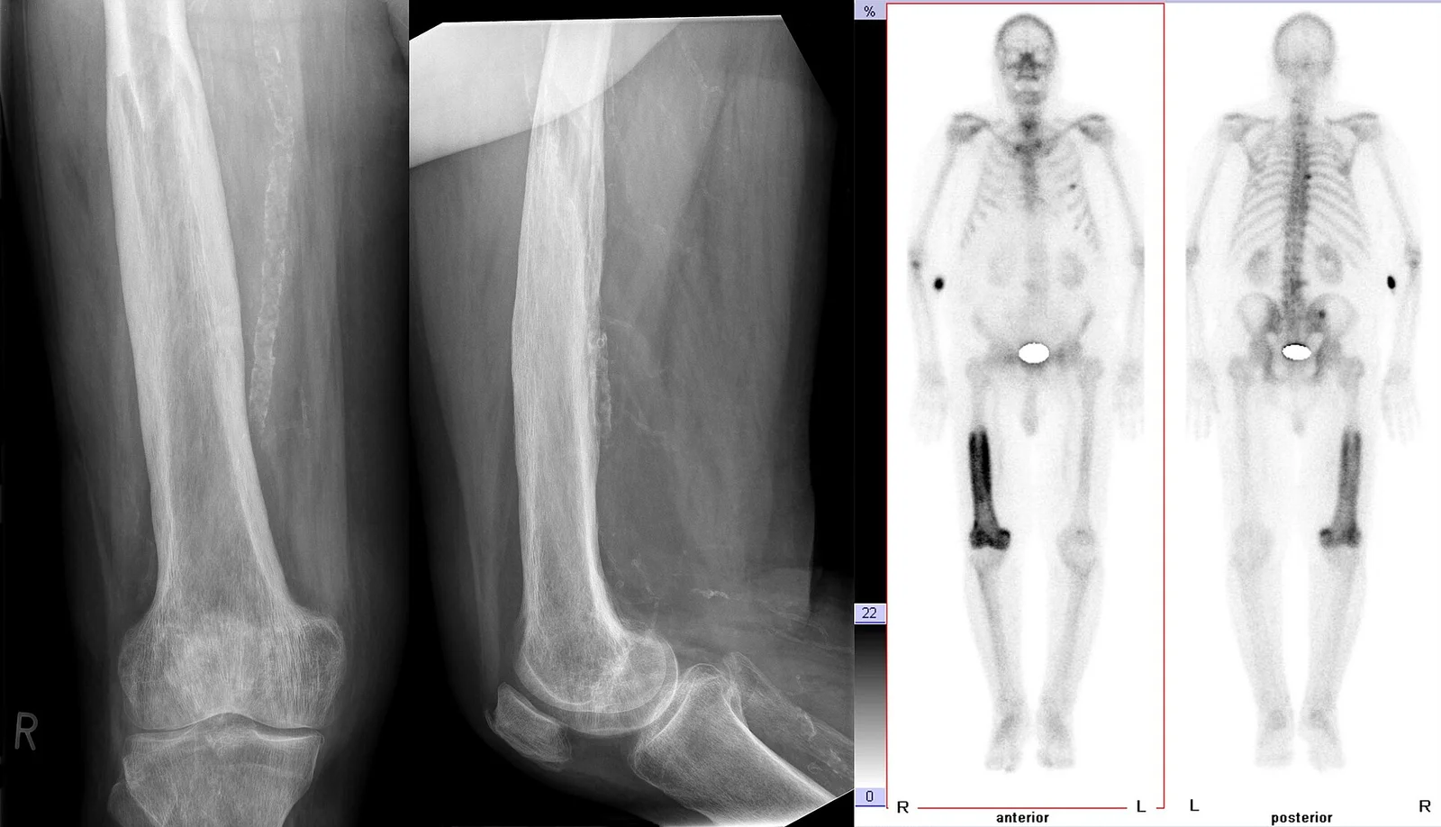
Long description
Radiographs of the femur in two planes and whole-body bone scintigraphy in Paget disease of the femur, with increased tracer uptake in the affected bone.

Figure 2Morbus Paget des Beckens rechts 77W - CR ap - 001
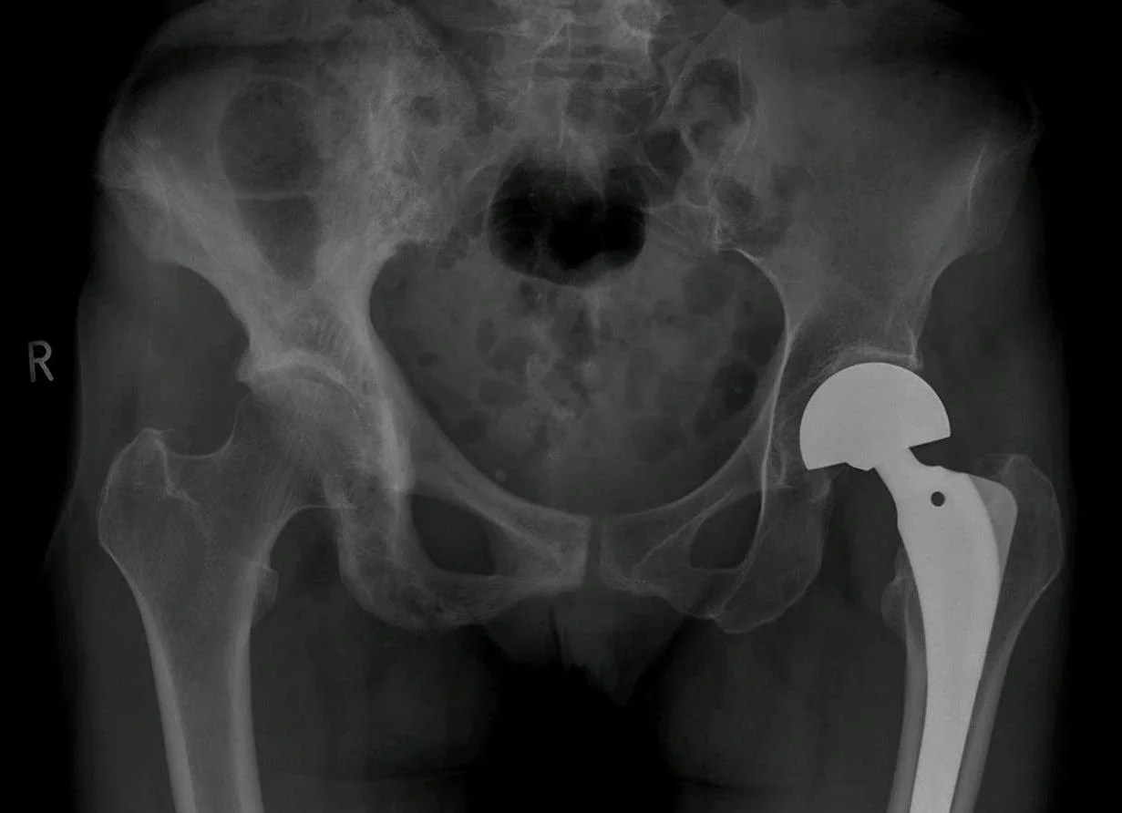
Long description
Pelvic radiograph in Paget disease of the right hemipelvis, with thickened cortex and coarse trabeculae; a left hip replacement is also visible.

Serum total alkaline phosphatase (ALP) is the recommended first-line biochemical screen, performed with liver function tests [2] [4] [5]. It is raised in most cases of active disease and correlates with skeletal extent on scanning [3]. It is a screening marker, not a diagnostic test: many other conditions raise ALP (liver, bone), and ALP can be normal in 42 to 58% of patients with limited (especially monostotic) disease [4].
Calcium, phosphate, creatinine, liver enzymes, and usually 25-hydroxyvitamin D and PTH, are checked alongside ALP before imaging [4]. These exclude the alternative diagnoses above and prepare for treatment.
Plain radiographs of the painful or suspected bone are the first imaging step [1] [2] [4]. The radiological phases and features are described in the next section. X-rays are available almost everywhere, are cheap, and identify stress fractures.
A whole-body radionuclide bone scan with technetium-99m labelled methylene diphosphonate (99mTc-MDP) is recommended to map all metabolically active disease [2] [4] [5]. The scan is more sensitive than radiographs: only about 3.7% of sites are visible on X-ray but scan-negative [2]. Typical pagetic appearances include "clover" or "Mickey mouse" signs in the spine and the "heart sign", which help distinguish the disease from metastases, although false positives occur [2] [4].
If a bone scan is not available, plain X-rays of the abdomen (including the lower ribs and femoral heads), both tibiae, the skull and facial bones detect Paget disease in about 93% of cases (79% for an abdominal film alone) [2] [4].
Targeted X-rays of the hot spots then confirm the diagnosis radiographically, because uniquely pagetic sclerotic areas can be present where the scan shows no uptake (burned-out disease).
CT and MRI are not recommended for diagnosing Paget disease [2] [4]. They are valuable for complications: basilar invagination, spinal stenosis, fracture, and suspected sarcoma. CT also helps in biopsy planning and preoperative planning for joint replacement.
When ALP is normal but clinical suspicion is high (for example monostotic disease of a small bone), more specific markers are useful [1] [3] [4]:
- Bone-specific alkaline phosphatase (bone ALP)
- Procollagen type 1 N-terminal propeptide (P1NP)
- Urinary N-telopeptide of type I collagen (uNTX)
BSALP is raised in about 90% of untreated patients and P1NP in 94%, versus total ALP in 74 to 76% [2]. Bone ALP and P1NP are also the markers to use when the liver or biliary tract is abnormal, because total ALP then becomes unreliable [3] [4].
Genetic testing for SQSTM1 (and, in early polyostotic disease, PFN1 and ZNF687) can be considered in familial disease or early-onset (under 50 years) polyostotic disease, and first-degree relatives of carriers can be screened [4]. Penetrance is incomplete, so a negative or positive test cannot be read in isolation.
There is no single gold-standard test; diagnosis is clinical plus radiological, supported by biochemistry. The current criteria used by the Reclast label, adopted widely, are a diagnosis of Paget disease with an ALP at least 2 times the upper limit of the age-specific normal range, or symptomatic disease, or disease at risk of complications [6].
The SIOMMMS position paper describes three phases, which also appear on the scan and explain the imaging [4]:
- Lytic phase: large, well-defined areas of osteolysis, sometimes flame-shaped, without peripheral sclerosis.
- Mixed phase: most cardinal features together; the most common presentation.
- Blastic or sclerotic phase: extensive osteosclerosis with loss of the cortex-medulla distinction, bone enlargement and deformity; the "burnt-out" phase on bone scan.
Characteristic radiological features, alone or in combination, are osteolytic areas, cortical thickening, loss of distinction between cortex and medulla, trabecular thickening, osteosclerosis, bone expansion and bone deformity [2]. The asymmetric distribution of lesions is a further clue to polyostotic disease.
Paget disease of the skull shows patchy sclerosis producing a "cotton wool" appearance, with thickening of the inner and outer tables. Circumscribed osteolytic lesions (osteoporosis circumscripta) occur in the frontal or occipital bone.
Diagnostic approach to suspected Paget disease of bone
- Suspected Paget diseaseBone pain, deformity, fracture, deafness, or an incidental raised ALP with normal liver tests
- First-line testsTotal ALP with liver function tests, plus serum calcium, phosphate, creatinine, PTH and 25-hydroxyvitamin D
- Question: Is ALP raised or clinical suspicion strong?
Yes - likely active disease
- Plain radiograph of the symptomatic boneConfirms Paget disease; shows phase, fracture, deformity
- Whole-body bone scan (99mTc-MDP)Maps all metabolically active disease
- Targeted X-rays of hot spotsConfirms pagetic changes; excludes burned-out or alternative lesions
No - ALP normal but scan clinically indicated
- Plain radiograph firstIf equivocal, add bone ALP, P1NP or uNTX
- Consider bone scan if suspicion remains high
- Complications, or atypical or equivocal lesionsCT or MRI for basilar invagination, spinal stenosis, fracture and suspected sarcoma; biopsy if imaging is not diagnostic
The management aim is to control symptoms and prevent or manage complications. Not every patient needs drug treatment: an asymptomatic patient with mild, stable, inactive disease can be watched. Guidelines differ on how aggressively to treat asymptomatic active disease (see below).
- The Endocrine Society 2014 suggests a bisphosphonate for most patients with active disease who are at risk of future complications, and a single 5 mg infusion of intravenous zoledronic acid as the treatment of choice where there is no contraindication [1].
- The Paget's Association 2019 clinical guideline recommends bisphosphonates for bone pain, with zoledronic acid the bisphosphonate most likely to give a favourable pain response, and recommends treatment aimed at improving symptoms rather than a "treat-to-target" strategy aimed at normalising ALP. It also recommends hip or knee replacement when osteoarthritis becomes severe [5].
- The SIOMMMS 2024 position paper recommends (strongly) treating symptomatic disease, and suggests treating most if not all cases at diagnosis, because of the safety and long-term efficacy of a single course of intravenous zoledronic acid [4].
These positions genuinely differ, and the difference reflects uncertainty: there are no good randomised trials showing that treating asymptomatic disease prevents complications such as fracture, deformity or deafness [4]. The honest summary is that all guidelines agree on treating symptomatic disease with a potent bisphosphonate, and that for an asymptomatic patient the decision should be individualised.
Treatment is indicated for patients with Paget disease who are:
- Symptomatic (bone pain from active disease).
- At risk of future complications: active disease in a weight-bearing long bone, lytic disease, skull-base disease threatening hearing or other cranial nerves, spinal involvement risking stenosis, progressive deformity, vertebral body involvement.
- About to undergo orthopaedic or other surgery on pagetic bone.
- Re-treated after biochemical relapse (rising ALP or recurrent pain).
The Reclast label puts it practically: treatment is indicated with an ALP at least 2 times the upper limit of the age-specific normal range, or symptomatic disease, or disease at risk for complications [6].
Where pain is arthritis pain from secondary osteoarthritis rather than active pagetic pain, analgesics, physiotherapy and joint replacement are the appropriate treatments, not a second bisphosphonate course.
- Ensure the patient is vitamin D replete (25-hydroxyvitamin D at least 50 nmol/L) with adequate calcium intake and normal serum calcium before giving a bisphosphonate. Hypocalcaemia can be induced otherwise, especially with zoledronic acid [3].
- Check creatinine and calculate clearance: zoledronic acid is contraindicated if creatinine clearance is under 35 mL/min [6].
- Optimise dental health before zoledronic acid because of the small risk of osteonecrosis of the jaw [6].
- Plan surgery only after biochemical control has been achieved, to reduce bleeding from vascular bone [3].
Zoledronic acid, as a single 5 mg intravenous infusion over at least 15 minutes [6]. It is the most potent, the longest-acting, the most likely to relieve pain and the most likely to normalise biochemistry. In the two registration trials comparing it with risedronate 30 mg daily for 2 months, 96% of zoledronic acid-treated patients achieved a therapeutic response (normal ALP or at least a 75% fall in ALP excess) versus 74% with risedronate, and 89% achieved normal ALP versus 58% [6]. Pain responds over weeks. Biochemical remission often lasts at least 6 years after a single infusion [3]. [5]
Concomitant with the infusion, give elemental calcium 1500 mg daily in divided doses and vitamin D 800 IU daily, particularly during the 2 weeks after treatment [6].
Re-treatment is not governed by a fixed schedule. Consider re-treatment if ALP rises again, normalisation was not achieved, or symptoms recur [6].
Adverse effects: a transient acute-phase reaction (fever, myalgia, influenza-like symptoms) for a day or two is common; renal monitoring is required; hypocalcaemia and (rarely) jaw osteonecrosis are the serious concerns.
- Risedronate 30 mg orally once daily for 2 months, possibly repeated if needed [5].
- Pamidronate intravenously: common practice is 60 mg infused on each of 3 consecutive days. Largely superseded by zoledronic acid, still useful where zoledronic acid is unavailable [5].
- Alendronate and tiludronate have been used historically and published in older randomised trials; they are not first-line today.
Calcitonin (salmon calcitonin, given by injection) is now rarely used. It can help bone pain but daily subcutaneous injections are required and relapse is rapid on stopping [3]. It has a place where bisphosphonates are not tolerated or contraindicated.
Denosumab is an osteoclast inhibitor used in other bone disease; it has been reported to lower markers in Paget disease but has not been studied in randomised trials for this condition [2].
Treat residual pain with analgesics or NSAIDs, physiotherapy, orthotics, occupational therapy and walking aids [3]. Neuropathic pain agents may help nerve root pain. None of these has been tested specifically in Paget disease, but all are used in practice.
- Total hip or knee replacement is recommended for osteoarthritis that is not controlled medically [2] [5].
- Realignment osteotomy, fracture fixation, prophylactic intramedullary nailing of a fissure fracture in a bowed weight-bearing bone, and nerve decompression (for spinal stenosis) are the other common operations.
- Bring pagetic bone under biochemical control before surgery, to reduce vascularity and bleeding [3].
- Surgery can be difficult because pagetic bone is vascular, deformity complicates alignment, and implants may be hard to seat. Outcomes for hip replacement in Paget disease are generally good when the disease is controlled preoperatively.
This is worth stating plainly because it sets expectations with patients:
- Bisphosphonates relieve pain and normalise bone markers; they usually do not reverse established deformity.
- Hearing loss generally does not respond to treating the Paget disease [3] [4].
- High-output cardiac failure may respond to treatment [3].
- Paraplegia from spinal pagetic bone may sometimes be reversed [1].
- Osteosarcoma requires referral to a sarcoma centre and has a poor prognosis [3].
- Clinical and biochemical review 3 to 6 months after treatment to confirm response [3] [5].
- Annual review thereafter for biochemical relapse and complications [3].
- Total ALP is the standard monitoring marker; use bone ALP or P1NP (and, less often, uNTX) when ALP is normal at baseline or the liver is affected [3] [4].
- Clinical relapse of pain and biochemical relapse are separate: in one extension study, bone pain recurred in 14 of 152 (9.2%) zoledronic acid-treated patients while biochemistry stayed normal [2]. [4]
- Bone scanning is occasionally used to assess response when markers are normal [1].
Management outline after a confirmed diagnosis
- Confirmed Paget diseaseClinical, radiographic and biochemical diagnosis
- AssessSymptoms; extent on bone scan; ALP or bone marker; renal function; 25-hydroxyvitamin D; dental status
- Question: Are symptoms or risk features present?
Yes
- Treat with a bisphosphonateFirst line: 5 mg zoledronic acid IV once; alternatives risedronate 30 mg daily for 2 months, or pamidronate regimes
- PreparationVitamin D replete; calcium normal; creatinine clearance at least 35 mL/min for zoledronic acid
- MonitorResponse at 3 to 6 months; annual review thereafter
Asymptomatic only (guidelines differ)
- Watch or treat selectivelyEndocrine Society: treat active disease at risk of complications; SIOMMMS: consider treating most cases; pain- or cosmetic-driven plans preferred under UK guidance
- If untreated, monitorAnnual ALP and clinical review
- ComplicationsJoint replacement, decompression, fissure fracture nailing, and pre-surgical biochemical control
Paget disease is often benign, but the complications below are what bring patients to hospital. The frequency estimates come from series of clinically identified patients and differ between studies.
Musculoskeletal (common):
- Fracture of pagetic bone (about 10 to 30% of patients) [3].
- Deformity and limb shortening or angulation, limiting gait.
- Secondary osteoarthritis at adjacent joints (a UK primary-care database study found a roughly 3-fold increase in hip arthroplasty for osteoarthritis among diagnosed patients) [2].
- Acetabular protrusion from pelvis involvement.
Neurological:
- Deafness and tinnitus (about 5.9 to 8.9% in large reviews) [4] [2].
- Basilar invagination, brainstem or cerebellar compression, cranial nerve palsies, obstructive hydrocephalus.
- Spinal stenosis and cauda equina syndrome; paraparesis or quadriparesis.
Cardiovascular (rare):
- High-output cardiac failure from shunting through vascular bone (reported but extremely rare) [2] [4].
- Aortic stenosis, endocardial calcification and accelerated atherosclerosis [3] [4].
Metabolic (rare):
- Immobilisation hypercalcaemia.
- Hyperuricaemia and nephrolithiasis.
Neoplastic (very rare):
- Osteosarcoma (about 0.3% of patients [2]) and other sarcomas, usually in long-standing disease.
- Giant cell tumour complicating pagetic bone (less than 1% overall; classically in southern Italy) [4].
Surgical and perioperative:
- Heavy bleeding if operations are done on vascular, biochemically active bone; this is why control is advised first.
- Refractory pain despite biochemical remission, when the problem is deformity or arthritis rather than active disease.
The prognosis is good for most patients. A single course of zoledronic acid produces biochemical remission that lasts at least 6 years, and survival in uncomplicated disease is normal [3]. Recurrence of biochemical activity after zoledronic acid was seen in only about 14% of elderly patients at 9 years in one series [4]. Without treatment, active disease can be slowly progressive in extent and severity but new bones rarely become involved [3].
The features that worsen prognosis are extensive polyostotic disease, early-onset familial disease, progressive deformity, and any sarcomatous change. A second (metachronous) pagetic focus or disease in a young adult should trigger a search for a familial mutation and genetic counselling.
There is no population screening programme. Primary prevention is not possible because the cause is unknown. Secondary prevention currently has two forms in research: (1) screening first-degree relatives of affected patients with ALP or, in mutation-carrying families, genetic testing followed by zoledronic acid, and (2) prophylactic zoledronic acid in asymptomatic SQSTM1 mutation carriers. Results so far are promising (in one trial, zoledronic acid stopped new lesions from developing and resolved most existing lesions on scan) [4], but this is not yet standard care.
Children: Classic Paget disease of bone is an adult disorder; significant disease before the age of 40 is very unusual. Early-onset polyostotic disease in a young person should raise the possibility of a familial mutation (PFN1, ZNF687) or a PDB-like syndrome, and should be referred [4]. Juvenile Paget disease is a different condition and is not covered here.
Older adults: The prevalence of polyostotic disease rises with age and renal function often declines. Before giving zoledronic acid, check creatinine clearance because the drug is contraindicated below 35 mL/min; adjust for body weight where applicable [6]. Dental review and falls prevention are wise.
Pregnancy: No specific guidance appears in the sources used here. Bisphosphonates are generally avoided in pregnancy; any pregnant patient should be managed jointly by her obstetric and specialist teams.
East Africa
Diagnosis relies on two inexpensive tools: an ALP (ideally with liver enzymes) and a plain radiograph. Serology and advanced imaging can often be avoided:
- Suspect Paget disease in a patient over 40 with bone pain and a raised ALP but normal calcium and phosphate. If scanning is unavailable and the scan cannot be done, X-rays of the abdomen (including the lower ribs and femoral heads), both tibiae, the skull and facial bones detect about 93% of cases, against 79% for an abdominal film alone [2] [4].
- Before concluding Paget disease, check plasma and urinary calcium and phosphate, renal function, liver function and protein electrophoresis, and, where available, PTH and 25-hydroxyvitamin D, to exclude osteomalacia, myeloma and metastases [4].
- CT and MRI are reserved for suspected sarcoma, spinal compression or basilar invagination, where the result would change management.
Treatment:
- Oral risedronate 30 mg daily for 2 months is inexpensive, oral and effective, and is the most practical option where infusion facilities are limited.
- If infusion services exist, a single 5 mg dose of zoledronic acid is the most effective treatment and it is inexpensive as a drug, though it needs an infusion set, nursing time and monitoring.
- Where neither bisphosphonate is available, salmon calcitonin injections may still relieve bone pain [3].
- Ensure adequate calcium and vitamin D (1500 mg elemental calcium and 800 IU vitamin D daily around treatment, and a sensible intake otherwise) [6].
- Surgery (hip replacement, fracture fixation, decompression) requires referral to a centre with the necessary orthopaedic and anaesthetic capacity; control bone activity biochemically first when possible.
- Remember immobilisation and fracture as causes of hypercalcaemia in these patients.
- Paget disease of bone is a focal disorder of remodelling: overactive osteoclasts drive disorganised, vascular, mechanically weak bone in one or more bones.
- It is the second most common metabolic bone disease. It usually presents after age 40, is slightly commoner in men, and clusters in north-west European ancestry; most cases are asymptomatic.
- Prevalence is roughly 1% over 55 in the UK and up to 5 to 8% by the eighth decade in high-incidence areas; incidence is falling.
- Cause is unknown but genetic (especially SQSTM1) plus an unidentified environmental factor, possibly viral, underlie it.
- Defining laboratory pattern: raised ALP with normal calcium and phosphate.
- Radiological phases: lytic, mixed (most common) and sclerotic (burned-out).
- Confirmatory imaging: plain radiographs; extent by 99mTc-MDP bone scan; CT/MRI for complications only.
- Main differential diagnoses: bone metastasis and multiple myeloma (not pagetic bone enlargement), osteomalacia (generalised, with vitamin D deficiency), and primary hyperparathyroidism.
- Complications: fracture, deformity, osteoarthritis, deafness, very rarely high-output heart failure, and osteosarcoma in about 0.3%.
- Treatment only for symptomatic or at-risk active disease: single 5 mg intravenous zoledronic acid, or risedronate 30 mg orally daily for 2 months. Bisphosphonate medical control before surgery.
- Hearing loss seldom responds; pain from secondary arthritis needs analgesia, physiotherapy or joint replacement rather than more anti-Paget treatment.
Answer each one in your head first, then open the answer.
A 68-year-old man with mild bowing of the right tibia has an isolated raised ALP. Calcium and phosphate are normal and he has no symptoms. What is the most likely diagnosis and what two tests would confirm it and define its extent?
Show answerHide answer
Paget disease of bone. A plain radiograph of the tibia will show the phase (lytic, mixed or sclerotic), cortical thickening and a fissure fracture; a 99mTc-MDP bone scan is then used to map all metabolically active disease, since further bones (often the pelvis) are frequently involved.
Which laboratory finding best separates Paget disease from osteomalacia and primary hyperparathyroidism, and what should you check alongside it?
Show answerHide answer
In Paget disease ALP is raised while calcium and phosphate are usually normal. Alongside it check liver enzymes, 25-hydroxyvitamin D and PTH. Osteomalacia shows low vitamin D and often low calcium or phosphate; primary hyperparathyroidism shows raised PTH with raised calcium and low phosphate.
How does the radiographic appearance of Paget disease differ from that of bone metastases?
Show answerHide answer
Pagetic bone is enlarged and widened, with coarsened trabeculae and cortical thickening; it bends and may contain both lytic and sclerotic areas. Metastases are usually ill-defined lytic or sclerotic lesions within a normal-sized bone, without expansion. The technetium bone scan pattern is also usually recognisable as pagetic.
What is the first-line drug treatment, the dose, and the concomitant supplementation?
Show answerHide answer
A single 5 mg intravenous infusion of zoledronic acid over at least 15 minutes. Give elemental calcium 1500 mg daily in divided doses and vitamin D 800 IU daily, especially in the 2 weeks after treatment, and recheck renal function first (contraindicated if creatinine clearance is under 35 mL/min). [6]
What can a patient expect bisphosphonate treatment to change, and what is it unlikely to help?
Show answerHide answer
Bisphosphonates relieve pagetic bone pain and normalise bone markers, and remission often lasts years. They generally do not reverse established deformity, and hearing loss from skull involvement seldom responds. Pain from secondary osteoarthritis needs analgesia, physiotherapy or joint replacement.
Before elective surgery on pagetic bone, what should be done and why?
Show answerHide answer
Bring the disease under biochemical control with a bisphosphonate first, because pagetic bone is hypervascular and more bleeding occurs when it is biochemically active. Operate with blood-loss precautions and plan for distorted anatomy.
No MedGrasp topics are currently listed as prerequisites for this page. As further metabolic bone topics are published (for example osteoporosis and disorders of calcium metabolism), they will be linked here in plain text. This page already depends on your familiarity with basic bone remodelling, alkaline phosphatase and bone scintigraphy.
- Singer FR, Bone HG III, Hosking DJ, et al. Paget's disease of bone: an endocrine society clinical practice guideline. J Clin Endocrinol Metab 2014;99:4408-4422. Endocrine Society. Accessed .
- Ralston SH, Corral-Gudino L, Cooper C, et al. Diagnosis and management of Paget's disease of bone in adults: a clinical guideline. J Bone Miner Res 2019;34:579-604. doi:10.1002/jbmr.3657. Accessed .
- Tuck SP, Walker J. Adult Paget's disease of bone. Clin Med (Lond) 2020;20:568-571. doi:10.7861/clinmed.20.6.page. Accessed .
- Rendina D, Falchetti A, Diacinti D, et al. Diagnosis and treatment of Paget's disease of bone: position paper from SIOMMMS. J Endocrinol Invest 2024;47:1335-1360. doi:10.1007/s40618-024-02318-1. Accessed .
- Paget's Association (UK). Clinical management of Paget's disease. Paget's Association. Accessed .
- Novartis Pharmaceuticals Corporation. Reclast (zoledronic acid) injection, solution: full prescribing information. DailyMed (US National Library of Medicine). Accessed .
Figure credits
Educational content for medical students and doctors. It is not a substitute for clinical judgement or local guidelines.