Thyroid function tests
How thyroid testing works: the HPT axis, interpreting TSH, free T4 and free T3, antibodies, imaging, drugs and practice in low-resource settings.
- Thyroid function tests (TFTs) measure how well the thyroid, pituitary and hypothalamus are working, usually through thyroid-stimulating hormone (TSH), free thyroxine (free T4) and sometimes free tri-iodothyronine (free T3).
- TSH is the most sensitive first test in most adults; if it is abnormal, free T4 (and free T3 when hyperthyroidism is suspected) is added to show whether disease is overt or subclinical.
- Most thyroid disease is primary: the gland itself fails or overacts, giving an opposite TSH. A low free T4 with a low or normal TSH suggests a pituitary or hypothalamic problem instead.
- In acute illness, drugs, pregnancy and antibody or biotin interference can all make TFTs misleading; always interpret them in the clinical context and repeat when recovery is complete.
- Antibodies (TPO, thyroglobulin, TSH receptor) and imaging (ultrasound and radioiodine uptake) answer the "why", not the "whether".
By the end of this page you should be able to:
- Explain the hypothalamic-pituitary-thyroid (HPT) axis and the synthesis, secretion and peripheral conversion of thyroid hormones.
- State what TSH, free T4 and free T3 each reflect, and choose the sensible next test when one is abnormal.
- Read a Table of patterns and classify primary and secondary hypo- and hyperthyroidism, subclinical disease, sick euthyroid (nonthyroidal illness) syndrome, pregnancy effects and assay interference.
- Explain what TPO, thyroglobulin and TSH receptor antibodies mean and when to request them.
- Describe the roles of thyroid ultrasound and radioiodine uptake scanning, including safety limits.
- Adjust interpretation for drugs such as amiodarone, glucocorticoids, oestrogen and heparin.
- Apply a stepwise algorithm to an abnormal TFT result, and adapt testing and management when resources are limited.
Thyroid function testing is one of the most commonly requested sets of blood tests, because the thyroid hormones set the metabolic pace of most organs: heart, brain, muscle, gut, bone and reproductive system. [1] A test is only as good as the question it answers, so the first skill is to know what question you are asking: a steady-state measurement of gland activity, a test of the pituitary feedback loop, or (later) a test of autoimmune damage or structure.
This page treats thyroid function testing as a clinical procedure. It is not a single test, but a process: deciding when to test, preparing the patient, choosing the assay, interpreting the result, confirming the pattern, finding the cause, and knowing when not to test again.
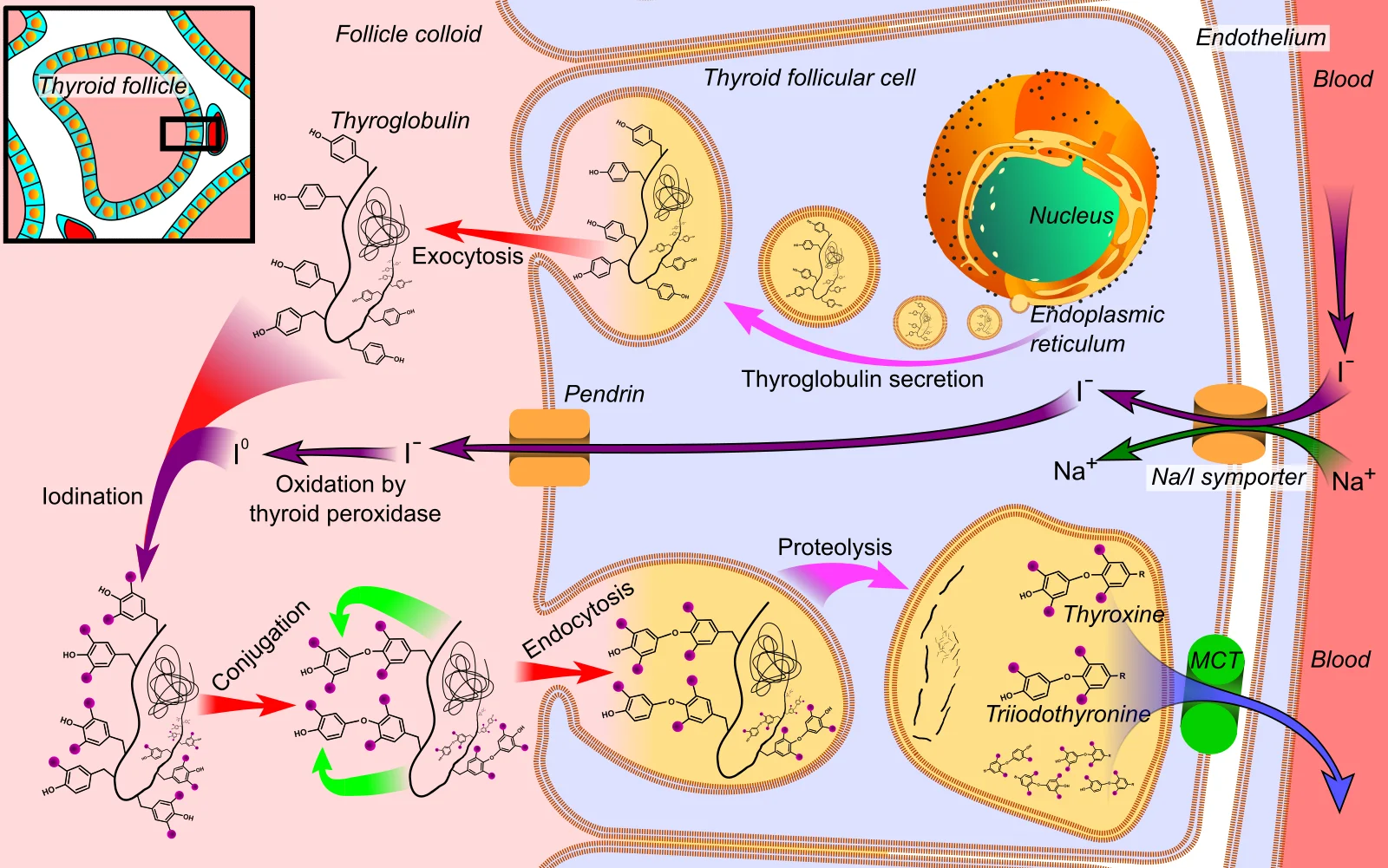
Figure 1Thyroid hormone synthesis
Long description
Thyroid hormone synthesis in the follicular cell: iodide enters through the sodium-iodide symporter, is oxidised and bound to thyroglobulin, taken back into the cell and cleaved to release thyroxine and triiodothyronine into the blood.

The thyroid gland sits in the lower anterior neck and is the only endocrine organ that stores its product outside the cell. [1] Hormone production proceeds in steps, and a block at any step explains a different test result.
Iodine uptake and organification. Iodide is concentrated in the follicular cell by the sodium-iodide symporter on its basolateral membrane, then moved across the apical membrane into the colloid. There, thyroid peroxidase (TPO) oxidises iodide and attaches it to tyrosine residues on thyroglobulin, producing mono-iodotyrosine (MIT) and di-iodotyrosine (DIT). [1] This step is blocked by thionamides and depends on adequate iodine intake.
Coupling. TPO also couples iodotyrosines: two DIT molecules form thyroxine (T4), and one MIT plus one DIT form tri-iodothyronine (T3). These hormones remain bound to thyroglobulin in the colloid until the cell takes the colloid back (endocytosis) and digests it, releasing T4 and T3 into the blood. [1]
Secretion and storage. T4 is the main secreted hormone (about 80% of secreted thyroid hormone); T3 is the more biologically active hormone but only about 20% is secreted directly. [1] The colloid store buffers short-term changes in iodine supply.
Peripheral conversion. Outside the thyroid, deiodinases in liver, kidney, muscle and other tissues remove an iodine atom from the outer ring of T4 to produce active T3, or from the inner ring to produce reverse T3 (rT3), which is inactive. In illness this balance shifts away from T3 toward rT3. [1]
Transport. Only a very small free fraction of each hormone circulates unbound; the rest is bound to thyroxine-binding globulin (TBG, the principal binder), transthyretin and albumin. Only the free fractions enter cells and suppress TSH. [1] Total concentrations therefore change when binding proteins change (pregnancy, oestrogen, liver disease, heparin), even when the patient is euthyroid. [1][2]
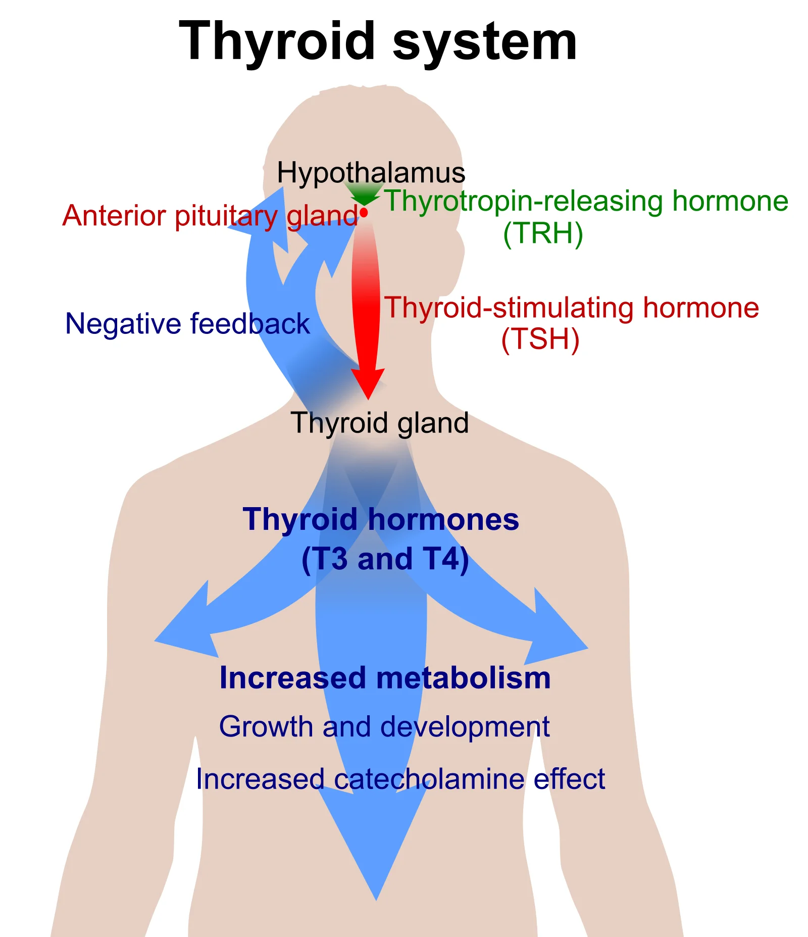
Figure 2Thyroid system
Long description
The thyroid axis: thyrotropin-releasing hormone from the hypothalamus drives thyroid-stimulating hormone from the anterior pituitary, which drives thyroid hormones T3 and T4 from the thyroid gland; the thyroid hormones feed back negatively on the pituitary and hypothalamus and raise metabolism.

Hypothalamic thyrotropin-releasing hormone (TRH) stimulates the anterior pituitary to secrete thyroid-stimulating hormone (TSH), a glycoprotein. TSH drives every step of thyroid hormone synthesis and release. T4 and T3 exert negative feedback on both hypothalamus and pituitary, with intra-pituitary conversion of T4 to T3 being important. The loop is exquisitely sensitive: a small fall in free T4 (within the normal range) can raise TSH several-fold. This is why TSH is the best single screening test for primary thyroid disease in stable outpatients. [1]
There are important exceptions. In central hypothyroidism the low free T4 is not matched by a raised TSH, because the pituitary or hypothalamus is the problem. [1] In TSH-secreting pituitary adenoma or thyroid hormone resistance (rare, mentioned for completeness), TSH is inappropriately normal or high despite high free hormones. [2] Severe nonthyroidal illness suppresses the axis, so axis logic fails in the critically ill. [1] [3]
- TSH reflects the integrated sensitivity of the pituitary to circulating free T4 and T3, plus TRH drive. It is usually normal in central disease "inappropriately" for the free hormones. [1]
- Free T4 is the biologically available hormone and the right test for hypothyroidism and for pregnancy monitoring. [2]
- Free T3 is the active form and is useful when hyperthyroidism is suspected (T3-toxicosis, where T4 is normal). [2]
- Total T4 / total T3 largely track binding proteins and are not preferred when TBG changes. [2]
- Free T4 index (T7) is an older estimate combining total T4 with T3 uptake (a crude measure of TBG); immunoassay free T4 has largely replaced it.
- Thyroid uptake and imaging test function (iodine trapping) and structure, not simply blood concentration. [4]
Normal ranges vary by laboratory. MedlinePlus cites a TSH reference range of 0.4 to 4.8 microunits/mL, with experts disagreeing on the upper limit, and some laboratories using up to 7 microunits/mL in older adults. Always use the reference interval printed on the report and the method-specific range. [3]
Test when there is clinical reason, not as a blanket screen:
- Symptoms or signs of hypothyroidism (fatigue, cold intolerance, weight gain, constipation, menorrhagia, dry skin, bradycardia, delayed relaxation of reflexes) or of hyperthyroidism (weight loss, heat intolerance, sweating, palpitations, tremor, anxiety, proximal weakness). [1]
- Unexplained atrial fibrillation, worsening heart failure, altered mental status, severe electrolyte disturbance (for example hyponatraemia/SIADH), or cardiovascular instability. [1]
- Known thyroid disease that is decompensated, or a drug known to affect the thyroid. [1]
- Women planning pregnancy or pregnant women with risk factors; see pregnancy below. [2]
- Monitoring of replacement (levothyroxine) or suppression therapy, with the dose assessed by TSH except in pituitary disease. [3]
Do not test routinely in every hospital admission or in mild, early symptoms that have not changed; a large share of abnormal results in hospital reflect NTIS rather than thyroid disease. [1]
Testing is blood sampling: the usual cautions around bleeding, haematoma and infection apply. [3] The real contraindications are interpretive:
- Pregnancy and breastfeeding contraindicate radioactive iodine uptake and scanning (and iodinated contrast for diagnostic thyroid purposes). [4]
- Recent iodine load (contrast scans within about two weeks, kelp supplements, iodine-containing drugs) distorts both uptake and hormone measurements. [4]
- Biotin (vitamin B7) in supplements can distort TSH, and possibly other thyroid assays, depending on platform. Patients should tell staff and stop biotin before testing as advised locally. [3]
- Acute systemic illness makes TFTs unreliable; if not urgent, defer. [1]
- Oestrogen or androgen states and drugs (see below) change binding proteins; a measured total hormone in these states is unhelpful. [1][2]
Preparation is part of the test:
- Note the patient's age, pregnancy status, medicines (including biotin-containing supplements, amiodarone, glucocorticoids, oestrogen, heparin, anticonvulsants), recent contrast, and whether they are systemically unwell. [1][3]
- Sample in the morning where possible; TSH varies through the day. [3]
- If the patient takes thyroid hormone, note dose and timing; morning fasting is not required for TFTs. [3]
- Tell the patient that a venepuncture is needed and that results may change with timing and illness. Consent must include the possibility of needing repeat testing.
- Venepuncture for TSH (first-line screen) and, if needed, free T4 and/or free T3. [3]
- In stable patients: if TSH is normal, stop; if TSH is abnormal, add free T4 (and free T3 if hyperthyroidism suspected or if TSH is suppressed). [1]
- During pregnancy use pregnancy-specific TSH and free T4 intervals; free T4 by immunoassay is less reliable, and mass-spectrometry methods (where available) are preferred. [2]
- Assay handling: mark the request with method, age, pregnancy data and relevant drugs, so the laboratory can interpret binding-protein effects and (if needed) re-run with a different platform. [1][2]
- Where a discordant result is suspected to reflect interference, the laboratory can change assay platforms, dilute, block heterophile antibodies, or use a different hormone measurement, and is best consulted directly. [5]
Read TSH first, then free T4 and (when relevant) free T3. The table summarises the common patterns. Values and cut-offs vary by laboratory and guideline; the categories are clinical patterns, not single numbers. [1][2][3]
| Pattern | TSH | Free T4 | Free T3 | Typical explanation |
|---|---|---|---|---|
| Primary hypothyroidism | Raised | Low | Low or normal | Gland failure (e.g. autoimmune thyroiditis, iodine deficiency) |
| Subclinical hypothyroidism | Raised (usually under 10 mIU/L) | Normal | Normal | Early or mild gland failure; often transient |
| Central (secondary) hypothyroidism | Low or inappropriately normal | Low | Low | Pituitary or hypothalamic disease |
| Primary hyperthyroidism | Suppressed | Raised | Raised (often T3-predominant) | Graves disease, toxic nodules, thyroiditis with release |
| Subclinical hyperthyroidism | Suppressed | Normal | Normal | Mild autonomic overactivity; often transient |
| T3-toxicosis | Suppressed | Normal | Raised | Early or nodular hyperthyroidism; conversion-dominant state |
| Nonthyroidal illness (sick euthyroid) syndrome | Low, normal or mildly raised (recovery phase) | Low or normal | Low (earliest change) | Acute illness; adaptive, not true thyroid disease |
| Pregnancy, first trimester | Lower than non-pregnant, often under 1 mIU/L; upper limit about 2.5 mIU/L before confirmatory testing | Raised (binding protein-dependent, method matters) | Variable | hCG thyrotropic effect, rising TBG |
| Assay interference (e.g. biotin, heterophile antibodies, macro-TSH) | Discordant with clinical state | Discordant | Discordant | Method-specific antibody or small-molecule interference |
| Thyroid hormone resistance or TSH-secreting adenoma | Inappropriately normal or raised | Raised | Raised | Rare receptor or pituitary pathology |
Notes on the numbers:
- Subclinical hypothyroidism is usually defined as a raised TSH with normal free T4; about nine in ten affected patients have a TSH under 10 mIU/L. [1]
- A treatment decision for overt hypothyroidism is often made when TSH is above 10 mIU/L if free T4 is low (or even within reference range if the clinical picture fits). [1] [2]
- In acute illness, mild TSH abnormalities (about 0.1 to 0.6 or 6.7 to 20 mIU/L) have very low positive likelihood for true thyroid disease; only markedly abnormal values under 0.01 or above 20 mIU/L increase the probability of intrinsic disease. [1]
- Nonthyroidal illness may show a low total T4 under 4 micrograms/dL in association with about 50% mortality, and under 2 micrograms/dL with more than 80%, but such patients are severely ill and should not be treated with thyroid hormone on the basis of these levels alone. [1] In hospital, this syndrome is present in up to 60 to 75% of patients. [1] [2]
Antibodies are tests of cause, usually added once biochemical dysfunction is established (or when infertility, miscarriage or a goitre is present). A positive result confirms autoimmunity but does not by itself mean treatment is needed. [5][6]
- Thyroid peroxidase antibodies (TPOAb, anti-TPO). TPO is the enzyme used to iodinate and couple thyroglobulin in the follicular cell. [1] In the blood, antibodies to TPO indicate autoimmune damage to the thyroid and are the hallmark of Hashimoto (autoimmune) thyroiditis. They also occur in Graves disease and can follow a viral thyroiditis. [5] Raised TPO antibodies are linked with an increased risk of miscarriage, pre-eclampsia, prematurity and in-vitro fertilisation failure. [5]
- Thyroglobulin antibodies (TgAb). Thyroglobulin leaks in small amounts from thyroid cells and the immune system may react against it. These are found in Hashimoto disease, Graves disease, subacute thyroiditis and some autoimmune diseases. [6] A positive Tg antibody also interferes with thyroglobulin measurement used in thyroid cancer follow-up, so it must be reported with it. [6]
- TSH receptor antibodies (TRAb, TSI). These bind and may stimulate (or block) the TSH receptor. They are the diagnostic test for Graves disease and are monitored in pregnancy to assess risk to the fetus and neonate. [2]
About 10 to 20% of pregnant women in the first trimester are TPO or thyroglobulin antibody positive while remaining euthyroid; of these, a substantial minority develop overt hypothyroidism by the third trimester or postpartum thyroiditis. [2] When Hashimoto disease is suspected, TPO antibody is the most efficient single test; when Graves disease is suspected, TRAb and TPO/thyroglobulin tests together help distinguish autoimmune from non-autoimmune causes. [5][6][2]
Thyroid ultrasound is the first-line structural test. [4] It sizes the gland and nodules, characterises echogenicity (diffuse hypoechogenicity supports thyroiditis), identifies cystic or solid nodules, guides fine-needle aspiration, and measures goitre or nodal disease. It involves no radiation and is safe in pregnancy. [4] Doppler can show vascularity. It should not be requested to explain abnormal biochemical results alone, but it helps when a goitre, nodule or neck mass is present.
Radioiodine uptake scan (RAIU) and thyroid scintigraphy assess how vigorously the gland traps iodine. [4] A small oral dose of radioactive iodine is taken; imaging and percentage uptake are measured at several hours (often 4 to 6 hours) and at 24 hours. [4]
- High uptake is expected when the gland is overproducing: diffuse uptake suggests Graves disease; a single "hot" nodule suggests a toxic adenoma; patchy uptake suggests multinodular goitre. [4]
- Low uptake suggests destructive or inflammatory thyroiditis (hormone leaks from damaged cells) or iodine contamination. [4]
- Technetium (99mTc-pertechnetate) can be used instead of iodine but is trapped only, not organified. [4]
- Pregnancy and breastfeeding are contraindications because of fetal/neonatal thyroid exposure. [4]
- Recent diarrhoea or iodine exposure (including contrast within about two weeks) reduces uptake and can be misleading. [4]
These scans and uptakes should not be requested for every biochemical abnormality; they answer anatomical/functional questions when the clinical picture supports it. [4]
Always review the drug list before diagnosing thyroid disease: drugs change TSH, synthesis, conversion, binding proteins and (with high-dose biotin) the assay itself. [1][3]
- Amiodarone contains large amounts of iodine and acts at several sites: it can block iodine uptake and hormone release (causing hypothyroidism or destructive thyroiditis) and inhibit peripheral T4 to T3 conversion. Always re-check thyroid tests in patients on amiodarone, but remember that its changes often represent true drug-induced disease rather than assay artefact. [1]
- Glucocorticoids (prednisone/dexamethasone) suppress TSH to low but detectable levels and reduce T4 to T3 conversion; dopamine agonists, octreotide and amphetamines do likewise. Dopamine antagonists such as metoclopramide can mildly raise TSH. [1] [3]
- Oestrogen and selective oestrogen receptor modulators increase thyroxine-binding globulin, raising total (not free) thyroid hormones and often requiring higher replacement doses in treated patients; methadone, heroin, mitotane and fluorouracil also raise TBG. [1]
- Heparin and drugs such as NSAIDs, furosemide, phenytoin and carbamazepine displace thyroid hormones from binding globulin, producing a transient rise in free T4/T3 and a fall in TSH without true disease. [1]
- Other agents affect synthesis (propylthiouracil, methimazole, lithium, iodinated contrast), cause destructive thyroiditis (amiodarone, tyrosine kinase inhibitors), accelerate conversion/clearance (barbiturates, phenytoin, carbamazepine, rifampicin, phenobarbital) or alter TBG. [1]
- Always record the drug and the clinical state on the request and repeat testing before changing long-term treatment. [1]
Pregnancy doubles the metabolic demand on the thyroid. hCG is weakly thyrotropic and suppresses TSH in the first trimester (frequently below the classical lower limit of 0.4 mIU/L, and to 0.2 or below at hCG above 400,000 IU/L); TBG rises by 6 to 8 weeks. [2] If trimester-specific TSH ranges are unavailable, the following are recommended: first trimester 0.1 to 2.5 mIU/L; second 0.2 to 3.0; third 0.3 to 3.0. [2] Free T4 assays are altered by binding protein changes; TSH is a more reliable index than many free T4 methods in pregnancy, and equilibrium dialysis/mass spectrometry free T4 is preferred where available. [2]
Classification during pregnancy (ATA 2011): overt hypothyroidism = TSH raised (>2.5) with low free T4, or any TSH 10 or above; subclinical hypothyroidism = TSH 2.5 to 10 with normal free T4; isolated hypothyroxinaemia = normal TSH with free T4 in the lowest 5-10% of the range. [2] Thyroid antibodies are present in 10 to 20% of women in the first trimester; many will develop overt hypothyroidism by the third trimester or postpartum thyroiditis. [2]
Radioiodine imaging is contraindicated in pregnancy; ultrasound is the structural tool and is safe. [2][4]
- Over-treating transient abnormalities, especially in hospital: mild TSH changes are common and rarely represent new disease; treatment with levothyroxine in this setting can produce iatrogenic thyrotoxicosis. [1]
- Missing central hypothyroidism because TSH is not raised; always check free T4 when the clinical picture is suggestive (pituitary disease, amenorrhoea, headache, visual field defect). [1]
- Binding-protein misinterpretation: acting on a total hormone result during pregnancy or on oestrogen or heparin. Use free hormones. [1][2]
- Assay interference from biotin or antibodies; discordant results in a healthy-feeling patient should prompt method review. [3]
- Venepuncture risks (haematoma, syncope) are small with good technique. [3]
- If a transient abnormality (intercurrent illness, a starting drug) is suspected, repeat testing after recovery, often in 4 to 6 weeks. [1]
- In pregnancy, TSH is monitored at least each trimester and more frequently with treatment or antibody positivity. [2]
- In postpartum thyroiditis, repeat TSH after resolution of the thyrotoxic phase. [2]
- Document why the test was taken, the pattern and the plan, so the next clinician does not repeats it without reason.
In settings where only TSH (or even total T4) is available:
- Screen with TSH if at all possible; it is the most sensitive first test and is widely available. [1]
- If free T4 is unavailable, total T4 interpreted with awareness of pregnancy (which raises TBG) may still guide a clinical decision. [2]
- Interpret mild abnormalities in the context of intercurrent illness, pregnancy and drugs, and defer re-testing until the patient has recovered; avoid starting treatment for borderline TSH values. [1]
- A thorough drug and symptom history costs nothing and often resolves otherwise puzzling results. [1]
- Radioiodine scanning may be unavailable; ultrasound (often more widely accessible) can evaluate nodules and goitre, and clinical assessment plus follow-up can substitute in many cases. [4]
- Iodine nutrition matters: populations with deficiency have higher goitre and hypothyroidism; correction of iodine is prevention, not treatment of established autoimmune disease. [2]
- Refer (where possible) when results are deeply discordant, there are signs of central or pituitary disease, thyroid eye disease, or suspected thyroid cancer/pressure symptoms.
Approach to an abnormal thyroid function test
- Start: measure TSHFirst test in a stable out-patient. If pregnancy or suspected central disease, assess more fully.
- TSH highMeasure free T4 and (if hyperthyroidism suspected) free T3.
- TSH suppressedMeasure free T4 and free T3 before concluding.
- TSH normal but result does not match the patientReview drug list, biotin, pregnancy, illness; consider assay interference and consult the laboratory.
- Sick euthyroid syndrome suspectedAcute illness; avoid treatment; retest after recovery (often 4-6 weeks).
- The thyroid is controlled by the HPT axis: TRH drives TSH, which drives iodine uptake, organification (TPO), coupling on thyroglobulin and release; T4 and T3 feed back to turn off TRH and TSH. [1]
- T4 is the main secreted hormone; most active T3 comes from peripheral de-iodination of T4. Only free hormone is physiologically active. [1]
- TBG and other binding proteins change total hormone markedly in pregnancy, with oestrogen, and after heparin or anticonvulsants, without changing thyroid status. [1][2]
- TSH is the best first test in a stable adult, because of the steep feedback curve. [1]
- Primary hypothyroidism gives high TSH and low free T4; primary hyperthyroidism gives suppressed TSH and high free hormones; subclinical states have normal free hormones. [1]
- Low free T4 with low or inappropriately normal TSH means central (secondary) disease, not primary thyroid failure. [1]
- Nonthyroidal illness is present in up to 60 to 75% of hospital patients: low T3 earliest, free T4 low in severe illness, TSH varies and can be transiently raised in recovery; treat the underlying illness, not the numbers. [1]
- Mild TSH abnormalities (about 0.1 to 0.6 or 6.7 to 20 mIU/L) add little diagnostic likelihood; under 0.01 or over 20 adds more. [1]
- TPO, thyroglobulin and TSH receptor antibodies explain autoimmune cause; a positive antibody does not always require treatment. [5][6][2]
- About 10 to 20% of women in the first trimester are TPO or thyroglobulin antibody positive while euthyroid. [2]
- Pregnancy-specific TSH ranges should be used (1st trimester 0.1 to 2.5; 2nd 0.2 to 3.0; 3rd 0.3 to 3.0 if needed), plus TRAb monitoring in Graves disease. [2]
- Free T4 assays are unreliable in pregnancy because of binding protein changes; TSH is often more informative. [2]
- Radioiodine uptake is high in Graves and toxic nodules, low in thyroiditis; it is high in structural questions, not in every biochemical abnormality. [4]
- Pregnancy and breastfeeding contraindicate radioactive iodine scanning. [4]
- Amiodarone, glucocorticoids, oestrogen and heparin are common reasons for misleading TFTs; include the drug history with the request. [1][3]
- Biotin can interfere with TSH assays; ask about supplements. [3]
- Where resources are limited, TSH-first screening with careful clinical interpretation and re-testing after recovery is more reliable than acting on borderline results. [1]
Answer each one in your head first, then open the answer.
A 52-year-old woman has tiredness, weight gain and a TSH of 7.2 mIU/L. Her free T4 is normal. What is the pattern, and what is your next step?
Show answerHide answer
Subclinical hypothyroidism: a raised TSH with a normal free T4. Next, look for anti-TPO antibodies, re-check thyroid function after an appropriate interval to confirm it is persistent rather than transient (especially after illness), and balance treatment (usually considered when TSH stays above 10, or at lower values with TPO positivity or pregnancy) against the mild, often self-limiting nature of the finding.
A 68-year-old man is admitted with pneumonia. On day 3 his TSH is 0.15 mIU/L and his free T4 is low. How do you interpret this, and what should you do?
Show answerHide answer
This is most likely nonthyroidal illness (sick euthyroid) syndrome: acute illness suppresses T3 and (in severe illness) T4, with variable TSH. Do not start levothyroxine on the basis of these results. Treat the pneumonia, reassess thyroid function after recovery (often 4 to 6 weeks later), and only investigate or treat if markedly and persistently abnormal values or strong clinical suspicion exists.
Why can a pregnant woman in her first trimester have a TSH below 1.0 and still be euthyroid?
Show answerHide answer
hCG is weakly thyrotropic and suppresses TSH early in pregnancy, while TBG rises. Therefore first-trimester TSH ranges are lower than outside pregnancy (about 0.1 to 2.5 mIU/L), and a suppressed TSH in this setting is often physiologic rather than evidence of hyperthyroidism.
A 70-year-old man on amiodarone presents with a suppressed TSH and a raised free T4. Before concluding hyperthyroidism, what do you need to consider?
Show answerHide answer
Exclude destructive thyroiditis (amiodarone can damage thyroid tissue and release hormone), recent iodine load and assay effects; confirm the pattern with free T3 (often raised) and, if the aetiology is unclear, a low uptake on a radioiodine scan (destructive thyroiditis) versus a high uptake (Graves/toxic nodules). Amiodarone-induced disease is true thyroid dysfunction, but its treatment and prognosis differ.
A patient with goitre, weight loss and a normal free T4 but a suppressed TSH has what pattern, and which test distinguishes Graves disease from thyroiditis?
Show answerHide answer
Subclinical or early hyperthyroidism, with possible T3-toxicosis if free T3 is raised. To distinguish causes, request TRAb for Graves disease and (where available) a radioiodine uptake scan: high uptake supports Graves/multinodular goitre, low uptake supports destructive thyroiditis. Ultrasound adds structure information.
How does high-dose biotin interfere, and how should a surprising result be handled?
Show answerHide answer
Biotin (vitamin B7) in supplements can distort TSH assay results on some platforms. Ask every patient about supplements, repeat after an appropriate wash-off period, and consult the laboratory about platform interference; do not change thyroid medication on a single surprising result. [3]
This page sits within the measurement of thyroid disease. Planned linked topics include hypothyroidism, hyperthyroidism, thyroid nodules and goitre, thyroid cancer, thyroiditis, iodine deficiency, and pituitary/hypothalamic disease.
- Shapoo N, Boma N. Thyroid Function Testing in Hospitalized Patients: A Practical Framework for Clinicians. Cureus 2026;18(4):e106544. Accessed .
- Stagnaro-Green A, Abalovich M, Alexander E, et al. Guidelines of the American Thyroid Association for the Diagnosis and Management of Thyroid Disease During Pregnancy and the Postpartum. Thyroid 2011;21(10):1081-1125. Accessed .
- MedlinePlus. TSH test. National Library of Medicine, MedlinePlus Medical Encyclopedia, updated 2026-01-25. Accessed .
- MedlinePlus. Thyroid scan. National Library of Medicine, MedlinePlus Medical Encyclopedia, updated 2026-01-25. Accessed .
- MedlinePlus. Thyroid peroxidase antibody. National Library of Medicine, MedlinePlus Medical Encyclopedia, updated 2026-01-25. Accessed .
- MedlinePlus. Antithyroglobulin antibody. National Library of Medicine, MedlinePlus Medical Encyclopedia, updated 2026-01-25. Accessed .
Figure credits
Educational content for medical students and doctors. It is not a substitute for clinical judgement or local guidelines.