Acute cholecystitis
How gallstone obstruction inflames the gallbladder, how we recognise, grade and image it, and how we treat it safely.
- Acute cholecystitis is acute inflammation of the gallbladder. In most patients it starts when a gallstone blocks the cystic duct, so bile cannot drain and the gallbladder becomes distended and inflamed.
- The characteristic illness is a persistent right upper quadrant or epigastric pain lasting more than six hours, with fever, nausea and vomiting. The classic bedside sign is Murphy sign: inspiratory arrest when the examiner's hand presses under the right costal margin during a deep breath.
- Ultrasound is the first imaging test. It looks for stones, a wall thicker than 3 mm, pericholecystic fluid and a sonographic Murphy sign. A HIDA scan helps when the clinical picture is strong but ultrasound is inconclusive.
- The diagnosis and severity are graded by the Tokyo Guidelines 2018, from grade I (mild) to grade III (severe, with organ dysfunction).
- Treatment is nil by mouth, intravenous fluids, analgesia, antibiotics that cover Gram-negative enteric organisms and anaerobes, and early laparoscopic cholecystectomy, ideally within 7 days of hospital admission and 10 days of symptom onset.
- Alternatives for patients who cannot safely have early surgery include delayed cholecystectomy or percutaneous cholecystostomy. Acalculous cholecystitis, which occurs without stones, is particularly dangerous in critically ill, burned, diabetic and post-surgical patients and in patients with HIV or CMV.
By the end of this page you should be able to:
- Explain how a stone obstructing the cystic duct produces acute cholecystitis and its symptoms.
- Recognise the presentation of acute cholecystitis and elicit Murphy sign correctly.
- Interpret ultrasound findings and decide when to use a HIDA scan, CT or MRCP.
- State the Tokyo Guidelines 2018 diagnostic criteria and grade the severity of a case.
- Plan initial treatment, including antibiotics and the timing of cholecystectomy, and tailor it for high-risk, elderly and pregnant patients.
- Recognise and manage the important complications, including gangrenous, perforated and emphysematous cholecystitis, Mirizzi syndrome and gallstone ileus.
- Outline management when endoscopy, imaging or surgery is limited, as in low-resource settings.
DefinitionAcute cholecystitis
Acute cholecystitis is acute inflammation of the gallbladder. It is most often caused by a gallstone impacted in the cystic duct or gallbladder neck (calculous cholecystitis); less often it occurs without stones in critically ill or post-operative patients (acalculous cholecystitis).
Acute cholecystitis should be distinguished from biliary colic. Biliary colic is a transient obstruction of the cystic duct that resolves when the stone falls back; the pain usually settles within one to a few hours. Once inflammation develops, the pain persists, fever develops and the patient is systemically unwell. The two share the same starting stone, and a biliary colic attack can precede the first episode of cholecystitis.
Severity is classified by the Tokyo Guidelines 2018 as grade I (mild), grade II (moderate) or grade III (severe), as shown below. [2]
Gallstones are common: their overall prevalence is about 10 to 15% in the general population, varying between countries. Between 20 and 40% of people with gallstones develop a gallstone-related complication, with an annual incidence of 1 to 3%; acute cholecystitis is the first clinical presentation in 10 to 15% of these cases. [1]
Cholecystectomy is one of the most common operations performed, and cholecystitis accounts for a large share of hospital admissions for abdominal pain. There is no robust sex or age restriction, although gallstone disease is more common with increasing age and in multiparous women. A calculous cholecystitis is the typical adult form; acalculous cholecystitis accounts for about 10% of acute cholecystitis and affects sicker patients. [4]
Outcomes after treatment are generally good where surgery is available early. Cholecystectomy for acute cholecystitis is associated with a death rate of about 0.1 to 1%, bile duct injury in about 0.2 to 1.5% of cases and major complications in 6 to 9% of cases. [1]
Calculous cholecystitis follows gallstone disease, and its risk factors are those of gallstone formation: female sex, increasing age, pregnancy and parity, obesity and rapid weight change, certain diets, diabetes mellitus and a family history. The relevant event is mechanical: a stone that was mobile becomes impacted in the cystic duct or the neck of the gallbladder, usually at a narrowing such as a fold or the valves of Heister.
Acalculous cholecystitis occurs when gallbladder stasis and ischaemia, usually in a critically ill patient, produce necro-inflammation without a stone. Recognised triggers and associated conditions include:
- major trauma, burns, major elective surgery and cardiopulmonary resuscitation;
- sepsis and shock, with hypotension and hypoperfusion of the gallbladder wall;
- prolonged fasting, mechanical ventilation and total parenteral nutrition, which reduce cholecystokinin stimulation and cause bile stasis;
- diabetes mellitus, peripheral vascular disease and atherosclerosis;
- end-stage renal disease, congestive heart failure and immunosuppression;
- infection-related causes, including immunosuppression, AIDS, salmonellosis, Staphylococcus aureus infection and cytomegalovirus;
- systemic lupus erythematosus and vasculitides.
These associations come together in acalculous disease: a critically ill, fasted, post-operative patient with TPN is at particular risk. [4]
Acalculous cholecystitis is clinically indistinguishable from the calculous form. It has higher morbidity and mortality because the patients are already critically ill, and perforation occurs in roughly 10% of cases. Mortality has historically been reported around 30%. [4]
The central mechanism is cystic duct obstruction by a stone. Each stage is predictable, and it explains the clinical picture and the urgency.
- Obstruction and distension. A stone impacts in the cystic duct or gallbladder neck. Bile already in the gallbladder cannot leave, while the mucosa continues to secrete mucus and water. Intraluminal pressure rises and the gallbladder distends. This distension produces visceral pain perceived in the right upper quadrant or epigastrium, often radiating to the right shoulder blade or scapula.
- Chemical inflammation. Trapped bile, particularly the lecithin-bile salt complex, is injurious to the mucosa. Lysolecithin and other bile components are generated, and phospholipase A, activated by mucosal damage, produces further lysolecithin. The mucosa becomes inflamed and oedematous, and may ulcerate, before any significant bacterial infection. This chemical phase accounts for the early changes on imaging and at laparoscopy.
- Venous and lymphatic obstruction. Rising intraluminal pressure compresses the thin-walled veins and lymphatics in the gallbladder wall. Mucosal ischaemia and oedema follow, weakening the wall's defences and increasing permeability to bacteria.
- Bacterial superinfection. Secondary infection is common once the wall is damaged. The usual organisms are the Gram-negative enteric bacteria such as Escherichia coli and Klebsiella pneumoniae, plus anaerobes such as Bacteroides fragilis. [1] Infection converts chemical inflammation into a septic process and can lead to pus in the gallbladder (empyema).
- Spread of inflammation. As the gallbladder becomes acutely inflamed it irritates the overlying parietal peritoneum and the adjacent liver, producing localised guarding and right upper quadrant tenderness. Inflammation spreading toward the porta hepatis or common bile duct can produce mild jaundice or abnormal liver tests, even without a common bile duct stone.
- Progression if unchecked. Sustained ischaemia leads to gangrene of part or all of the gallbladder wall, then perforation into the peritoneal cavity or into an adjacent organ. The sequence explains why duration of symptoms matters and why delayed surgery is riskier.
In acalculous disease the starting point is different but the end is similar: stasis and hypomotility cause increased intraluminal pressure and ischaemia, with bacterial colonisation following. [4]
The defining symptom is pain. It is usually in the right upper quadrant or epigastrium, constant rather than colicky, and lasts more than six hours, often 12 to 24 hours. It may follow a fatty meal and may radiate to the right scapula or shoulder. Patients often describe nausea and vomiting, anorexia and intolerance of food. Fever and rigors suggest infection. Mild jaundice or dark urine can occur, particularly if a stone is also present in the common bile duct. In older or immunocompromised patients the story may be muted: fever, confusion or a general deterioration may be the only clues. [1]
Look for systemic signs first. Fever, tachycardia and tachypnoea indicate inflammation and possible sepsis. The patient may lie still with shallow breathing to limit diaphragmatic movement.
On abdominal examination there is right upper quadrant tenderness, often with guarding. Two signs are characteristic:
- Murphy sign. The examiner places a hand below the right costal margin and asks the patient to breathe in deeply. As the diaphragm descends it pushes the inflamed gallbladder against the hand, causing pain and an abrupt halt in inspiration (inspiratory arrest). A positive sign supports, but does not by itself prove, acute cholecystitis. [1]
- Boas sign. Hyperaesthesia or referred pain over the right scapula or shoulder girdle produced by irritation of the phrenic nerve. It is a supporting rather than a diagnostic finding.
In some patients a distended gallbladder can be palpated as a tender mass in the right upper quadrant, especially in late presentation or when the cystic duct is obstructed but the bile is sterile initially. Generalised peritonitis, hypotension and reduced urine output mark progression to complicated or severe disease.
The clinical picture is atypical. Fever, leucocytosis, unexplained sepsis or a general deterioration in a ventilated or unconscious patient is common. The signs of peritoneal inflammation are often absent, and Murphy sign cannot reliably be elicited in the sedated patient. A high index of suspicion in anyone with the risk factors above is what saves lives. [4]
Acute cholecystitis can mimic several other acute abdominal and thoracic conditions, and the reverse is also true. The table summarises the main mimics and the features that separate them.
| Condition | Features that point to it | How it is separated |
|---|---|---|
| Cholangitis | Fever, jaundice, right upper quadrant pain (Charcot triad); rigors | Dilated common bile duct, stones and biliary dilatation on ultrasound or MRCP; often markedly abnormal cholestatic enzymes |
| Acute hepatitis | Jaundice, fatigue, hepatomegaly; transaminases greatly raised | Viral serology; no stones; ultrasound shows no gallbladder inflammation |
| Acute pancreatitis | Epigastric pain radiating to the back, vomiting; pain worse lying flat | Raised amylase or lipase; contrast CT changes; gallstones may be the common cause |
| Perforated peptic ulcer | Sudden severe epigastric pain, rigid abdomen, pneumoperitoneum | Free air under the diaphragm; surgery; amylase and lipase not raised by ulcer |
| Right basal pneumonia | Cough, fever, pleuritic pain; elderly may present with abdominal pain | Consolidation on chest X-ray; no biliary findings on ultrasound |
| Myocardial infarction | Crushing chest pain, dyspnoea, ECG changes | Cardiac troponins raised; ECG ischaemia; RUQ examination otherwise normal |
| Fitz-Hugh-Curtis syndrome | Young sexually active woman with right upper quadrant pain and fever | Pelvic inflammatory disease; "violin-string" adhesions; chlamydial or gonococcal testing |
| Right renal colic or pyelonephritis | Loin pain radiating to groin, haematuria, dysuria | Urine microscopy and culture; renal ultrasound; no gallbladder inflammation |
| Appendicitis (high or subhepatic) | Migrating pain, central then right sided | Ultrasound or CT of appendix; normal gallbladder |
- White cell count is usually raised. Inflammation may also produce a neutrophil leucocytosis.
- C-reactive protein rises and is useful for monitoring.
- Liver tests are often slightly abnormal: a mild elevation of bilirubin, alkaline phosphatase, gamma-glutamyl transferase or transaminases can occur even without common bile duct stones, because inflammation spreads near the porta. Marked cholestatic abnormality points to a common bile duct stone and prompts further imaging.
- Amylase or lipase are checked to exclude pancreatitis.
Figure 1Ultrasound of acute cholecystitis
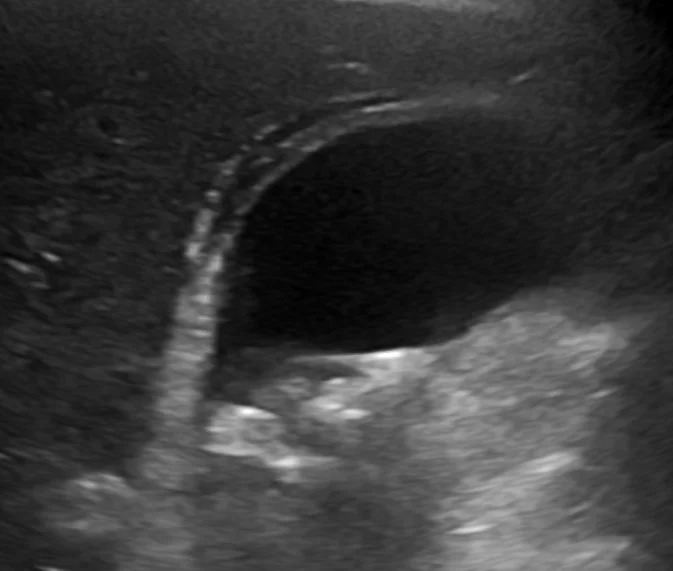
Long description
Ultrasound of a distended gallbladder with a thickened wall, stones with acoustic shadowing and a dark strand of fluid around the gallbladder. Such findings support the diagnosis when the clinical picture fits.

Ultrasound is the recommended first imaging test because it is available, safe, inexpensive and accurate for gallstones. [1] It should be performed in every patient with suspected cholecystitis. Key findings are:
- gallstones, often in the neck or cystic duct;
- gallbladder wall thickening beyond 3 mm (normal wall is 3 mm or less); [3]
- pericholecystic fluid or oedema;
- distension of the gallbladder;
- a positive sonographic Murphy's sign: tenderness directly over the visualised gallbladder with the ultrasound probe, with little or no tenderness elsewhere. [3]
Ultrasound has a reported sensitivity of about 81% and specificity of about 83% for acute cholecystitis, but performance varies between operators and departments. A negative or equivocal ultrasound never excludes the diagnosis when clinical suspicion is strong. [1]
- HIDA scan (hepatobiliary scintigraphy). The patient is given a technetium-labelled iminodiacetic acid derivative that is taken up by hepatocytes and excreted into bile. The gallbladder is usually seen within about 30 to 60 minutes; if it is not, delayed imaging up to 3 to 4 hours (or morphine augmentation) is used, and persistent non-visualisation means the cystic duct is obstructed so tracer never enters the gallbladder. [3] A HIDA scan has the highest sensitivity and specificity of the imaging modalities for acute cholecystitis and is the test of choice when the clinical picture is strong but ultrasound is equivocal. [1]
- CT of the abdomen. CT is less accurate than HIDA for the primary diagnosis and is not the first test. It is valuable for complications: suspected gangrenous or emphysematous cholecystitis, perforation, abscess, Mirizzi syndrome and other causes of acute abdomen. It is also useful in patients who are post-operative or have altered anatomy. Reported CT sensitivity for acute cholecystitis is around 85%, with specificity near 100%. [3]
- MRCP (magnetic resonance cholangiopancreatography). MRCP is as accurate as abdominal ultrasound for the gallbladder and is excellent for the bile ducts. It is used for suspected common bile duct stones, Mirizzi syndrome and when endoscopic access is planned.
Common bile duct stones accompany acute cholecystitis in roughly 5 to 15% of cases. LFTs and ultrasound can screen for them; patients at high or intermediate risk benefit from MRCP, endoscopic ultrasound or intra-operative cholangiography, and stones are removed endoscopically or at surgery. [1]
The Tokyo Guidelines 2018 (TG18) define diagnostic criteria and a three-grade severity classification that many hospitals use to guide management. [2][5]
Three groups of findings are combined:
- A. Local signs of inflammation: Murphy sign, or right upper quadrant mass, pain or tenderness.
- B. Systemic signs of inflammation: fever, raised C-reactive protein, or raised white cell count.
- C. Imaging findings: imaging characteristic of acute cholecystitis.
A suspected diagnosis is one item from A plus one item from B. A definite diagnosis is one item from A plus one item from B plus C. [2]
| Grade | Criteria |
|---|---|
| Grade I (mild) | Does not meet the criteria for grade II or grade III acute cholecystitis |
| Grade II (moderate) | Associated with at least one of: white cell count greater than 18,000 per mm3; palpable tender mass in the right upper quadrant; duration of symptoms more than 72 hours; marked local inflammation (gangrenous, emphysematous or purulent cholecystitis, pericholecystic or hepatic abscess, or biliary peritonitis) |
| Grade III (severe) | Associated with dysfunction of one or more organ systems: cardiovascular (hypotension requiring dopamine at least 5 micrograms per kg per minute, or any dose of norepinephrine); neurological (reduced level of consciousness); respiratory (PaO2/FiO2 ratio under 300); renal (oliguria or creatinine above 2.0 mg per dL, about 177 micromol per L); hepatic (INR above 1.5); haematological (platelet count below 100,000 per mm3) |
TG18 itself bases treatment decisions on severity together with an age-adjusted Charlson comorbidity score and the ASA physical status: early cholecystectomy is offered to fitter patients with grade I or II disease, while patients with severe grade III disease are stabilised first. [5]
Approach to suspected acute cholecystitis
- Right upper quadrant pain lasting more than 6 hours with fever or leucocytosisNausea, vomiting, or Murphy sign support the diagnosis
- Clinical assessment plus blood testsWhite cell count, CRP, liver tests, amylase or lipase
- Question: Abdominal ultrasound
Stones plus wall thickening, pericholecystic fluid, or sonographic Murphy sign
- Definite acute cholecystitis by Tokyo criteriaGrade the severity (grade I, II or III)
- Grade severity and surgical riskUse TG18 severity plus age-adjusted Charlson score and ASA status
- Acute treatmentNil by mouth, IV fluids, analgesia, IV antibiotics covering Gram-negative enterics and anaerobes
- Definitive careEarly laparoscopic cholecystectomy when feasible; percutaneous cholecystostomy or delayed surgery for high-risk patients
Normal or equivocal ultrasound, high clinical suspicion
- HIDA scanNon-visualisation of the gallbladder confirms cystic duct obstruction
- Or MRCP, endoscopic ultrasound, or CT for bile duct stones or complicationsChoose by local availability and suspected complication
- Grade and treat as above
Alternative diagnosis or low suspicion
- Treat the identified conditionCholangitis, pancreatitis, perforated ulcer, pneumonia, hepatitis, myocardial infarction, renal colic
While the diagnosis is being confirmed:
- Nil by mouth. This rests the gut and prepares for possible surgery.
- Intravenous fluids to correct dehydration from vomiting and to maintain urine output and perfusion.
- Analgesia, with paracetamol or an anti-inflammatory if not contraindicated, and opioids when needed. Non-steroidal anti-inflammatory drugs reduce pain and may lower the inflammatory response; they are avoided in renal impairment, gastric ulcer disease and pregnancy.
- Antiemetics.
- Address precipitants and complications. Review fluids, oxygenation, electrolytes and clotting, particularly in older patients. [1]
- Early surgical referral. The decision about surgery is made with the surgical team from the outset, not after a failed trial of conservative treatment.
Antibiotics are started promptly when infection is suspected, because the disease is bacterial in most patients. The choice should match the likely organisms (Gram-negative aerobes such as Escherichia coli and Klebsiella pneumoniae, and anaerobes such as Bacteroides fragilis) and local patterns of resistance. [1]
- Uncomplicated (calculous) disease after source control. For uncomplicated acute cholecystitis the World Society of Emergency Surgery recommends not giving routine postoperative antibiotics once cholecystectomy has controlled the infection focus. [1] Regimens otherwise follow local policy and the patient's risk profile.
- Community-acquired infection. Options recommended in standard guidance include a second or third generation cephalosporin such as cefuroxime or ceftriaxone, ampicillin-sulbactam, piperacillin-tazobactam, or a fluoroquinolone plus metronidazole, depending on allergy and resistance. [2][4]
- Severe, high-risk or immunocompromised patients. Broader regimens such as a carbapenem or piperacillin-tazobactam, each combined with metronidazole, are appropriate; vancomycin is added for healthcare-associated infection. [4]
- Enterococci are not routinely covered in community-acquired infection; coverage is added for immunosuppressed patients such as transplant recipients. [1]
- Duration. Four to seven days is typical. If source control is achieved, stopping four to five days later is reasonable. [4]
- Dosing example from a current trial protocol. Intravenous cefuroxime 1.5 g every 8 hours, ciprofloxacin 400 mg every 12 hours for penicillin allergy, or ertapenem 1 g every 24 hours for ESBL carriage, with postoperative continuation guided by the findings at surgery. [2]
- Postoperative discontinuation. Antibiotics can usually be stopped after surgery in uncomplicated disease unless the gallbladder was gangrenous, perforated or empysematous. [2]
The standard treatment is laparoscopic cholecystectomy. [1]
- First-line. Laparoscopic cholecystectomy is the recommended first-line treatment for patients with acute cholecystitis who are fit for surgery. [1]
- Timing. Early laparoscopic cholecystectomy performed as soon as possible, within 7 days of hospital admission and within 10 days of symptom onset, is recommended in the presence of adequate surgical expertise. [1] It shortens hospital stay and has lower serious adverse event rates than intermediate or delayed surgery. Early surgery should be performed by surgeons experienced in difficult gallbladders.
- Conversion and bail-out. Conversion to open surgery is recommended for severe inflammation, adhesions, bleeding from Calot's triangle or suspected bile duct injury. If a safe critical view of safety cannot be obtained, subtotal cholecystectomy is a safe bail-out. [1]
- Delayed surgery. If early cholecystectomy cannot be performed, delayed laparoscopic cholecystectomy is usually carried out after 6 weeks, once perioperative risk has been reduced. [1]
- Non-operative management. Observation with antibiotics and supportive care is considered for patients who refuse surgery or are not suitable for surgery, but recurrent gallstone disease is common. [1]
- High-risk patients. Randomised data (the CHOCOLATE trial) show that immediate laparoscopic cholecystectomy is superior to percutaneous drainage in high-risk patients. [1]
- Percutaneous (or endoscopic) gallbladder drainage. This is recommended for patients who are not suitable for surgery, converting a septic patient into a non-septic one. [1] Percutaneous transhepatic drainage, endoscopic transpapillary drainage and ultrasound-guided transmural drainage with a lumen-apposing metal stent are options depending on local expertise. [1][4]
- Older than 80 years. Early laparoscopic cholecystectomy is supported even in patients over 80, with attention to frailty and close postoperative monitoring. [1]
- Cirrhosis. Laparoscopic cholecystectomy is recommended for Child A and B cirrhosis; child C or uncompensated cirrhosis is more controversial and often managed conservatively unless cholecystitis is refractory. [1]
- Pregnancy. The same diagnostic tools apply, although leucocytosis and Murphy sign can be difficult to interpret late in pregnancy. Surgery is preferred as first-line when feasible to avoid complications and drug toxicity; cholecystectomy is safe in any trimester when needed. [1]
- Gallbladder in an immobile, high-risk patient. For patients never likely to undergo surgery, endoscopic ultrasound-guided drainage with a lumen-apposing metal stent can avoid a percutaneous drain and its maintenance. [4]
Acute cholecystitis can progress locally and systemically if untreated:
- Gangrenous cholecystitis. Necrosis of the gallbladder wall from sustained ischaemia, more common in older and diabetic patients and in prolonged obstruction. It requires urgent surgery.
- Perforation and biliary peritonitis. From gangrene. Can be free (general peritonitis) or contained (pericholecystic or hepatic abscess). Mortality rises steeply.
- Gallbladder empyema (suppurative cholecystitis). Pus in the gallbladder following bacterial superinfection, presenting with rigors and sepsis.
- Emphysematous cholecystitis. Gas in the wall or lumen from gas-forming organisms infecting an ischaemic gallbladder. Diabetes mellitus with hyperglycaemia is a major risk factor. Ultrasound shows hyperechoic foci with "dirty shadowing"; CT is used when images are unclear. It is an emergency needing intravenous antibiotics and surgery. [3]
- Cholecystoenteric fistula and gallstone ileus. Chronic inflammation can erode into duodenum or colon, creating a fistula. A large stone may then pass and obstruct the small bowel, usually at the terminal ileum. Classically the patient has small bowel obstruction without prior abdominal surgery, sometimes with pneumobilia or a stone seen on imaging. Treatment is surgical.
- Mirizzi syndrome. A stone becomes impacted in the cystic duct or gallbladder neck and compresses the adjacent common hepatic duct. It occurs in under 1% of patients with gallstones and presents with jaundice and cholestasis; preoperative recognition matters because it increases the risk of bile duct injury at surgery. [1][3]
- Bile duct injury and bile leak during surgery. The major surgical hazards (bile leak, bile duct injury in about 0.2 to 1.5%, major complications in 6 to 9%) are reduced by operating before severe inflammation develops, by obtaining the critical view of safety and by not hesitating to convert or perform subtotal cholecystectomy. [1]
- Subhepatic or hepatic abscess secondary to perforation.
With early laparoscopic cholecystectomy, recovery is usually rapid and hospital stay short. Delay increases operative difficulty, conversion, complications and recurrent admissions. In patients treated non-operatively, recurrent gallstone disease and repeated admissions are common. For critically ill patients with acalculous cholecystitis, outcome depends mainly on the underlying illness.
- Pregnancy. Surgery is the preferred first-line treatment when feasible; medical treatment carries a higher rate of miscarriage, threatened abortion and premature birth. Ultrasound, and MRCP without gadolinium if needed, are the imaging tools of choice. [1]
- Children. Acute cholecystitis is uncommon in children and usually associated with haemolytic disease, cystic fibrosis or congenital biliary anomalies; management follows the same surgical principles.
- Older adults. Outcomes are good with early surgery, even beyond 80 years, provided frailty is assessed and the patient is referred to a high-volume centre. [1][5]
- Immunocompromised patients. Acalculous disease and CMV-associated cholecystitis occur in HIV and transplant settings; broad-spectrum antibiotics and low thresholds for drainage are appropriate. [4]
Where imaging, endoscopy or surgery is limited:
- Clinical diagnosis matters. A patient with right upper quadrant pain lasting more than six hours, fever and a positive Murphy sign almost certainly has acute cholecystitis; blood tests for leucocytosis support it.
- Ultrasound is the most cost-effective test. Even a basic ultrasound can show stones and wall thickening. If ultrasound is unavailable, the clinical diagnosis should guide treatment.
- A HIDA scan is often unavailable. Empirical treatment and early referral for surgery are then appropriate; equivocal cases can be observed closely and transferred.
- Antibiotics and fluids come first. Start IV antibiotics covering enteric Gram-negative organisms and anaerobes, along with fluids and analgesia, while arranging surgery.
- Surgery is the cure. Only processes such as gangrene, perforation or empyema mandate emergency operation; otherwise early cholecystectomy within available resources is best. Where laparoscopy is not available, open cholecystectomy by an experienced surgeon is safe.
- Drainage as a bridge. Percutaneous cholecystostomy, even with simple ultrasound or fluoroscopic guidance, can stabilise a high-risk or septic patient and convert them to a surgical candidate later. [1][4]
- Referral. Transfer early for suspected perforation, empyema, severe sepsis, common bile duct stones or any patient who cannot be supported locally.
- Acute cholecystitis is usually acute inflammation caused by a gallstone impacted in the cystic duct or gallbladder neck; 10 to 15% of people with gallstones ever develop it, and it is the first presentation in a substantial minority of gallstone disease.
- Acalculous cholecystitis, 10% of cases, occurs in critically ill, burned, diabetic, post-surgical and TPN-fed patients and in HIV or CMV infection, with higher morbidity, mortality and perforation rates.
- Persistent right upper quadrant pain beyond six hours with fever and Murphy sign characterise the condition; Murphy sign is inspiratory arrest on palpation under the right costal margin.
- Ultrasound is first-line: stones, wall thicker than 3 mm, pericholecystic fluid and a sonographic Murphy sign. A HIDA scan confirms cystic duct obstruction when ultrasound is equivocal; CT and MRCP are for complications and bile duct problems.
- Tokyo Guidelines 2018 combine local signs, systemic inflammation and imaging into a suspected or definite diagnosis, and grade severity as I (mild), II (moderate) or III (severe, with organ dysfunction).
- Early laparoscopic cholecystectomy within 7 days of admission and 10 days of symptom onset is the standard treatment when expertise is available.
- High-risk patients are better served by early laparoscopic cholecystectomy than by percutaneous drainage; drainage is reserved for those unsuitable for surgery.
- Uncomplicated disease does not need routine postoperative antibiotics once source control is achieved; empiric antibiotics must cover enteric Gram-negative organisms and anaerobes.
- Complications include gangrenous, perforated and emphysematous cholecystitis, empyema, cholecystoenteric fistula with gallstone ileus, Mirizzi syndrome and bile duct injury.
- Common bile duct stones occur in 5 to 15% of cases and require imaging and removal.
- In low-resource settings, clinical and ultrasound diagnosis, early antibiotics and fluids, and referral for surgery or drainage save lives.
Answer each one in your head first, then open the answer.
A 58-year-old woman has constant right upper quadrant pain for 14 hours, fever to 38.2 degrees, nausea and a positive Murphy sign. Ultrasound shows gallstones, a 5 mm gallbladder wall and pericholecystic fluid. What is the diagnosis and what is the immediate plan?
Show answerHide answer
Acute calculous cholecystitis (definite by Tokyo criteria: local and systemic signs plus characteristic ultrasound). Plan: nil by mouth, intravenous fluids, analgesia, empiric antibiotics covering Gram-negative enterics and anaerobes, and early surgical referral for laparoscopic cholecystectomy within 7 days of admission.
What are the four moderate (grade II) criteria in the Tokyo Guidelines 2018?
Show answerHide answer
White cell count over 18,000 per mm3; palpable tender mass in the right upper quadrant; symptoms present for more than 72 hours; or marked local inflammation such as gangrenous, emphysematous or purulent cholecystitis, pericholecystic or hepatic abscess, or biliary peritonitis.
How can acute cholecystitis be distinguished from biliary colic?
Show answerHide answer
Biliary colic is transient cystic duct obstruction whose pain usually resolves within a few hours, without fever or leucocytosis. Acute cholecystitis is persistent inflammation with pain over six hours, fever, leucocytosis and ultrasound signs of inflammation.
A diabetic patient with cholecystitis has air in the gallbladder wall on ultrasound. What is this called, why is it dangerous, and what must be done?
Show answerHide answer
Emphysematous cholecystitis, usually caused by gas-forming organisms in an ischaemic gallbladder; diabetes is a major risk factor. It is an emergency because of high risk of gangrene and perforation. The patient needs intravenous antibiotics and urgent surgery, with a CT if imaging is unclear.
An 82-year-old with severe cholecystitis, confusion and creatinine 200 micromol per L. How is this graded, and how does it alter initial management?
Show answerHide answer
Grade III severe cholecystitis (renal and neurological dysfunction). The first step is stabilisation and organ support; surgery is considered once the patient is fit, with percutaneous drainage as an option for those not suitable for operation.
A critically ill ventilated patient has fever and worsening organ function, with ultrasound showing a distended gallbladder without stones and wall thickening. What is likely, and how should the finding be interpreted?
Show answerHide answer
Acalculous cholecystitis is likely, but ultrasound findings are common and non-specific in critically ill patients. The diagnosis is made by combining risk factors (trauma, burns, sepsis, TPN, fasting), clinical deterioration and imaging, looking for strongly supportive findings such as gas in the gallbladder on CT.
- Gallstone disease (cholelithiasis)
- Jaundice (hyperbilirubinaemia)
- Acute pancreatitis
- Cholangitis
- Intra-abdominal sepsis and antibiotic therapy
- Laparoscopic cholecystectomy
- Bile duct injury and its repair
- Pisano M, Allievi N, Gurusamy K, et al. 2020 World Society of Emergency Surgery updated guidelines for the diagnosis and treatment of acute calculus cholecystitis. World Journal of Emergency Surgery 2020;15:61. Accessed .
- Terho P, Sallinen V, Lampela H, et al. Preoperative antibiotics for mild or moderate acute cholecystitis (POMMAC): study protocol for a non-inferiority, open-label, randomized controlled trial. Trials 2026;27:617. Accessed .
- Ummadi S, Kroeger TP, Prasad A, et al. Acute Emergent Biliary Pathologies Identified Using Point-of-Care Ultrasound. Diagnostics 2026;16:3023. Accessed .
- Balmadrid B. Recent advances in management of acalculous cholecystitis. F1000Research 2018;7:F1000 Faculty Rev-1660. Accessed .
- Kobayashi Y, Hata S, Oikawa R, et al. Reappraisal of Real-World Management of Acute Cholecystitis in Elderly Patients Based on the Adherence to Tokyo Guidelines 2018 (TG18). Annals of Gastroenterological Surgery 2026;10:1250-1260. Accessed .
Figure credits
Educational content for medical students and doctors. It is not a substitute for clinical judgement or local guidelines.