Cholelithiasis and biliary colic
Gallstones: types, risks, biliary colic, complications, ultrasound diagnosis, treatment, and low-resource guidance.
- Gallstones are solid deposits that form in the gallbladder or, less often, in the bile ducts. They are mostly cholesterol, sometimes pigment.
- Most people who have gallstones never know it: about four out of five are symptom-free for life.
- The classic symptom is biliary colic, a steady severe upper abdominal pain that peaks within 15 minutes to 1 hour, lasts under 6 hours, and often spreads to the back or right shoulder.
- Ultrasound of the abdomen is the best first test: it detects gallbladder stones with about 95% sensitivity and specificity.
- Symptomatic stones are treated by laparoscopic removal of the gallbladder; asymptomatic stones are usually left alone, with exceptions for very large stones, a calcified (porcelain) gallbladder, and selected high-risk patients.
By the end of this page you should be able to:
- Describe how bile is made, concentrated and emptied, and how an imbalance of cholesterol, bile salts and bilirubin produces each type of stone.
- Recognise the three stone types and the conditions that favour each one.
- Weigh the risk factors, including why a young thin man can still have stones.
- Take a focused history of biliary colic and tell it from cholecystitis, pancreatitis and peptic ulcer disease.
- Interpret a gallbladder ultrasound report and know what normal liver tests add.
- Plan management for asymptomatic and symptomatic stones, including when to dissolve stones with ursodeoxycholic acid and when to offer prophylactic cholecystectomy.
- Work up a patient with postcholecystectomy pain.
- Outline the cancer risk of porcelain gallbladder and gallbladder polyps.
Cholelithiasis is the presence of one or more stones in the gallbladder. When a stone migrates and blocks the cystic duct temporarily, it produces biliary colic; prolonged or complete obstruction leads to cholecystitis, choledocholithiasis, cholangitis, gallstone pancreatitis, or gallstone ileus. The topics overlap, so this page concentrates on gallbladder stones and biliary colic and explains the whole spectrum in a single table.
A stone is not necessarily a disease. More than three out of four patients with stones have no symptoms at all and will die of something else. The job of the clinician is to decide which patients will benefit from treatment today and which should simply be reassured and left alone.
Bile is primarily water containing bile salts, cholesterol, phospholipids (mainly phosphatidylcholine, or lecithin), bilirubin and electrolytes. The liver secretes it continuously; between meals it is stored and concentrated in the gallbladder, which acts as a reservoir and concentrates bile several-fold by absorbing water and electrolytes.
When a meal reaches the duodenum, especially one containing fat and protein, the hormone cholecystokinin is released from duodenal I cells. It contracts the gallbladder and relaxes the sphincter of Oddi, so bile enters the duodenum to emulsify fat. The bile salts then form mixed micelles with cholesterol and phospholipids, keeping cholesterol dissolved.
After a meal, 95% of bile salts are reabsorbed in the terminal ileum and returned to the liver through the portal vein. This enterohepatic circulation means that the bowel is as important as the liver in keeping bile balanced: if the terminal ileum is diseased or resected, bile salts are lost, the liver compensates by synthesising less cholesterol excretion and more cholesterol secretion, and the bile becomes saturated.
Gallstones form when the delicate solubility balance breaks. Cholesterol stones need three things: supersaturation of bile with cholesterol, precipitation of solid cholesterol microcrystals, and growth of those crystals into stones, helped by mucin that acts as a scaffold. Supersaturation usually comes from excessive secretion of cholesterol, as in obesity or diabetes, but can come from reduced bile salt or lecithin secretion. Black and brown pigment stones form when there is too much bilirubin for the bile to hold in solution, or when infection and inflammation alter the bilirubin chemically so it precipitates.
Most stones grow 1 to 2 mm a year. They take 5 to 20 years to become large enough to cause symptoms, which is why stones are rarely a disease of young adults unless there is haemolysis, infection, or a strong family history.
Cholesterol stones account for more than 85% of gallstones in the Western world[1]. They are hard, often pale yellow to greenish, and usually multiple. Rich bile becomes cloudy with cholesterol microcrystals first; mucin then binds them into a stone. Obesity, diabetes, pregnancy, and rapid weight loss all push cholesterol secretion up or bile salt secretion down, so cholesterol stones are the stone of modern metabolic disease.
Black pigment stones are small and hard, made of calcium bilirubinate plus inorganic calcium salts such as calcium carbonate and calcium phosphate. They form in a sterile gallbladder when there is chronic haemolysis (sickle cell disease, hereditary spherocytosis, thalassaemia) that floods the bile with bilirubin, or when there is cirrhosis or advanced age[1]. Haemolytic stones can begin in childhood. They are usually many and small, and they may be dismissed on ultrasound as sludge or not seen at all because they are not always brightly echogenic.
Brown pigment stones are soft and greasy, made of bilirubinate and calcium palmitate or stearate. They form in ducts, not the gallbladder, during infection, inflammation or parasitic infestation such as liver flukes[1]. Because they form in response to an infected, poorly draining system, they are more common in East and Southeast Asia and in anyone with a biliary stricture or previous biliary surgery. Brown pigment stones are the ones most likely to be missed before surgery and to recur after cholecystectomy.
Many real-world stones are mixed. Biliary sludge (microlithiasis) is a gel of mucin, cholesterol microcrystals and calcium bilirubinate that acts as a precursor of stones. Sludge develops in pregnancy, during prolonged fasting, on total parenteral nutrition, during octreotide therapy and during rapid weight loss. Most sludge resolves when the trigger resolves; some of it hardens into stones.
Gallstones are the commonest gastrointestinal reason for hospital admission in Europe. About 20% of adults develop gallstones, and a little over 20% of those go on to develop symptoms or complications[6]. In the United States, prevalence is estimated at 14% of people over 20 years of age, and that figure doubled between 1988 and 2020[1]. In some northern European countries the prevalence reaches about 20%[1].
Prevalence rises with age and may plateau in the sixth decade, and women are affected more often than men[1]. Genetic factors matter: twin studies and population studies attribute about a quarter of the total risk to heredity, with common variants in the cholesterol transporter ABCG8 as the leading contributors[6].
American Indian populations have by far the highest prevalence of gallstone disease of any ethnic group studied[6]. This explains why family history and ethnicity are asked about routinely.
The old medical-school shorthand is five F's: female, fat, fertile, forty, and fair. It is a useful memory aid but it is not a rule. Men develop stones, thin people develop stones, and stones in patients under 30 are not rare when there is an underlying cause.
- Female. Women are affected more often, mostly through oestrogen, which increases hepatic cholesterol secretion and reduces bile salt secretion. [1][6]
- Fat. Obesity, particularly central obesity, is a strong and dose-dependent risk factor for cholesterol stones. [1][6]
- Fertile. Pregnancy promotes stasis and cholesterol supersaturation; sludge and stones can appear in any trimester and often resolve postpartum.
- Forty. Prevalence rises steeply after the third decade and plateaus around the sixth.
- Fair. Fair skin does not itself cause stones; the link is with Northern European and especially American Indian ancestry, both genetically high-risk groups[6].
Oestrogen increases risk through pregnancy, the oral contraceptive pill, and hormone replacement therapy. All three raise biliary cholesterol secretion. Insulin resistance, diabetes, dyslipidaemia (especially low HDL and high triglycerides), and metabolic syndrome cluster with cholesterol stones[6]. The liver of a person with insulin resistance secretes more cholesterol into bile, which is why diabetes is a risk factor independent of body weight.
A diet high in refined carbohydrate and low in fibre, combined with physical inactivity, favours stones. Conversely, regular exercise and a high-fibre diet are protective[5]. Obesity contributes, but the rate of weight change matters as much as body weight: very low calorie diets, rapid weight loss and jejuno-ileal bypass-style surgery all precipitate stones because the gallbladder empties less often during fasting and the liver secretes more cholesterol during fat mobilisation.
Disease or resection of the terminal ileum (Crohn disease, ileal resection, coeliac disease) causes bile salt loss and cholesterol stone formation. Somatostatin analogues (octreotide) reduce gallbladder contractility and cause sludge. Ceftriaxone is excreted into bile and can precipitate as a calcium-ceftriaxone salt, producing reversible sludge or stones in children and adults on prolonged high-dose therapy. Total parenteral nutrition produces biliary stasis and sludge within weeks; a proportion of patients on long-term parenteral nutrition develop symptomatic stones.
Family history of gallstones is a recognised risk factor. Atherosclerotic heart disease, cirrhosis, and haemolytic disorders favour pigment stones. A calcified (porcelain) gallbladder, although uncommon, is a special case discussed below. In young adults with stones, think first of haemolysis, cystic fibrosis or ileal disease rather than lifestyle.
The most important number in this topic is how rarely an asymptomatic stone becomes a problem. About 1 to 4% of people with previously asymptomatic stones develop symptoms each year[1], and the long-term cumulative risk is roughly 10% at 5 years[1]. Most patients who do become symptomatic get biliary colic rather than a serious complication.
Once symptoms begin, they tend to recur: biliary pain returns in 20 to 40% of patients per year[1], and complications (cholecystitis, choledocholithiasis, cholangitis, pancreatitis) develop in 1 to 2% per year[1][5]. Symptomatic stones carry an annual complication rate of about 1 to 3%, whereas complications in asymptomatic stones occur in less than 0.3% a year[5].
Almost all stones that cause trouble do so within 5 years of becoming symptomatic; after a 5-year silence, the risk drops sharply. This is the rationale for offering cholecystectomy early to symptomatic patients and for observing the truly asymptomatic.
Biliary colic is not colicky in the true sense (the pain does not come in waves). It is a steady, intense pain that builds to a plateau. Typically it:
- begins in the right upper quadrant or epigastrium and may be poorly localised, especially in older adults and in people with diabetes;
- often radiates into the back, the right scapula or down the arm;
- begins suddenly, reaches full intensity within 15 minutes to 1 hour;
- stays at a steady severe level for usually under 6 hours, though it may last up to 12 hours in a small minority;
- eases over 30 to 90 minutes, leaving a dull ache;
- is brought on by eating, classically fatty meals, but equally by any meal or by nothing at all;
- is accompanied by nausea and sometimes vomiting, but not fever or rigors (those point to cholecystitis or cholangitis);
- leaves the patient feeling well between episodes.
Biliary colic does not cause dyspepsia, bloating or postprandial fullness on its own; those symptoms have the same prevalence in people with and without gallstones, and attributing them to stones is a common error. Fatty food is also an unreliable trigger, so the diagnosis rests on the pain pattern, not on what the patient ate.
On examination between attacks there is little to find beyond mild right upper quadrant tenderness. During an attack the patient may be restless and unable to get comfortable. Fever, rigors, tachycardia or peritonism indicate a complication and not simple colic.
- Pain lasting longer than 12 hours, especially with vomiting or fever: suspect acute cholecystitis or pancreatitis.
- Fever with rigors and jaundice: Charcot triad for ascending cholangitis.
- Severe epigastric pain with vomiting in a patient with known stones: gallstone pancreatitis.
- Colicky pain in an elderly patient followed by intestinal obstruction: gallstone ileus (a stone eroding through into the small bowel).
The table compares the six clinical syndromes that a single stone can produce. It is the single most useful revision table in the topic.
| Disorder | Definition and mechanism | Typical presentation | Laboratory findings | Diagnosis | Management |
|---|---|---|---|---|---|
| Cholelithiasis | Stone in the gallbladder; often asymptomatic | None, or biliary colic | Normal unless a complication develops | Ultrasound (stones, shadowing) | Observation if asymptomatic; laparoscopic cholecystectomy if symptomatic |
| Acute cholecystitis | Cystic duct obstruction longer than a few hours, leading to gallbladder inflammation; bacterial infection later | Constant RUQ pain, fever, positive Murphy sign | Raised white cell count and CRP; maybe mildly raised bilirubin or ALP | Ultrasound (thickened wall, pericholecystic fluid, stones); ASGE/Tokyo criteria | Fluids, analgesia, antibiotics for severe disease; early laparoscopic cholecystectomy within 24 hours |
| Choledocholithiasis | Stone in the common bile duct, passed from the gallbladder or formed in situ | RUQ or epigastric pain, jaundice, dark urine, pale stools; may be asymptomatic | Cholestatic pattern: raised bilirubin, ALP and GGT; ALT/AST raised | Ultrasound (dilated duct), then MRCP or EUS; ERCP if therapeutic | Bile duct clearance (ERCP or laparoscopic bile duct exploration) plus cholecystectomy |
| Acute cholangitis | Bacterial infection of an obstructed biliary tree | Fever, rigors, jaundice and pain (Charcot triad); possible hypotension and confusion (Reynolds pentad) | Raised bilirubin and ALP; high CRP and white cell count; positive blood cultures | Ultrasound and liver tests; MRCP/CT if needed; clinical diagnosis | Urgent antibiotics plus biliary decompression by ERCP, percutaneous transhepatic drainage or surgery |
| Gallstone pancreatitis | Stone temporarily obstructing the ampulla, triggering pancreatic inflammation | Severe epigastric pain radiating to the back, vomiting; mild or severe course | Raised amylase and lipase; bilirubin may be raised | Ultrasound, liver tests, CT if severe | Supportive care; urgent ERCP within 24 hours if cholangitis; cholecystectomy before discharge or within 2 weeks |
| Gallstone ileus | Stone erodes through the gallbladder wall into the bowel and obstructs, usually at the terminal ileum | Intermittent small-bowel obstruction in an older adult; may be preceded by unexplained pain | Variable; renal and electrolyte abnormalities from vomiting | CT showing ectopic stone and pneumobilia | Resuscitation; enterotomy to remove the stone; cholecystectomy and fistula repair sometimes delayed |
Diagnosis rests on clinical suspicion confirmed by imaging. Several points from above and below guide the sequence.
Figure 1Ultrasonography of sludge and gallstones
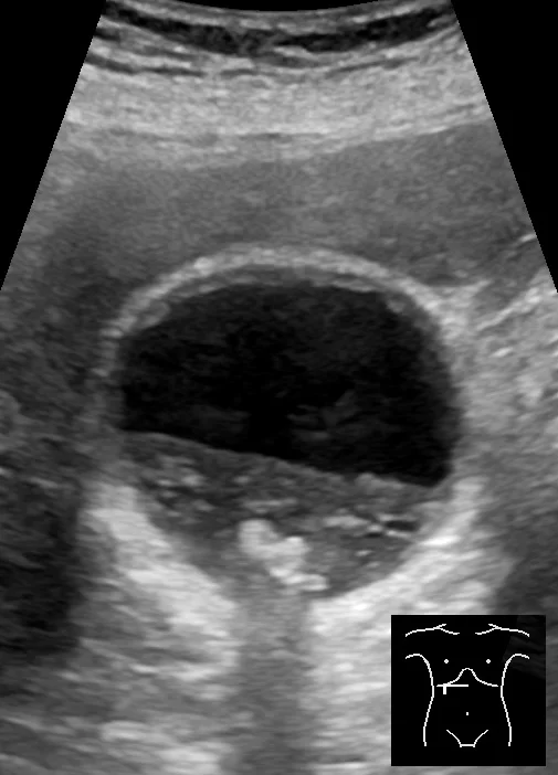
Long description
Abdominal ultrasonography of an 83-year-old man who had biliary colic two weeks prior, showing a gallbladder wall about 3 mm thick, biliary sludge, and gallstones that create acoustic shadowing, described as gallstones without current cholecystitis.

Transabdominal ultrasound is the initial and best test for gallbladder stones: sensitivity and specificity are about 95%[1]. A stone appears as a mobile echogenic focus with posterior acoustic shadowing, and it should move when the patient changes position; this mobility distinguishes it from polyps and from the wall. Ultrasound also shows wall thickness, pericholecystic fluid, and the calibre of the bile duct, and it detects sludge and polyps. It is cheap, safe, available at the bedside, and involves no radiation. A negative scan does not completely exclude tiny (under 3 mm) or radiolucent stones; endoscopic ultrasound detects stones under 3 mm when the first scan is equivocal.
Routine blood tests are usually normal in uncomplicated cholelithiasis[1]. The point of ordering them is not to find the stone but to catch a complication:
- Full blood count and CRP: raised white cells support cholecystitis or cholangitis.
- Liver function tests (bilirubin, ALP, GGT, ALT/AST): a cholestatic rise suggests choledocholithiasis; mild transaminase rises are common during an attack of colic even without duct stones.
- Amylase and lipase: raised in gallstone pancreatitis.
- Renal function and electrolytes in vomiting patients.
If the ultrasound shows no duct stone but the common bile duct is dilated or liver tests are abnormal, NICE guidance advises MRCP[4]; endoscopic ultrasound is used when MRCP is inconclusive or unavailable[4].
CT is poor at seeing non-calcified stones but is excellent for complications (abscess, perforation, gallbladder cancer, pancreatitis severity) and for gallstone ileus. MRI/MRCP is the best non-invasive test for the bile duct. Cholescintigraphy (HIDA) assesses cystic duct patency and is used for acute cholecystitis when ultrasound is equivocal. Plain abdominal radiographs show only the 10 to 15% of stones that are calcified[1], but sometimes show air in the biliary tree.
Approach to a patient with suspected gallstone disease
- History and examinationBiliary colic pattern, fever, jaundice, peritonism; red flags
- Ultrasound abdomen + bloodsFBC, CRP, LFT, amylase/lipase if pain suggests a complication
- Question: Stone seen in gallbladder?
No
- Look for other causesPeptic ulcer disease, reflux, pancreatitis, liver disease
Yes
- Question: Symptoms?
No (asymptomatic)
- Observe; reassure (with exceptions)Prophylactic cholecystectomy if stone >3 cm, porcelain, or bariatric surgery context
Biliary colic
- Analgesia now (NSAIDs first)Then laparoscopic cholecystectomy when well
Acute cholecystitis or complications
- Admit: fluids, analgesia, antibiotics if severeEarly laparoscopic cholecystectomy, usually within 24 hours
- Question: Is choledocholithiasis suspected?
Yes: dilated duct or abnormal LFT, jaundice
- MRCP or EUS to confirmThen ERCP with clearance, followed by cholecystectomy within 72 hours
No
- Proceed with management of the gallbladder stoneAs above
Non-steroidal anti-inflammatory drugs are first-line for biliary colic[5]. Spasmolytics or nitrates can be added and opioids used for severe pain. Antibiotics have no role in uncomplicated biliary colic; treat the pain and arrange definitive care.
The default for a stone found by accident is reassurance and observation. More than 80% of people never develop symptoms, and the annual complication rate of asymptomatic stones is under 0.3%[1][5]. NICE guidance puts the same principle in words: patients with asymptomatic gallbladder stones in a normal gallbladder and normal biliary tree need no treatment unless they develop symptoms[4].
Offer prophylactic cholecystectomy despite the absence of symptoms when any of the following apply:
- stone larger than 3 cm (tenfold higher risk of gallbladder cancer);
- porcelain (heavily calcified) gallbladder (controversial; risk up to 62% in historical series);
- gallbladder polyp over 1 cm (risk of cancer around 50%);
- patients undergoing bariatric surgery, when stones are already known, because rapid weight loss precipitates new stones;
- patients with sickle cell disease or other haemolytic disease, when prophylactic cholecystectomy avoids later emergency surgery;
- immunosuppressed or transplant patients, in whom elective removal is safer than emergency removal;
- incidental gallbladder carcinoma found on imaging or at surgery.
In porcelain gallbladder, opinion has shifted: several recent studies argue for careful observation in selected patients because a proportion of "porcelain" gallbladders turn out to be intramural calcification without increased cancer risk. The decision should be made with a surgeon and recorded.
For symptomatic gallbladder stones, laparoscopic cholecystectomy is the treatment of choice. It prevents further biliary colic and removes the risk of stones migrating into the duct. Conversion to open surgery occurs in 4 to 8% of cases[1], usually because the anatomy cannot be identified safely. Elective day-case surgery is appropriate for most patients. Gallbladder removal does not cause nutritional problems and does not require long-term dietary restriction; a few patients develop bile acid diarrhoea, which usually settles but may need cholestyramine.
In acute cholecystitis, early laparoscopic cholecystectomy within 24 hours has better outcomes than delayed surgery, with lower morbidity and shorter hospital stay. NICE recommends within 1 week of diagnosis[4]; German guidelines and the ACDC study support within 24 hours of admission[5]. Either way, the rule is the same: treat early rather than waiting for inflammation to settle.
Asymptomatic duct stones (choledocholithiasis) are cleared plus cholecystectomy even without symptoms, because they can precipitate cholangitis at any time[4]. Clearance is by ERCP or laparoscopic bile duct exploration, followed ideally by cholecystectomy within 72 hours.
UDCA is a bile acid that can dissolve tiny cholesterol stones under 0.5 cm by making bile less supersaturated, but the success rate for larger stones is low and stones recur in about 50% of patients within 5 years of dissolution[1]. Reserve UDCA for:
- patients with very small radiolucent cholesterol stones in a functioning gallbladder who decline or are unfit for surgery;
- prevention during rapid weight loss, for example after bariatric surgery or on a very low calorie diet, where a randomised trial showed a marked reduction in stone formation;
- selected inherited or recurrent stone formers (for example low-phospholipid-associated cholelithiasis, LPAC), where it can prevent new stones.
General pharmacological prevention with UDCA is not recommended for everyone with risk factors. It does not dissolve pigment stones. Where UDCA is used, agree monitoring and a stop point with the patient; imaging guides both.
Simultaneous prophylactic cholecystectomy is not advised for every bariatric patient. Up to 20% of patients after laparoscopic Roux-en-Y bypass develop biliary symptoms within 3 years, yet guidelines still favour observation plus UDCA for high-risk periods[5]. If stones are known beforehand, cholecystectomy can be performed at the same time. In pregnancy, symptomatic stones are treated surgically; laparoscopic cholecystectomy is safe in all trimesters when clinically necessary.
The complications of gallstones are the same as the table above. Two deserve emphasis here: ascending cholangitis can deteriorate from Charcot triad to Reynolds pentad (hypotension and confusion) within hours, and gallstone pancreatitis ranges from mild and self-limited to fatal. Both require urgent biliary decompression. About one third of patients managed conservatively for acute cholecystitis develop choledocholithiasis or pancreatitis within 2 years, which is why early cholecystectomy is recommended once the diagnosis is secure[5].
Postcholecystectomy syndrome is the persistence or development of abdominal pain and dyspepsia after gallbladder removal. It occurs in 5 to 47% of patients[3] and most often consists of vague dyspepsia, bloating or discomfort rather than true biliary colic. The gallbladder is gone, so the symptoms are rarely "genuine gallbladder pain".
Recognised causes include:
- retained or new bile duct stones;
- strictures;
- sphincter of Oddi dysfunction (functional or stenotic), which explains biliary pain in up to 30% of patients;
- papillary stenosis after instrumentation or passage of a stone;
- bile acid diarrhoea;
- extrabiliary disorders that were present before surgery but misattributed (gastro-oesophageal reflux disease, functional dyspepsia, irritable bowel syndrome, pancreatic disease);
- post-surgical adhesions.
Workup should be directed and stepwise:
- Take a careful history: is the pain true biliary colic (steady, lasting over 15 minutes, radiating, with nausea)? Most postcholecystectomy pain is not.
- Exclude extrabiliary causes clinically.
- Measure alkaline phosphatase, bilirubin, ALT, amylase and lipase when pain suggests biliary origin.
- Ultrasound of the abdomen to look for a dilated duct or residual stone.
- If biliary pain persists, cross-sectional imaging (CT or MRCP), or CT/MRCP to detect stones, strictures or leaks.
- ERCP with biliary manometry is the most sensitive test for sphincter of Oddi dysfunction, but it can induce pancreatitis in up to 30%, so it is reserved for patients with objective abnormalities; manometry shows raised biliary pressure during pain.
- Biliary scintigraphy can demonstrate delayed hilum-to-duodenum transit.
Treatment is cause-directed: bile acid binders for diarrhoea, a stepwise approach for functional disorders, and endoscopic sphincterotomy for objectively proven sphincter of Oddi dysfunction, especially papillary stenosis. Empirical sphincterotomy without objective findings is not supported by evidence.
A porcelain gallbladder is one whose wall is extensively calcified. Historically it was considered a strong indication for prophylactic cholecystectomy because of an association with cancer. More recent opinion is more cautious: extensive intramural calcification is only weakly associated with gallbladder carcinoma, and observation is acceptable in selected patients. Older teaching put the risk as high as 62% in some series; modern reviews consider that figure an overestimate[2][5]. Either way, the gallbladder still needs to be removed when symptomatic, and calcification itself is an indication to discuss surgery with a hepatobiliary surgeon rather than to operate automatically.
Gallbladder carcinoma is uncommon (worldwide incidence 1.4 per 100,000) but aggressive[2]. It is more common in older patients, in women, in people with gallstones, and in American Indian populations. Most patients (69 to over 95%) also have gallstones, and large stones over 3 cm increase risk roughly tenfold compared with small stones[5]. Median survival is about 6 months and 5-year survival about 5%; cure is possible only when the tumour is found early, often incidentally at cholecystectomy[2]. Manifestations range from an incidental finding at surgery to advanced disease with constant pain, weight loss and jaundice.
Polyps are usually asymptomatic mucosal projections found in about 5% of adults[2]. Most are under 10 mm and are cholesterol deposits (cholesterolosis); these need surveillance ultrasound with a frequency based on size and risk factors. Polyps over 10 mm carry a roughly 50% risk of neoplasia and should be removed by cholecystectomy[5]. Other, much rarer benign polyps include adenomas and inflammatory polyps.
Acute cholecystitis, the most frequent complication, is usually diagnosed on three of four clinical findings (right upper quadrant pain, Murphy sign, leukocytosis, fever) plus either cholelithiasis or ultrasound signs of inflammation; ultrasound then confirms the diagnosis. Scores and formal criteria exist (Tokyo guidelines), but this page deliberately does not reproduce staging systems; consult the local acute-abdomen protocol.
- Pregnancy. Symptomatic cholecystectomy should be considered; laparoscopic cholecystectomy is safe in all trimesters when indicated.
- Children and adolescents. Stones suggest an underlying cause: haemolytic disease, cystic fibrosis, ileal resection or total parenteral nutrition.
- Older adults. Symptoms are often atypical (poorly localised pain), operative risk is higher, and gallbladder cancer is more common.
- People with sickle cell disease. Stones often develop in childhood; prophylactic cholecystectomy is common practice to avoid emergency surgery during a crisis.
- Patients on total parenteral nutrition or receiving octreotide. Maintain enteral feeding where possible; ultrasound surveillance; UDCA may prevent sludge.
- A plain bedside or clinic ultrasound is the single most useful tool: it finds stones, sludge and duct dilatation, and it costs little. Knowing how to scan for gallstones and how to recognise free fluid is a high-value skill.
- Where MRCP, ERCP and endoscopic ultrasound are not available, refer patients with suspected choledocholithiasis or cholangitis early to a centre that can clear the duct, rather than waiting until they are septic.
- UDCA for dissolution is expensive and requires months of therapy and follow-up; for most symptomatic patients surgery is the better use of resources.
- Delayed presentation is common. Explain plainly that biliary pain is not life-threatening but recurrent, and that earlier elective surgery is safer and cheaper than emergency surgery.
- In rural settings, transport and consent logistics, not just the operation, determine outcome. Family education about warning symptoms (fever, jaundice, prolonged pain) should accompany any discharge.
The European Association for the Study of the Liver published clinical practice guidelines on the prevention, diagnosis and treatment of gallstones in 2016 (J Hepatol 2016;65:146-181)[7]. The points on which most guidelines (EASL 2016, NICE CG188, the German guidelines, WSES/Tokyo) agree are:
- asymptomatic stones in a normal gallbladder and biliary tree should be left alone;
- symptomatic gallbladder stones should be treated with laparoscopic cholecystectomy;
- ultrasound is the first-line diagnostic test;
- ERCP is for therapy (clearing the duct), not a diagnostic fishing trip;
- early cholecystectomy is preferred for acute cholecystitis;
- ERCP plus cholecystectomy is preferred for choledocholithiasis;
- prophylactic cholecystectomy is reserved for large stones, polyps over 1 cm, porcelain gallbladder, selected bariatric patients and high-risk (haemolytic, immunosuppressed) patients.
Where guidelines differ, the most relevant for day-to-day practice is the timing of surgery in acute cholecystitis (within 24 hours versus within 1 week) and whether to drain the gallbladder percutaneously before surgery in very ill patients. State the local protocol on the ward.
- A stone is not necessarily a disease: more than 80% of people with gallstones are symptom-free for life.
- Gallstones form from an imbalance between cholesterol, bile salts, phospholipids and bilirubin in gallbladder bile; cholesterol stones dominate in the West.
- Black pigment stones come from excess bilirubin (haemolysis, cirrhosis); brown pigment stones from infection and inflammation of the biliary tree.
- The classic risk profile is female, fat, fertile, forty and fair-skinned Northern European or American Indian ancestry, but ileal disease, TPN, octreotide, ceftriaxone, rapid weight loss and family history are equally important.
- Only about 1 to 4% of asymptomatic people become symptomatic each year; pain returns in 20 to 40% a year after the first attack; complications occur in 1 to 2% a year.
- Biliary colic is a steady, severe pain that builds within an hour, lasts under 6 hours, and radiates to the back or right shoulder; it is not truly colicky and is not triggered only by fat.
- Ultrasound detects gallbladder stones with about 95% sensitivity and specificity; labs are usually normal in uncomplicated cholelithiasis.
- Asymptomatic stones need no treatment unless they are over 3 cm, the gallbladder is porcelain, there is a polyp over 1 cm, or the patient is high-risk.
- Symptomatic stones are treated with laparoscopic cholecystectomy; UDCA dissolves only tiny cholesterol stones and is best used for prevention during rapid weight loss.
- Porcelain gallbladder is now considered only weakly associated with cancer; counsel and decide with a surgeon.
- Postcholecystectomy pain is common and usually non-biliary; retained stones, strictures, sphincter of Oddi dysfunction and extrabiliary causes must be excluded stepwise.
- Gallbladder polyps over 10 mm carry substantial cancer risk and should be removed.
Answer each one in your head first, then open the answer.
A 42-year-old woman has three months of identical episodes of severe epigastric pain lasting 2 hours, radiating to the right scapula, beginning 30 minutes after dinner. She has no fever. What is the most likely diagnosis and the best first test?
Show answerHide answer
Biliary colic from gallstones. The pain is steady and severe, reaches a plateau, radiates to the scapula, lasts under 6 hours and is postprandial. The best first test is an abdominal ultrasound, which will show hyperechoic stones with posterior acoustic shadowing that move when the patient changes position.
A 28-year-old man with sickle cell disease has a routine ultrasound that shows multiple small stones. He has no pain. Does he need surgery?
Show answerHide answer
Usually yes, a prophylactic laparoscopic cholecystectomy is considered. Haemolytic stones are black pigment stones, they begin in childhood, they generate repeated episodes once symptomatic, and elective surgery in a stable patient is far safer than emergency surgery during a crisis.
Why are people on prolonged total parenteral nutrition or octreotide at risk of gallstones, and what kind of stones or sludge do they form?
Show answerHide answer
Biliary stasis and increased cholesterol secretion produce sludge and cholesterol stones (octreotide) or rapidly appearing cholesterol-rich sludge (TPN). Keeping some enteral feeding and considering ursodeoxycholic acid can reduce the risk.
In the table of gallstone disorders, what distinguishes biliary colic from acute cholecystitis, and which investigation best separates them?
Show answerHide answer
Biliary colic is episodic pain under 6 hours with normal temperature and usually normal bloods; acute cholecystitis is constant pain over 12 hours with fever, leukocytosis and often a thickened gallbladder wall on ultrasound. Ultrasound is the key test.
A 35-year-old man has had laparoscopic cholecystectomy for stones. He returns 6 months later with identical epigastric pain. List the main causes and the first-line investigations.
Show answerHide answer
Causes: retained bile duct stone, sphincter of Oddi dysfunction, papillary stenosis, and extrabiliary disorders such as reflux or functional dyspepsia. First-line tests: LFT (ALP, bilirubin, ALT), amylase, lipase, and abdominal ultrasound. If pain is biliary and recurrent, MRCP and possibly ERCP with manometry follow.
A calcified (porcelain) gallbladder is found incidentally. Should it always be removed?
Show answerHide answer
No longer automatically. Historical data linked porcelain gallbladder to cancer risk up to 62%, but modern studies show the association is weaker, and intramural calcification alone is not strong evidence of cancer. Discuss elective removal with a hepatobiliary surgeon, and remove it if there are symptoms, large stones, polyps, or a growing lesion.
A 50-year-old man on a prolonged course of high-dose ceftriaxone develops right upper quadrant pain. Ultrasound shows echogenic sludge. What is the likely cause and expected course?
Show answerHide answer
Ceftriaxone precipitates with calcium in bile and can form sludge or reversible stones. Stop or reduce the drug where possible, and the sludge usually resolves; operate only if complications develop.
Cholecystitis; choledocholithiasis and cholangitis; acute pancreatitis; gallbladder cancer; sphincter of Oddi dysfunction.
(References are listed in the front matter; each is cited in the text by its number.)
- Saiman Y. Cholelithiasis (Full Review, Oct 2025). The Merck Manual, Professional Version. Rahway (NJ): Merck & Co.. Accessed .
- Saiman Y. Tumors of the Gallbladder and Bile Ducts (Full Review, Oct 2025). The Merck Manual, Professional Version. Rahway (NJ): Merck & Co.. Accessed .
- Saiman Y. Postcholecystectomy Syndrome (Full Review, Oct 2025). The Merck Manual, Professional Version. Rahway (NJ): Merck & Co.. Accessed .
- National Institute for Health and Care Excellence (NICE). Gallstone disease: diagnosis and management (CG188). NICE clinical guideline. Published 2014; last reviewed 2018.. Accessed .
- Gutt C, Schläfer S, Lammert F. The Treatment of Gallstone Disease. Dtsch Arztebl Int 2020;117(9):148-158. doi:10.3238/arztebl.2020.0148. Accessed .
- Lammert F, Gurusamy K, Ko CW, et al. Gallstones. Nat Rev Dis Primers. 2016;2:16024. doi:10.1038/nrdp.2016.24. Accessed .
- European Association for the Study of the Liver (EASL). EASL Clinical Practice Guidelines on the prevention, diagnosis and treatment of gallstones. J Hepatol. 2016;65(1):146-181. doi:10.1016/j.jhep.2016.03.005. Accessed .
Figure credits
Educational content for medical students and doctors. It is not a substitute for clinical judgement or local guidelines.