Acute pancreatitis
Acute pancreatitis: causes from gallstones to alcohol and drugs, Revised Atlanta diagnosis and severity, fluids, feeding, ERCP, and complications.
- Acute pancreatitis is an acute inflammatory injury of the pancreas in which digestive enzymes are activated inside the gland and start to digest it.
- The two commonest causes are gallstones (about 40 to 70% of cases) and alcohol (about 25 to 35% of cases). [4]
- Inflammation of the pancreas triggers a systemic inflammatory response, so the disease ranges from mild and self-limited to multi-organ failure and death.
- The diagnosis needs two of three things: typical epigastric pain going through to the back, lipase or amylase over 3 times the upper limit, or characteristic imaging. [1]
- Treatment is supportive: moderate intravenous fluid with lactated Ringer solution, good analgesia, early feeding, no prophylactic antibiotics, and drainage of infected necrosis by a step-up approach.
By the end of this page you should be able to:
- Explain how trypsinogen activation begins inside the acinar cell and drives autodigestion and the systemic inflammatory response.
- List the causes of acute pancreatitis, including the rare ones, and group them by mechanism.
- Apply the Revised Atlanta diagnostic criteria and distinguish interstitial from necrotising pancreatitis.
- Grade severity as mild, moderately severe or severe, and recognise organ failure using the modified Marshall criteria.
- Plan initial management: fluids, analgesia, nutrition, antibiotics, ERCP and cholecystectomy, with the timing each guideline gives.
- Describe the local collections, infected necrosis and step-up drainage.
- Outline prognosis, recurrent and chronic pancreatitis, and low-resource practice.
The pancreas digests fat, protein and carbohydrate for the rest of the body. In acute pancreatitis it begins to digest itself. The process starts inside the acinar cell, spreads to the whole gland, and activates circulating inflammatory signals that can injure lung, kidney and the circulation. Most patients recover within a week. About 1 in 5 develop a complicated course with necrosis or organ failure. [3]
This page covers the adult disease in full: definition, causes, pathophysiology, diagnosis, examination, severity, management, complications, prognosis, special situations and low-resource care. It is written for a first-time reader; abbreviations are expanded at first use.
Acute pancreatitis is acute inflammation of the pancreatic parenchyma and, frequently, the peripancreatic tissues, with elevation of pancreatic enzymes and abdominal pain. It is not the same as chronic pancreatitis, which is a fibrotic, atrophic end-stage disease; acute pancreatitis can be the first step toward it.
By type, acute pancreatitis is:
- Interstitial (oedematous) pancreatitis: diffuse enlargement of the gland with homogeneous enhancement on contrast-enhanced CT. It is the commoner form and usually resolves within the first week. [1]
- Necrotising pancreatitis: necrosis of pancreatic parenchyma, peripancreatic tissue, or both. It occurs in about 5 to 10% of patients and runs a longer, more severe course. [4]
Severity is graded separately (see below) as mild, moderately severe or severe.
Acute pancreatitis is one of the commonest gastrointestinal conditions requiring hospital admission. In reviewing series, overall incidence ranges from about 3.4 to 73.4 per 100 000 population per year in the ACG 2024 guideline and from about 10 to 90 per 100 000 in the Polish guideline, averaging about 34 per 100 000 where reported. [2][8] Incidence is rising, driven by obesity, gallstones and alcohol. Published global estimates put male incidence slightly higher than female. Overall mortality is about 5 to 10%, depending on severity. [8]
Gallstones account for about 40 to 70% of cases and alcohol for about 25 to 35%. [4] The rest is a long list of less common causes. The disease is rare in children, where trauma and infections feature more.
Any trigger that injures the acinar cell or obstructs the main pancreatic duct can release active enzymes into the interstitium and start autodigestion. The commonest mechanisms are duct obstruction by a stone, toxic metabolites of alcohol, and sustained hypertriglyceridaemia.
Gallstones are the commonest cause. [4] The mechanism is not fully settled. A stone (or oedema from a stone that has already passed) transiently obstructs the ampulla, raising pancreatic duct pressure and triggering enzyme activation. Bile acid reflux on to acinar cells is an alternative or additional mechanism. [4] Small stones under 5 mm and microlithiasis (biliary sludge) are enough to cause pancreatitis. In gallstone pancreatitis the pain often begins suddenly. [4]
Alcohol is the second commonest cause. Risk rises with dose and with years of intake, but there is wide individual variation, and some susceptible patients develop pancreatitis after a short heavy intake. [4] Acinar cells metabolise alcohol to toxic metabolites by oxidative and non-oxidative pathways; these destabilise zymogen and lysosomal granules, produce calcium overload, and activate pancreatic stellate cells. [4] Fewer than 5% of heavy drinkers ever develop pancreatitis, so cofactors matter. [4] About 10 to 30% of patients with alcoholic pancreatitis have recurrent attacks and about 35% of those progress to chronic pancreatitis. [4]
- Hypertriglyceridaemia: about 1 to 4% of all cases in the Merck manual and about 5% of all cases in ACG 2024 (higher in pregnancy). Risk is highest when fasting triglycerides are 1000 mg per dL (about 11.3 mmol per L) or higher. [4][2][8]
- Hypercalcaemia, including primary hyperparathyroidism. [4]
- Obesity and metabolic syndrome, mediated partly by visceral adipose tissue inflammation. [8]
Drugs are a recognised but uncommon cause. Examples supported by current reviews are azathioprine, 6-mercaptopurine, valproate, thiazides and other sulphonamides, furosemide, oestrogens (often with high lipid levels), tetracyclines, ACE inhibitors, asparaginase, pentamidine, GLP-1 receptor agonists and DPP-4 inhibitors, and 5-aminosalicylic acid. [4][8] Most drug-event associations are low quality evidence, and drug-induced pancreatitis is usually mild. [3]
- Post-ERCP pancreatitis occurs in about 5 to 8% of ERCPs, and up to about 16.5% in inpatient series. It is usually mild to moderate. [4]
- Abdominal trauma, both blunt and penetrating.
- Pancreas divisum, choledochal cyst, sphincter of Oddi stenosis.
- Pancreatic and periampullary tumours are important to exclude in patients over 40 with no clear cause. [2][8]
- Atheroembolism and ischaemia from hypotension.
Viruses include mumps, coxsackie B, cytomegalovirus, hepatitis A and E, Epstein-Barr virus, varicella, rubella and HIV. [4][8] Bacterial and parasitic causes include Campylobacter jejuni, Legionella, leptospirosis, mycobacterial disease, Ascaris lumbricoides and Cryptosporidium. [8] Most infection-related pancreatitis is mild.
Autoimmune pancreatitis, most often IgG4-related disease in older men, presents with painless jaundice or a pancreatic mass and responds to steroids. Autoimmune vasculitis (systemic lupus erythematosus, Sjogren syndrome, polyarteritis) can also be causative. [8]
Mutations in PRSS1 (cationic trypsinogen), SPINK1 (trypsin inhibitor) and CFTR (cystic fibrosis transmembrane conductance regulator) are the best known. An autosomal dominant PRSS1 mutation causes pancreatitis in about 80% of carriers. [4] CFTR mutation increases risk of recurrent acute and chronic pancreatitis. [4] Hereditary pancreatitis usually presents in childhood or adolescence.
Methanol, cigarette smoking, pregnancy and post-renal transplant states have all been reported. [4] Scorpion sting (Tityus trinitatis in the Caribbean) is a recognised but rare cause in endemic areas. If no cause is found after a full work-up (repeat ultrasound, MRCP or endoscopic ultrasound for stones, triglycerides, autoimmune and tumour screening), the case is called idiopathic; this is the label for up to about 10% of patients. [8]
A traditional mnemonic groups causes as Idiopathic, Gallstones, Ethanol, Trauma, Steroids, Mumps (and other infections), Autoimmune, Scorpion sting, Hyperlipidaemia and Hypercalcaemia, ERCP and Drugs. It is useful as a memory aid only; the list above is more complete and ordered by mechanism.
Pancreatic acinar cells synthesise digestive enzymes as inactive zymogens and store them in zymogen granules. Activation is normally confined to the duodenal lumen, where enterokinase turns trypsinogen into trypsin, and trypsin activates the rest.
In acute pancreatitis this control fails. The favoured modern model is a rise in cytosolic calcium within acinar cells, which causes lysosomes to fuse with zymogen granules and activates trypsinogen prematurely. [4] Trypsin then activates phospholipase A2, elastase and other enzymes, and the cell's own membrane and proteins are digested. This is the autodigestive injury that defines the disease. [4]
The activated enzymes and cytokines pour into the interstitium, the peritoneal cavity (causing a chemical burn and third spacing) and the systemic circulation (causing the systemic inflammatory response). Phospholipase A2 is thought to injure alveolar membranes and contributes to lung injury. [4]
Damaged acinar and stellate cells release damage-associated molecular patterns and cytokines (TNF-alpha, IL-1, IL-6, IL-8). These activate complement, recruit neutrophils and monocytes, and increase capillary permeability. The result is pancreatic oedema, haemorrhage and, in severe cases, necrosis. The systemic effect is loss of vascular tone and leaky capillaries in distant organs; this is why the severity of pancreatitis is defined by organ failure rather than by local pancreatic damage. [4][1]
- Early phase (days 1 to 7): systemic disturbances from the host response. Cytokine cascades produce SIRS. If SIRS persists, organ failure develops. [1]
- Late phase (weeks): persistence of systemic inflammation or evolution of local complications. Infected necrosis and multi-organ failure are the dominant causes of death late. [1]
Acute pancreatitis increases the risk of infection by compromising the gut barrier, allowing bacterial translocation from the gut lumen into the circulation. This underlies infected pancreatic necrosis and supports the preference for early enteral rather than parenteral nutrition. [4]
The pain is steady, severe and upper abdominal, classically epigastric and radiating straight through to the back. It is often so severe that parenteral opioids are required. It comes on suddenly in gallstone pancreatitis and evolves over days in alcoholic pancreatitis. Sitting up and leaning forward eases it; coughing, deep breathing and movement worsen it. Nausea and vomiting are common. The first attack may be the first sign of underlying chronic pancreatitis.
The patient looks acutely ill, sweaty and tachycardic. Common signs:
- Tachycardia and tachypnoea, sometimes hypotension, especially in moderate and severe disease. [4]
- Fever in over 60% of patients. [4]
- Blunted sensorium to obtundation in severe cases. [4]
- Scleral icterus from bile duct obstruction or oedema of the pancreatic head. [4]
- Ileus with distension, decreased bowel sounds. [4]
- Marked upper abdominal tenderness; rarely a rigid abdomen from severe peritonism. [4]
- Atelectasis or pleural effusion, usually left-sided or bilateral, on chest examination. [4]
- Cullen sign: bluish discoloration around the umbilicus from haemorrhagic exudate. Grey Turner sign: ecchymosis of the flanks. Both are uncommon (fewer than 3% of cases) and portend a poor prognosis. [4]
- Pancreatic ascites if the duct is disrupted. [4]
- Infection of the gland or a collection is suspected when fever and leucocytosis return or worsen after initial stabilisation. [4]
Severe upper abdominal pain with raised amylase has many mimics. The main ones to exclude are:
- perforated gastric or duodenal ulcer;
- mesenteric ischaemia (often pain out of proportion, with lactate rise);
- strangulating intestinal obstruction;
- aortic aneurysm or dissection;
- biliary colic and acute cholecystitis;
- acute hepatitis;
- appendicitis and diverticulitis;
- inferior wall myocardial infarction;
- rectus sheath or splenic haematoma; and
- right lower lobe pneumonia.
Clinical context, an ECG, troponin, imaging and the trend of lipase help separate them.
Every patient with suspected acute pancreatitis should have: full blood count, urea and electrolytes (BUN and creatinine), glucose, calcium, magnesium, liver function tests, triglycerides, amylase and lipase, C-reactive protein later, arterial blood gas, and a urinalysis. Chest radiograph to look for atelectasis or effusion. Abdominal ultrasound if gallstone disease is plausible. [4][2]
- Lipase and amylase: rise on day 1 and return to normal in 3 to 5 days. Lipase is more specific; amylase is also raised in salivary gland disease, macroamylasaemia, renal failure and various acute abdomens. Fractionation into pancreatic (p-type) and salivary (s-type) isoamylase improves accuracy. [4]
- A urine dipstick test for trypsinogen-2 has sensitivity and specificity over 90% for acute pancreatitis. [4]
- White cell count often rises to 12 000 to 20 000 per microlitre. [4]
- Haematocrit and BUN are useful markers of hypovolaemia and severity. Third-space losses can push the haematocrit to 50 to 55% and raise BUN. Persistent BUN elevation despite resuscitation predicts worse outcome. [4]
- Hypocalcaemia and hyperglycaemia are common. Check magnesium if calcium is low. Liver tests may be abnormal from a retained stone or pancreatic head oedema. [4]
- Amylase and lipase may stay normal in extensive chronic fibrosis or when a patient presents late. Hypertriglyceridaemic serum contains a circulating inhibitor that must be diluted before amylase elevation is detectable. [4]
Figure 1Contrast CT of acute pancreatitis
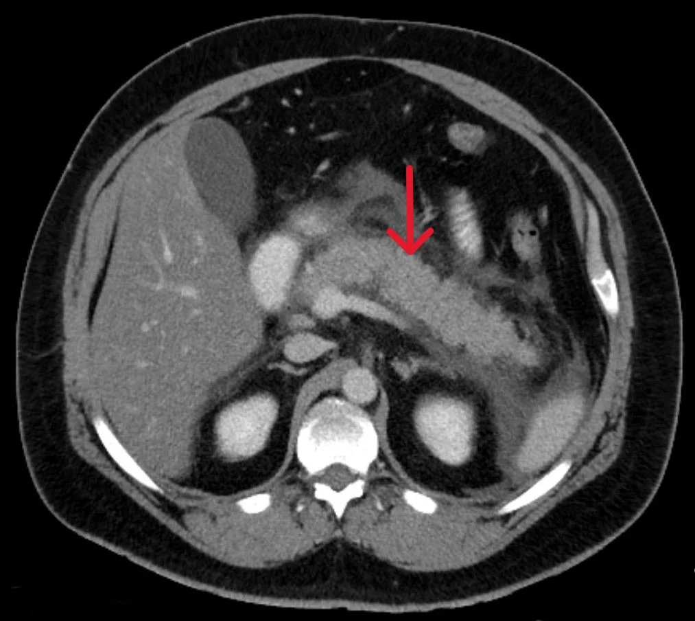
Long description
Axial contrast-enhanced CT through the upper abdomen. The body and tail of the pancreas are enlarged and poorly defined, and fluid tracks around the gland and in the surrounding fat. The red arrow points to the swollen pancreas.

- Abdominal ultrasound is the first imaging study when gallstone pancreatitis is suspected. It detects gallstones and common bile duct dilatation, and screens for other causes. Oedema of the pancreas may be seen but gas frequently obscures it. [4]
- CT with intravenous contrast is the diagnostic standard when the diagnosis is in doubt or to exclude other causes, and the staging scan of choice for necrosis and collections. Necrotic tissue does not enhance. It is usually deferred to 5 to 7 days after admission, unless there is diagnostic doubt or failure to improve within 48 to 72 hours: the ACG 2024 guideline reserves CT for patients who do not improve within 48 to 72 hours, while the Revised Atlanta consensus finds contrast-enhanced CT 5 to 7 days after admission more reliable for establishing the extent of pancreatic necrosis, because early CT underestimates necrosis. [2][1]
- Plain abdominal radiographs, if performed, may show calcified pancreatic duct stones, calcified gallstones, a sentinel loop in the left upper quadrant, or the colon cut-off sign (absence of air in the left colonic flexure or descending colon) in severe disease. [4]
- Chest radiographs may reveal atelectasis or pleural effusion. [4]
- MRCP is preferred to CT for detecting choledocholithiasis and pancreatic duct injury. It is used when liver tests, especially direct bilirubin and ALT, are abnormal and ultrasound does not exclude a common bile duct stone. Endoscopic ultrasound has comparable specificity for duct stones but MRCP is non-invasive. [4]
- ERCP is for therapy, not diagnosis: to relieve bile duct obstruction.
Approach to suspected acute pancreatitis
- Suspected acute pancreatitis: severe upper abdominal painNausea, vomiting, raised enzymes; risk factors for gallstones or heavy alcohol
- Diagnosis: 2 of pain, lipase or amylase over 3 times, imagingLabs on admission; ultrasound if gallstone cause likely
- Question: Diagnosis confirmed?
No
- Reconsider differentialsPerforated ulcer, mesenteric ischaemia, dissection, hepatitis, etc.
Yes
- Grade severity at the bedsideOrgan failure now, or within 24 h? SIRS? hypovolaemia?
- Start Ringer lactate 1.5 mL per kg per hourBolus 10 mL per kg if hypovolaemic; monitor response
- Question: Aetiology?
Gallstones likely
- Ultrasound all patientsRepeat if negative; MRCP if LFTs abnormal
- Cholecystectomy before discharge if mildERCP within 24 h only if cholangitis
Alcohol
- Counsel to stop; thiamine; assess dependencePlan for chronic pancreatitis
Triglycerides over 1000 mg per dL
- Insulin infusion; consider fibrates and plasma exchangeTreat the metabolic cause
No cause found
- Further work-up: EUS, MRCP, autoimmune and tumour screenLabel idiopathic only when negative
- Nutrition earlyOral low-fat diet as tolerated within 24 h; enteral tube if not oral
- AnalgesiaParenteral opioids and antiemetic
- Red flags?Persistent organ failure over 48 h, infected necrosis: ICU, step-up drainage
The diagnosis needs two of three features: [1]
- Pain consistent with acute pancreatitis: acute onset of persistent, severe epigastric pain, often radiating to the back.
- Serum lipase (or amylase) at least three times the upper limit of normal.
- Characteristic findings on contrast-enhanced CT, or less commonly MRI or ultrasound.
If pain is strongly suggestive but enzymes are under three times normal (as in late presentation), imaging is required to confirm. If diagnosis is made on pain and enzymes, CT on admission is usually not needed. [1]
Grading is based on organ failure and complications, not on how large the pancreas looks. [1]
- Mild: no organ failure, no local or systemic complications. Mortality is very rare; patients are usually discharged during the first week without pancreatic imaging.
- Moderately severe: transient organ failure that resolves within 48 hours, and/or local or systemic complications, without persistent organ failure.
- Severe: persistent organ failure lasting more than 48 hours (single or multiple).
About 20% of patients develop a complicated course. [3]
Organ failure is defined as a score of 2 or more in one system, using the modified Marshall score. [1] For a first-time reader the thresholds to remember are:
- Cardiovascular: systolic blood pressure under 90 mm Hg, not responsive to fluid.
- Renal: serum creatinine at least 1.9 mg per dL (about 170 micromol per L) with deterioration.
- Respiratory: a PaO2 to FiO2 ratio of 300 mm Hg or less.
A score of 2 or higher in any system indicates organ failure. Persistent organ failure beyond 48 hours defines severe disease. [1][4]
SIRS is defined by two or more of: [1][2][4]
- temperature above 38 C or below 36 C (Revised Atlanta 2012 and ACG 2024); some texts, including the Merck manual, use above 38.3 C or below 36.0 C;
- heart rate above 90 per minute;
- respiratory rate above 20 per minute or PaCO2 under 32 mm Hg; and
- white cell count above 12 000 per microlitre, below 4 000 per microlitre, or more than 10% band forms.
Persistent SIRS increases the risk of persistent organ failure; a patient with persistent SIRS is treated as potentially severe from the outset. [1]
Several scores are used to estimate risk; they are adjuncts to bedside assessment. The main ones are the BISAP score (calculated in the first 24 hours), Ranson criteria (five variables on admission and six more at 48 hours, from the original 1974 paper), APACHE II, the Harmless acute pancreatitis score, and imaging-based CT severity index (Balthazar). Severity scoring systems can delay management and most perform better at 48 hours than at admission. [4][7] No single score is clearly best; risk should be reassessed daily in the first week.
The old habit of keeping every patient fasting is not supported. Current guidance is:
- Resuscitate and monitor. Oxygen, pulse oximetry, strict fluid balance. Patients with multiple risk factors for severe disease should be managed in a monitored or intensive care setting. [2][3]
- Fluids: start with lactated Ringer solution at 1.5 mL per kg per hour. Give a 10 mL per kg bolus if the patient is hypovolaemic, unless contraindicated by cardiac or renal disease. Reassess frequently in the first 6 hours and for 24 to 48 hours, using heart rate, blood pressure, urine output, haematocrit and BUN. [4]
- Avoid aggressive boluses. The WATERFALL trial of aggressive versus moderate resuscitation showed that structured checkpoints and close monitoring matter more than giving large volumes. [2][3]
- Analgesia: adequate parenteral opioid (hydromorphone is often preferred to morphine because of the theoretical risk of sphincter of Oddi spasm), plus antiemetic. [4]
- Nutrition: the AGA guideline recommends early oral feeding within 24 hours as tolerated, rather than keeping the patient nil per os; use a low-residue, low-fat soft diet in mild disease. If oral feeding is not possible, enteral (nasogastric or nasojejunal) is preferred to parenteral, which carries more infectious complications. [5][4]
- Prophylactic antibiotics: not recommended in acute pancreatitis, regardless of severity. [2][5]
- Ultrasound in every patient to detect gallstones or common bile duct dilatation. A negative initial scan does not exclude a passed stone; repeat it or use MRCP. [2][4]
- ERCP: only for concurrent acute cholangitis, and within 24 hours. [2][4] Do not perform ERCP to fish for stones without cholangitis or rising bilirubin. [4]
- Cholecystectomy during the same admission for mild gallstone pancreatitis, before discharge, to prevent recurrence. [2][4][8] In moderate to severe disease with peripancreatic collections, defer cholecystectomy until inflammation and collections resolve. [8]
Start insulin infusion (especially in diabetic patients) and lipid-lowering therapy. For severe hypertriglyceridaemia above 1000 mg per dL (about 11.3 mmol per L) that does not respond to conservative therapy, therapeutic plasma exchange is considered. [8]
Counsel complete abstinence; offer thiamine, nutritional support and addiction services. Explain the risk of progression to chronic pancreatitis and of recurrent attacks. [4]
Prophylactic antibiotics are not used. They are started only for documented extrapancreatic infection (cholangitis, pneumonia, bloodstream or urinary infection) or for infected pancreatic necrosis. [4][5]
When infection is suspected, use antibiotics that penetrate pancreatic necrosis: carbapenems, fluoroquinolones combined with metronidazole. Infection is suspected when the patient deteriorates after 7 to 10 days, with fever, tachycardia, leucocytosis or organ failure. [4]
Drainage of infected necrosis uses a step-up approach: conservative medical care first, then percutaneous or endoscopic drainage, then minimally invasive retroperitoneal (VARD) or laparoscopic necrosectomy if needed; open surgery is reserved for failure. Drainage is delayed, preferably more than 4 weeks, in stable patients, to allow the collection to mature into walled-off necrosis. [4][8]
ICU care is needed for organ failure. Infected necrosis with persistent organ failure carries extremely high mortality; transfer to a centre with pancreatic expertise is appropriate. [3][8]
Survivors need follow-up because of the risks of recurrence, chronic pancreatitis and pancreatic cancer. One European guideline recommends follow-up ultrasound at 3 and 6 months and, for patients with a BISAP score of 2 or more or a CT severity index of 3 or more, a contrast CT at 1 to 3 months after discharge. [8] Exocrine insufficiency occurs in about 19% after mild disease and 33% after severe; endocrine (type 3c) diabetes develops in about 37% after a first attack. [8] Recurrence rates are about 10 to 30% in alcoholic disease; many go on to chronic pancreatitis. [4]
Local complications (Revised Atlanta definitions) [1]
- Acute peripancreatic fluid collection (APFC): homogeneous fluid, no wall, adjacent to the pancreas, within the first 4 weeks of interstitial pancreatitis. Most resolve.
- Pancreatic pseudocyst: encapsulated fluid collection with a well-defined wall, minimal necrotic material, maturation after more than 4 weeks.
- Acute necrotic collection (ANC): heterogeneous fluid and necrosis, no definable wall, within the first 4 weeks of necrotising pancreatitis.
- Walled-off necrosis (WON): mature encapsulated pancreatic or peripancreatic necrosis, usually after more than 4 weeks, heterogeneous with solid and liquid debris.
Other local complications include splenic and portal vein thrombosis, pseudoaneurysm (often splenic artery), gastric outlet obstruction, colonic ischaemia or necrosis, and abdominal compartment syndrome. Infected necrosis occurs in roughly one third of patients with necrosis and is rare in the first week. [1][4]
Acute single or multi-organ failure (respiratory, renal, cardiovascular), shock, acute respiratory distress syndrome, acute kidney injury, hypocalcaemia from fat necrosis (saponification), hyperglycaemia and diabetes, and hypovolaemic shock from third spacing. [4]
Mortality is rare in mild disease. In severe disease, mortality can be as high as 36 to 50% when persistent organ failure develops within the first few days. [1] The most severe necrotising forms complicated by infection carry mortality up to about 50%. [8] Mortality is bimodal: early from multi-organ failure, late from infected necrosis and iatrogenic complications.
- Pregnancy: gallstone pancreatitis can occur in any trimester but is most common in the third. Gallstone pancreatitis is rare in pregnancy and is managed with the same supportive care; definitive biliary treatment is planned with obstetric and surgical colleagues. [4]
- Children: pancreatitis suggests trauma, infections (mumps), hereditary or cystic fibrosis-related disease; genetic causes predominate.
- Older adults: atypical pain, higher operative risk, higher chance of a tumour cause over 40.
- Hypertriglyceridaemia: see above; recurrence risk is high without lipid control.
- Immunocompromised and transplant patients: atypical presentation; prophylactic cholecystectomy may be prudent in known gallstone disease.
| Feature | Acute pancreatitis | Chronic pancreatitis |
|---|---|---|
| Onset | Sudden; recurrent attacks possible | Insidious, progressive over years |
| Pain | Severe epigastric, radiating to back | Recurrent or continuous epigastric pain |
| Morphology | Inflammation and oedema or necrosis; gland can return to normal | Fibrosis, atrophy, duct stones and strictures, calcification |
| Exocrine function | Usually preserved | Profound insufficiency; steatorrhoea |
| Endocrine function | Transient hyperglycaemia | Diabetes (type 3c) common |
| Calcification | Absent (unless pre-existing chronic) | Pancreatic calculi on imaging |
| Lipase/amylase | Raised, usually over 3 times normal | Normal or mildly raised |
| Treatment | Supportive, fluids, feeding, drainage | Analgesia, enzyme replacement, alcohol and smoking cessation, surgery for complications and chronic pain |
| Progression | Can precipitate chronic pancreatitis, especially with alcohol and smoking | Progressive and irreversible |
- Clinical diagnosis plus inexpensive plasma amylase or lipase and ultrasound will handle most cases. A urine trypsinogen-2 dipstick is a cheap bedside test where available. [4]
- Lactated Ringer solution, oxygen, nasogastric feeding and parenteral opioids are the core; they are affordable and save lives.
- Reserve CT for diagnostic doubt or clinical deterioration at a week, not for every admission. Early CT adds cost without benefit.
- Infected necrosis and persistent organ failure need a centre with ICU and drainage capability; refer early rather than waiting.
- Alcohol counselling, thiamine and lipid management are cheap prevention. Smoking cessation after a first attack reduces recurrence and chronic pancreatitis.
- In endemic regions, ask about scorpion sting and parasites as causes.
- ACG 2024 supports routine ultrasound, imaging only when diagnosis is unclear or the patient does not improve by 48 to 72 hours, moderate fluid resuscitation, early enteral nutrition, no prophylactic antibiotics, and early cholecystectomy for mild biliary disease. [2][3]
- AGA 2018 supports goal-directed fluids without recommending saline versus Ringer, early oral feeding within 24 hours, enteral over parenteral nutrition, and against prophylactic antibiotics and routine urgent ERCP without cholangitis. [5]
- IAP/APA 2013 underpins the Revised Atlanta terminology, delayed drainage of necrosis and the early feeding strategy. [6]
- All agree: severity is better defined by organ failure and complications than by imaging, and persistent organ failure beyond 48 hours is the marker of severe disease. [1][4]
- Pancreatic enzymes activate inside the gland and digest it; inflammatory signals then drive remote organ failure.
- Gallstones and alcohol together account for about two thirds to four fifths of cases.
- Hypertriglyceridaemia becomes a cause when fasting levels are above 1000 mg per dL.
- Drugs, post-ERCP pancreatitis and infections are less common; exclude a tumour in anyone over 40 with no clear cause.
- Diagnosis needs 2 of pain, enzymes over 3 times normal, or characteristic imaging; lipase is more specific than amylase.
- Severity is graded mild, moderately severe, severe by organ failure and complications; persistent organ failure over 48 hours is severe.
- Initial treatment: lactated Ringer at 1.5 mL per kg per hour, adequate opioids, early feeding within 24 hours, no prophylactic antibiotics.
- ERCP within 24 hours only with acute cholangitis; cholecystectomy before discharge for mild gallstone pancreatitis.
- Infected necrosis gets antibiotics that penetrate necrosis and step-up drainage, preferably after 4 weeks.
- Mortality can reach 36 to 50% with early persistent organ failure; milder disease is rarely fatal.
Answer each one in your head first, then open the answer.
A 48-year-old man presents with sudden severe epigastric pain radiating to the back, amylase 1800 U per L and lipase 2400 U per L. Ultrasound shows gallstones. What is the most likely diagnosis and the first imaging and management steps?
Show answerHide answer
Acute gallstone pancreatitis. First step: confirm hypovolaemia and start lactated Ringer at 1.5 mL per kg per hour with reassessment. Ultrasound has already identified the cause; CT is not needed now. Plan cholecystectomy during the same admission if the course is mild, and perform ERCP within 24 hours only if there is acute cholangitis.
List the components of the Revised Atlanta diagnostic triad and the severity classes, and say what distinguishes moderate from severe disease.
Show answerHide answer
Diagnosis needs two of: typical epigastric pain radiating to the back; lipase or amylase at least three times the upper limit of normal; and characteristic imaging. Severity: mild has no organ failure or complications; moderately severe has transient organ failure under 48 hours or local or systemic complications without persistent organ failure; severe has persistent organ failure over 48 hours.
Why is early aggressive fluid therapy no longer recommended, and what is the evidence-based alternative?
Show answerHide answer
Goal-directed moderate resuscitation with lactated Ringer at 1.5 mL per kg per hour, a 10 mL per kg bolus if hypovolaemic, and structured checkpoints, replaces aggressive bolus therapy. The WATERFALL trial showed that close monitoring and moderate fluids matter more than giving large volumes. Avoid aggressive therapy especially in older or cardiac patients. [4]
A patient with acute pancreatitis deteriorates on day 9 with fever, leucocytosis and rising CRP. What complication is suspected and how is it managed?
Show answerHide answer
Infected pancreatic necrosis or an infected collection. Start antibiotics that penetrate necrosis (carbapenem, or fluoroquinolone plus metronidazole). Obtain contrast CT to look for gas in the collection. Plan step-up drainage, preferably delayed beyond 4 weeks, starting with percutaneous or endoscopic drainage and escalating as needed.
A patient has a triglyceride level of 1400 mg per dL during an attack of pancreatitis. What is the likely cause and treatment?
Show answerHide answer
Hypertriglyceridaemic pancreatitis, a recognised cause when levels are above 1000 mg per dL. Treat with insulin infusion and lipid-lowering therapy; consider therapeutic plasma exchange for severe, refractory hypertriglyceridaemia.
When should CT be performed in acute pancreatitis, and when should ERCP be performed?
Show answerHide answer
CT only when the diagnosis is uncertain, for another cause of pain, or when there is failure to improve at 48 to 72 hours or a suspected complication. CT for necrosis is more reliable at 5 to 7 days. ERCP only for acute cholangitis, within 24 hours.
Pancreatitis overview; chronic pancreatitis; cholelithiasis and biliary colic; choledocholithiasis and cholangitis; biliary colic; gallbladder disease; abdominal pain approach.
(References are listed in the front matter; each is cited in the text by number.)
- Banks PA, Bollen TL, Dervenis C, et al. Classification of acute pancreatitis—2012: revision of the Atlanta classification and definitions by international consensus. Gut. 2013;62(1):102–111. doi:10.1136/gutjnl-2012-302779. Accessed .
- Tenner S, Vege SS, Sheth SG, et al. American College of Gastroenterology Guidelines: Management of Acute Pancreatitis. Am J Gastroenterol. 2024;119(3):419–437. doi:10.14309/ajg.0000000000002645. Accessed .
- Marino D, Saini J, Tenner S. Management of Acute Pancreatitis. Am J Gastroenterol. 2025;120(1):2–4. doi:10.14309/ajg.0000000000003180. Accessed .
- Bartel M (reviewed by Nguyen M). Acute Pancreatitis. Merck Manual, Professional Version. Full review Aug 2026. Accessed .
- Crockett SD, Wani S, Gardner TB, et al. American Gastroenterological Association Institute Guideline on Initial Management of Acute Pancreatitis. Gastroenterology. 2018;154(4):1096–1101. doi:10.1053/j.gastro.2018.01.032. Accessed .
- Working Group IAP/APA Acute Pancreatitis Guidelines. IAP/APA Evidence-Based Guidelines for the Management of Acute Pancreatitis. Pancreatology. 2013;13(4 suppl 2):e1–15. doi:10.1016/j.pan.2013.07.063. Accessed .
- Ranson JH, Rifkind KM, Roses DF, et al. Prognostic signs and the role of operative management in acute pancreatitis. Surg Gynecol Obstet. 1974;139(1):69–81. Accessed .
- Szeliga J, Jagielski M, Sobocki J, et al. Diagnostic and therapeutic management of severe acute pancreatitis. Evidence based medicine (EBM) clinical practice guidelines. Videosurgery Miniinv. 2025;20(1):1–29. doi:10.20452/wiitm.2025.17941. Accessed .
Figure credits
Educational content for medical students and doctors. It is not a substitute for clinical judgement or local guidelines.